



Nevadensin

Based on 1 Customer Validation
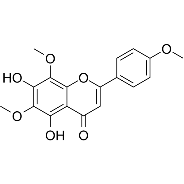
Nevadensin (Lysionotin), a natural flavonoid, is a selective human carboxylesterase 1 (hCE1) inhibitor with an IC50 of 2.64 μM. Nevadensin is more selective for hCE1 than hCE2 (IC50 of 132.8 μM). Nevadensin can induce apoptosis and DNA damage in cancer cells. Nevadensin has a variety of pharmacological effects such as anti-inflammatory, anti-tumour, anti-hypertensive, anti-tubercular, antitussive, antioxidant and anti-microbial activities.
Nur für Forschungszwecke. Wir verkaufen nicht an Patienten.
- Reinheit : 99.93%
- CAS. Nr.: 10176-66-6
- Formel: C18H16O7
- Molecular Weight:344.32
-
Speicherung:
4°C, protect from light
* In solvent : -80°C, 6 months; -20°C, 1 month (protect from light)

To place orders, for customer services and technical support, please contact: MedChemExpress USA
Tel: 609-228-6898 E-mail: [email protected] [email protected]


-
 Biologische Aktivität
-
Chemical Information
-
Lösungsmittel & Löslichkeit
- Protokoll
- Reinheit & Dokumentation
- Verweise
-
 Help & FAQs


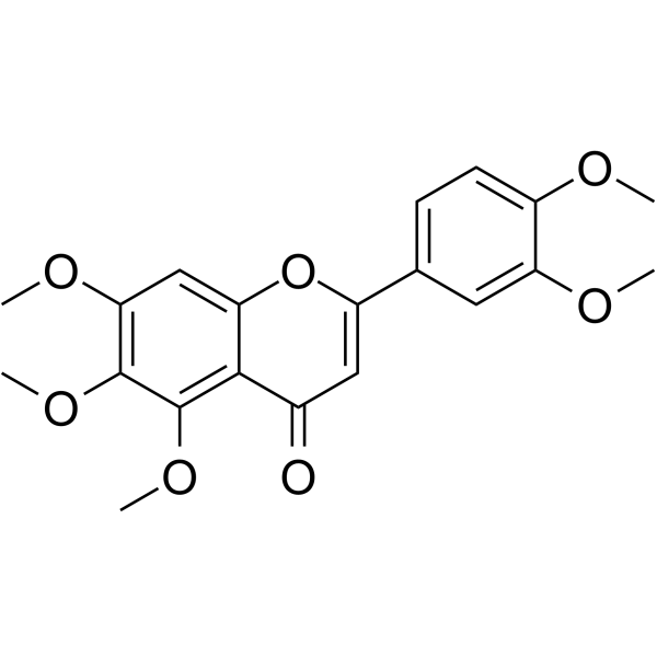

Alle DNA/RNA Synthesis Isoform-spezifische Produkte anzeigen
More

Biologische Aktivität
Beschreibung
IC50 & Target
IC50: 2.64 μM (hCE1), 132.8 μM (hCE2)[1]
Cellular Effect
|
Cell Line

|
Type | Value | Description | References |
|---|---|---|---|---|
| HCT-116 | IC50 |
5 μM
Compound: 61; Lys
|
Antiproliferative activity against human HCT-116 cells
Antiproliferative activity against human HCT-116 cells
|
[PMID: 38142509] |
| RAW264.7 | IC50 |
5.08 μM
Compound: 6
|
Inhibition of NO production in LPS-stimulated mouse RAW264.7 cells pre-incubated for 2 hrs before LPS stimulation for 24 hrs by Griess assay method
Inhibition of NO production in LPS-stimulated mouse RAW264.7 cells pre-incubated for 2 hrs before LPS stimulation for 24 hrs by Griess assay method
|
[PMID: 25176187] |
| SW480 | IC50 |
5 μM
Compound: 61; Lys
|
Antiproliferative activity against human SW480 cells
Antiproliferative activity against human SW480 cells
|
[PMID: 38142509] |
In Vitro
Nevadensin exhibits inhibition activity against Mycobacterium tuberculosis, with a MIC value of 200 μg/mL. Nevadensin exhibits antioxidant activity against the radical scavenging ability of DPPH, with the IC50 value of 7.7 μg/mL[2].
Nevadensin potently inhibits p40 protein tyrosine kinase activity with an IC50 value of 50 μg/mL[3].
Nevadensin shows DNA-intercalating properties, with IC50s of 31.63 µM and 296.91 µM for minor groove binding and DNA intercalation[3].
Nevadensin (500 μM; 1 h) in human colon carcinoma cell line HT29 cells, significantly increases the amount of topoisomerase I (TOPO I) bound to DNA, but has no stabilizing effect on the TOPO IIα/DNA complex[3].
Nevadensin (1-500 μM; 24-48 h) in HT29 cells, causes a slight but significant reduction in cell viability of about 20% at concentrations ≥250 μM after 24 h, and a significant decrease in cell viability but still higher than 50% after 48 h[3].
Nevadensin (1-500 μM; 24 h) in HT29 cells, increases the number of cells in the G2/M phase at concentrations ≥100 μM[3].
Nevadensin (50-500 μM; 24 h) in HT29 cells, increases the activities of caspase-3 and caspase-9 in a concentration-dependent manner and has no effect on the activity of caspase-8[3].
Nevadensin (12.5-50 µM; for 24 h) significantly inhibits growth of hepatocellular carcinoma (HCC) cells via inducing cell cycle arrest and apoptosis. Nevadensin regulates multiple functional signaling pathways associated with cancer including Hippo signaling. Nevadensin notably induces activation of the MST1/2-LATS1/2 kinase in HCC cells[4].
Nevadensin (1-10 μg/mL) inhibits the release of β-hexosaminidase and histamine in bone marrow-derived mast cells[5].
Nevadensin (2-20 μg/mL; 3 days) in BMMCs (bone marrow-derived mast cells) inhibits cell proliferation, decreases the expression of c-Kit and accelerates their apoptosis[5].
MedChemExpress (MCE) has not independently confirmed the accuracy of these methods. They are for reference only.
-
Cell Line:HT29 cells
-
Concentration:1 μM, 10 μM, 100 μM, 250 μM, 500 μM
-
Incubation Time:24 h, 48 h
-
Result:After 24 h, at concentrations ≥250 μM, cell viability was slightly but significantly reduced by about 20%.
-
Cell Line:HT29 cells
-
Concentration:1 μM, 10 μM, 100 μM, 250 μM, 500 μM
-
Incubation Time:24 h
-
Result:At concentrations ≥100 μM, the number of cells in the G2/M phase increased.
-
Cell Line:HT29 cells
-
Concentration:50 μM, 100 μM, 250 μM, 500 μM
-
Incubation Time:24 h
-
Result:Increased the levels of apoptosis-related proteins such as Cleaved Caspase 3, Cleaved-PARP, and Cleaved Caspase 9 in a concentration-dependent manner.
In Vivo
MedChemExpress (MCE) has not independently confirmed the accuracy of these methods. They are for reference only.
-
Animal Model:Female BALB/c mice (6-8 weeks) were sensitized through intraperitoneal injection of 100 μg of ovalbumin and 2 mg of alum in 200 μL of PBS on days 0 and 14, and then orally challenged with 50 mg of ovalbumin in 200 μL of PBS every 3 days from day 28 for a total of five challenge events[5].
-
Dosage:1, 5 or 10 mg/kg
-
Administration:Oral gavage, once a day, from day 27 to day 40
-
Result:Significantly attenuated oral ovalbumin-challenge-induced food allergy, as assessed by improvements in hypothermia, diarrhea, and anaphylaxis score.
Also led to significantly decreased levels of OVA-specific IgE, histamine, and mMCP-1 in the serum.


Chemical Information
-
CAS. Nr. 10176-66-6
-
Appearance Solid
-
Molecular Weight 344.32
-
Formel C18H16O7
-
Color Light yellow to yellow
-
SMILES
O=C1C=C(C2=CC=C(OC)C=C2)OC3=C(OC)C(O)=C(OC)C(O)=C13
-
Synonyms
Lysionotin
-
Structure Classification
-
Initial Source
-
Versand
Room temperature in continental US; may vary elsewhere.
-
Speicherung
4°C, protect from light
* In solvent : -80°C, 6 months; -20°C, 1 month (protect from light)


Lösungsmittel & Löslichkeit
In Vitro:
DMSO : 14.71 mg/mL (42.72 mM; Need ultrasonic; Hygroscopic DMSO has a significant impact on the solubility of product, please use newly opened DMSO)
Please refer to the solubility information to select the appropriate solvent. Once prepared, please aliquot and store the solution to prevent product inactivation from repeated freeze-thaw cycles.
Storage method and period of stock solution: -80°C, 6 months; -20°C, 1 month (protect from light). When stored at -80°C, please use it within 6 months. When stored at -20°C, please use it within 1 month.
Please refer to the solubility information to select the appropriate solvent. Once prepared, please aliquot and store the solution to prevent product inactivation from repeated freeze-thaw cycles.
Storage method and period of stock solution: -80°C, 6 months; -20°C, 1 month (protect from light). When stored at -80°C, please use it within 6 months. When stored at -20°C, please use it within 1 month.
Konzentration (Stammlösung) × Volumen (Stammlösung) = Konzentration (Ziellösung) × Volumen (Ziellösung)
In Vivo:
For the following dissolution methods, please prepare the working solution directly:
It is recommended to prepare fresh solutions and use them promptly within a short period of time.
The percentages shown for the solvents indicate their volumetric ratio in the final prepared solution. If precipitation or phase separation occurs during preparation, heat and/or sonication can be used to aid dissolution.
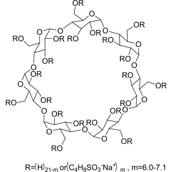
Add each solvent one by one: 0.5% CMC-Na/saline water
Solubility: 2.5 mg/mL (7.26 mM); Suspended solution; Need ultrasonic


Reinheit & Dokumentation
-
Data Sheet (281 KB)
-
SDS (394 KB)
- English - EN (394 KB)
- Français - FR (394 KB)
- Deutsch - DE (394 KB)
- Norwegian - NO (394 KB)
- Español - ES (394 KB)
- Swedish - SV (394 KB)
- Italian - IT (394 KB)
- Korean - KR (394 KB)
- Portuguese - PT (394 KB)
-
Handling Instructions (2659 KB)
Verweise
[1]. Ya-Qiao Wang, et al. Nevadensin is a naturally occurring selective inhibitor of human carboxylesterase 1. Int J Biol Macromol. 2018 Dec;120(Pt B):1944-1954. [Content Brief]
[2]. Suksamrarn A, et al. Antimycobacterial and antioxidant flavones from Limnophila geoffrayi. Arch Pharm Res. 2003 Oct;26(10):816-20. [Content Brief]
[3]. Müller L, et al. Topoisomerase poisoning by the flavonoid nevadensin triggers DNA damage and apoptosis in human colon carcinoma HT29 cells. Arch Toxicol. 2021 Dec;95(12):3787-3802. [Content Brief]
[4]. Shi H, et al. Activating the Hippo pathway by nevadensin overcomes Yap-drived resistance to sorafenib in hepatocellular carcinoma. Discov Oncol. 2023 May 27;14(1):83. [Content Brief]
[5]. Zhang YF , et al. Nevadensin relieves food allergic responses and passive cutaneous anaphylaxis in mice through inhibiting the expression of c-Kit receptors. Food Funct. 2020 Dec 1;11(12):10375-10385. [Content Brief]


Complete Stock Solution Preparation Table
Please refer to the solubility information to select the appropriate solvent. Once prepared, please aliquot and store the solution to prevent product inactivation from repeated freeze-thaw cycles.
Storage method and period of stock solution: -80°C, 6 months; -20°C, 1 month (protect from light). When stored at -80°C, please use it within 6 months. When stored at -20°C, please use it within 1 month.
| Optional Solvent | Concentration Solvent Mass | 1 mg | 5 mg | 10 mg | 25 mg |
|---|---|---|---|---|---|
| DMSO | 1 mM | 2.9043 mL | 14.5214 mL | 29.0428 mL | 72.6069 mL |
| 5 mM | 0.5809 mL | 2.9043 mL | 5.8086 mL | 14.5214 mL | |
| 10 mM | 0.2904 mL | 1.4521 mL | 2.9043 mL | 7.2607 mL | |
| 15 mM | 0.1936 mL | 0.9681 mL | 1.9362 mL | 4.8405 mL | |
| 20 mM | 0.1452 mL | 0.7261 mL | 1.4521 mL | 3.6303 mL | |
| 25 mM | 0.1162 mL | 0.5809 mL | 1.1617 mL | 2.9043 mL | |
| 30 mM | 0.0968 mL | 0.4840 mL | 0.9681 mL | 2.4202 mL | |
| 40 mM | 0.0726 mL | 0.3630 mL | 0.7261 mL | 1.8152 mL |


Nevadensin Related Classifications
HY-N1377 Related Classifications
 Powered by Bioz