



Polmacoxib

Based on 1 Customer Validation
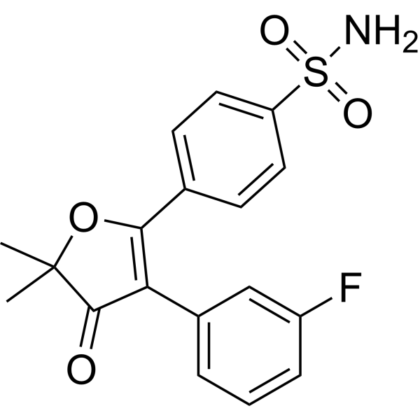
Polmacoxib (CG100649) is a first-in-class, orally active nonsteroidal anti-inflammatory agent (NSAID) which is a dual inhibitor of COX-2 (IC50 around 0.1 μg/ml) and carbonic anhydrase. Polmacoxib inhibits colorectal adenoma and tumor growth in mouse models.
For research use only. We do not sell to patients.
- Purity : 99.70%
- CAS No.: 301692-76-2
- Formula: C18H16FNO4S
- Molecular Weight:361.39
-
Storage:Powder -20°C, 3 years , 4°C, 2 years ; In solvent -80°C, 6 months , -20°C, 1 month

To place orders, for customer services and technical support, please contact: MedChemExpress USA
Tel: 609-228-6898 E-mail: [email protected] [email protected]


-
 Biological Activity
-
Chemical Information
-
Solvent & Solubility
- Protocols
- Purity & Documentation
- References
-
 Help & FAQs


-

HY-16726R
Polmacoxib (Standard)COX-2/Carbonic Anhydrase Inhibitor -
HY-13568
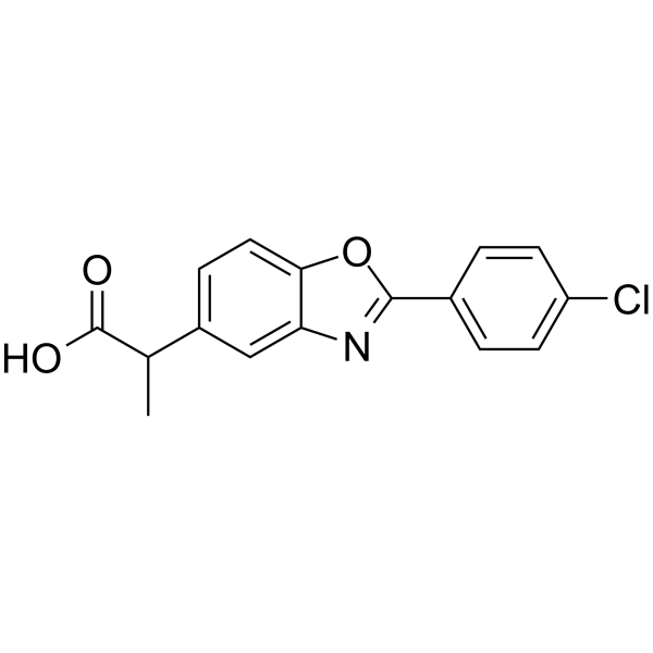
BenoxaprofenNonsteroidal Anti-inflammatory Agent -
HY-13513
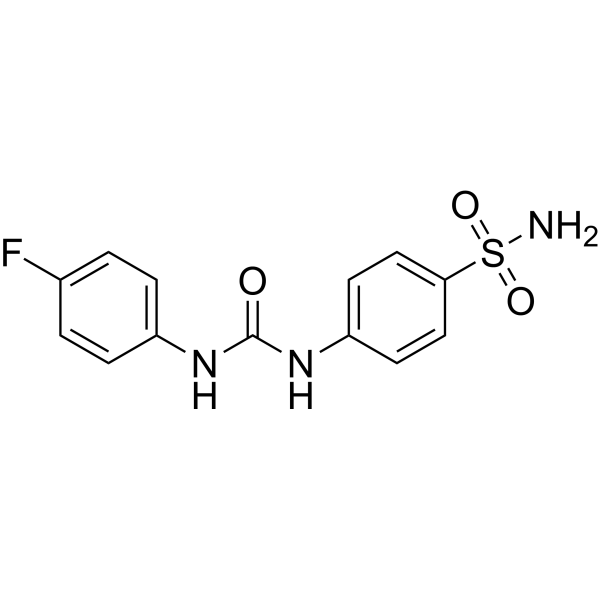
U-104CA IX/XII Inhibitor -
HY-76938
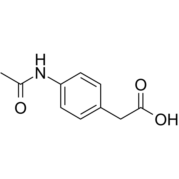
ActaritCAII Inhibitor -
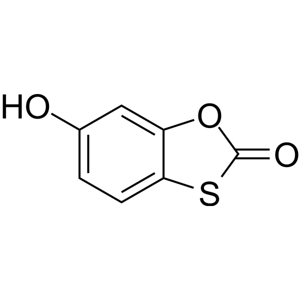
HY-B0483
TioxoloneCarbonic Anhydrase Inhibitor -
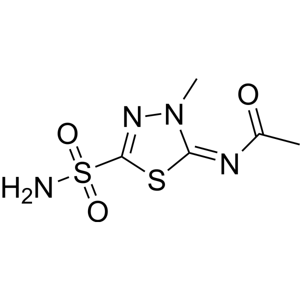
HY-B0553
MethazolamideCarbonic Anhydrase Inhibitor -
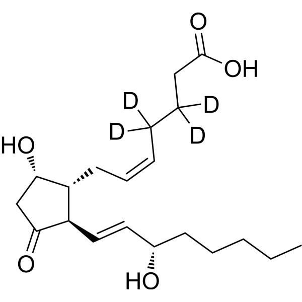
HY-101988S
Prostaglandin D2-d4Stable Isotope -

HY-131495
(S)-6-O-DesmethylnaproxenMetabolite -
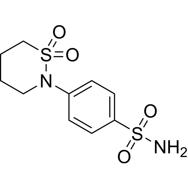
HY-108316
SultiameCarbonic Anhydrase Inhibitor -
HY-B0397
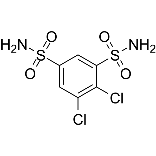
DichlorphenamideCarbonic Anhydrase Inhibitor -
HY-110243
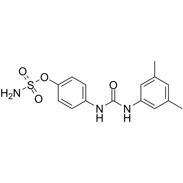
CAIX Inhibitor S4CA IX/XII Inhibitor -
HY-101087
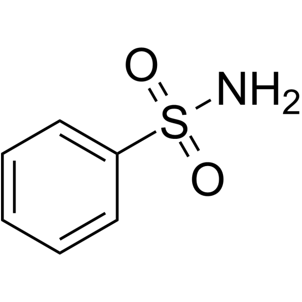
BenzenesulphonamideCA Inhibitor


Biological Activity
Description
IC50 & Target
[1]|
COX-2 0.1 μg/mL (IC50) |
carbonic anhydrase |
In Vitro
Polmacoxib (CG100649) (0-1 μg/ml; 24 hours; HCA-7 and HT-29 cells) can inhibit COX-2 activity and PGE2 production in human colon cancer cells, at lower concentrations compared to Celecoxib[2].
MedChemExpress (MCE) has not independently confirmed the accuracy of these methods. They are for reference only.
In Vivo
Polmacoxib (7-15 mg/kg; p.o.; daily from day 27 post-injection to day 111; athymic nude mice; subcutaneous xenograft mouse model) reduces tumor volume and tumor weight by 58% and 48%, respectively, compared to a 48% and 36% reduction following treatment with Celecoxib[2].
Polmacoxib (7-15 mg/kg; p.o.; began on day 14 and continued for 8 weeks; athymic nu/nu mice; orthotopic xenograft mouse model) inhibits CRC growth in an orthotopic xenograft mouse model, reducing tumor weight by 70% using 7 mg/kg or by 83% using 15 mg/kg, compared to a similar 70% reduction following treatment with 500 mg/kg of Celecoxib[2].
MedChemExpress (MCE) has not independently confirmed the accuracy of these methods. They are for reference only.
Clinical Trial
| NCT Number | Sponsor | Condition | Start Date |
Phase

|
|---|---|---|---|---|
| NCT01329991 | 2011-05 | PHASE1 |


Chemical Information
-
CAS No. 301692-76-2
-
Appearance Solid
-
Molecular Weight 361.39
-
Formula C18H16FNO4S
-
Color Light yellow to green yellow
-
SMILES
O=S(C1=CC=C(C(OC2(C)C)=C(C3=CC=CC(F)=C3)C2=O)C=C1)(N)=O
-
Synonyms
CG100649
-
Shipping
Room temperature in continental US; may vary elsewhere.
-
Storage
Powder -20°C 3 years 4°C 2 years In solvent -80°C 6 months -20°C 1 month


Solvent & Solubility
In Vitro:
DMSO : 250 mg/mL (691.77 mM; Need ultrasonic; Hygroscopic DMSO has a significant impact on the solubility of product, please use newly opened DMSO)
Please refer to the solubility information to select the appropriate solvent. Once prepared, please aliquot and store the solution to prevent product inactivation from repeated freeze-thaw cycles.
Storage method and period of stock solution: -80°C, 6 months; -20°C, 1 month. When stored at -80°C, please use it within 6 months. When stored at -20°C, please use it within 1 month.
Please refer to the solubility information to select the appropriate solvent. Once prepared, please aliquot and store the solution to prevent product inactivation from repeated freeze-thaw cycles.
Storage method and period of stock solution: -80°C, 6 months; -20°C, 1 month. When stored at -80°C, please use it within 6 months. When stored at -20°C, please use it within 1 month.
Concentration (start) × Volume (start) = Concentration (final) × Volume (final)
In Vivo:
Select the appropriate dissolution method based on your experimental animal and administration route.
- For the following dissolution methods, please ensure to first prepare a clear stock solution using an In Vitro approach and then sequentially add co-solvents:
- To ensure reliable experimental results, the clarified stock solution can be appropriately stored based on storage conditions. As for the working solution for In Vivo experiments, it is recommended to prepare freshly and use it on the same day.
- The percentages shown for the solvents indicate their volumetric ratio in the final prepared solution. If precipitation or phase separation occurs during preparation, heat and/or sonication can be used to aid dissolution.
Add each solvent one by one: 10% DMSO 90% (20% SBE-β-CD in Saline)
Solubility: ≥ 2.08 mg/mL (5.76 mM); Clear solution
This protocol yields a clear solution of ≥ 2.08 mg/mL (saturation unknown).
Taking 1 mL working solution as an example, add 100 μL DMSO stock solution (20.8 mg/mL) to 900 μL 20% SBE-β-CD in Saline, and mix evenly.
Preparation of 20% SBE-β-CD in Saline (4°C, storage for one week): 2 g SBE-β-CD powder is dissolved in 10 mL Saline, completely dissolve until clear.
In Vivo Dissolution Calculator
Please enter the basic information of animal experiments:
-
-
-
-
Recommended: Prepare an additional quantity of animals to account for potential losses during experiments.
Please enter your animal formula composition:
-
%DMSO +
Recommended: Keep the proportion of DMSO in working solution below 2% if your animal is weak.
-
%+
-
+%Tween-80 + +
-
%Saline +
The co-solvents required include: DMSO, . All of co-solvents are available by MedChemExpress (MCE). , Tween 80. All of co-solvents are available by MedChemExpress (MCE).
Working solution concentration: 0.22 mg/mL
Method for preparing stock solution: mg drug dissolved in μL DMSO. Stock solution concentration: mg/mL.
1. Take μL DMSO stock solution;
2. Add μL .
μL , mix evenly;
3. Then add μL Tween 80, mix evenly;
4. Then add μL
Please ensure that the stock solution in the first step is dissolved to a clear state, and add co-solvents in sequence. You can use ultrasonic heating (ultrasonic cleaner, recommended frequency 20-40 kHz), vortexing, etc. to assist dissolution.


Purity & Documentation
-
Data Sheet (282 KB)
-
SDS (393 KB)
- English - EN (393 KB)
- Français - FR (393 KB)
- Deutsch - DE (393 KB)
- Norwegian - NO (393 KB)
- Español - ES (393 KB)
- Swedish - SV (393 KB)
- Italian - IT (393 KB)
- Korean - KR (393 KB)
- Portuguese - PT (393 KB)
-
Handling Instructions (2659 KB)
References
[1]. Kim SH, et al. CG100649, a novel COX-2 inhibitor, inhibits colorectal adenoma and carcinoma growth in mouse models. Invest New Drugs. 2014;32(6):1105-1112. [Content Brief]
[2]. Flick AC, et al. Synthetic Approaches to the New Drugs Approved During 2015 [published correction appears in J Med Chem. 2017 Oct 26;60(20):8680]. J Med Chem. 2017;60(15):6480-6515. [Content Brief]


Complete Stock Solution Preparation Table
Please refer to the solubility information to select the appropriate solvent. Once prepared, please aliquot and store the solution to prevent product inactivation from repeated freeze-thaw cycles.
Storage method and period of stock solution: -80°C, 6 months; -20°C, 1 month. When stored at -80°C, please use it within 6 months. When stored at -20°C, please use it within 1 month.
| Optional Solvent | Concentration Solvent Mass | 1 mg | 5 mg | 10 mg | 25 mg |
|---|---|---|---|---|---|
| DMSO | 1 mM | 2.7671 mL | 13.8355 mL | 27.6709 mL | 69.1773 mL |
| 5 mM | 0.5534 mL | 2.7671 mL | 5.5342 mL | 13.8355 mL | |
| 10 mM | 0.2767 mL | 1.3835 mL | 2.7671 mL | 6.9177 mL | |
| 15 mM | 0.1845 mL | 0.9224 mL | 1.8447 mL | 4.6118 mL | |
| 20 mM | 0.1384 mL | 0.6918 mL | 1.3835 mL | 3.4589 mL | |
| 25 mM | 0.1107 mL | 0.5534 mL | 1.1068 mL | 2.7671 mL | |
| 30 mM | 0.0922 mL | 0.4612 mL | 0.9224 mL | 2.3059 mL | |
| 40 mM | 0.0692 mL | 0.3459 mL | 0.6918 mL | 1.7294 mL | |
| 50 mM | 0.0553 mL | 0.2767 mL | 0.5534 mL | 1.3835 mL | |
| 60 mM | 0.0461 mL | 0.2306 mL | 0.4612 mL | 1.1530 mL | |
| 80 mM | 0.0346 mL | 0.1729 mL | 0.3459 mL | 0.8647 mL | |
| 100 mM | 0.0277 mL | 0.1384 mL | 0.2767 mL | 0.6918 mL |


Polmacoxib Related Classifications
HY-16726 Related Classifications
 Powered by Bioz