



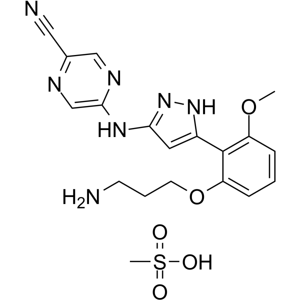
Prexasertib mesylate
Based on 39 publication(s) in Google Scholar
Prexasertib mesylate (LY2606368 mesylate) is a selective, ATP-competitive second-generation checkpoint kinase 1 (CHK1) inhibitor with a Ki of 0.9 nM and an IC50 of <1 nM. Prexasertib mesylate inhibits CHK2 (IC50=8 nM) and RSK1 (IC50=9 nM). Prexasertib mesylate causes double-stranded DNA breakage and replication catastrophe resulting in apoptosis. Prexasertib mesylate shows potent anti-tumor activity.
For research use only. We do not sell to patients.
- CAS No.: 1234015-55-4
- Formula: C19H23N7O5S
- Molecular Weight:461.49
-
Storage:
Please store the product under the recommended conditions in the Certificate of Analysis.

To place orders, for customer services and technical support, please contact: MedChemExpress USA
Tel: 609-228-6898 E-mail: [email protected] [email protected]


-
 Biological Activity
-
Chemical Information
- Protocols
- Purity & Documentation
- References
-
 Help & FAQs


-
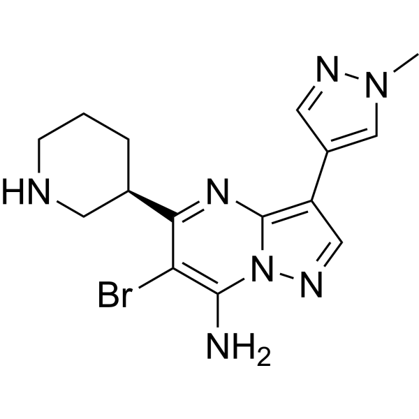
HY-15532
SCH900776CHK1 Inhibitor -
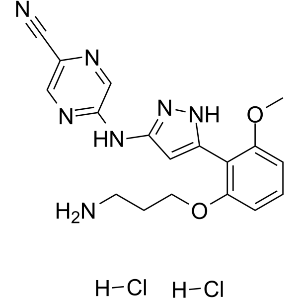
HY-18174A
Prexasertib dihydrochlorideCHK1 Inhibitor -
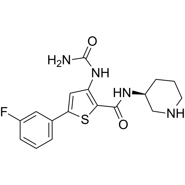
HY-10992
AZD-7762Chk Inhibitor -
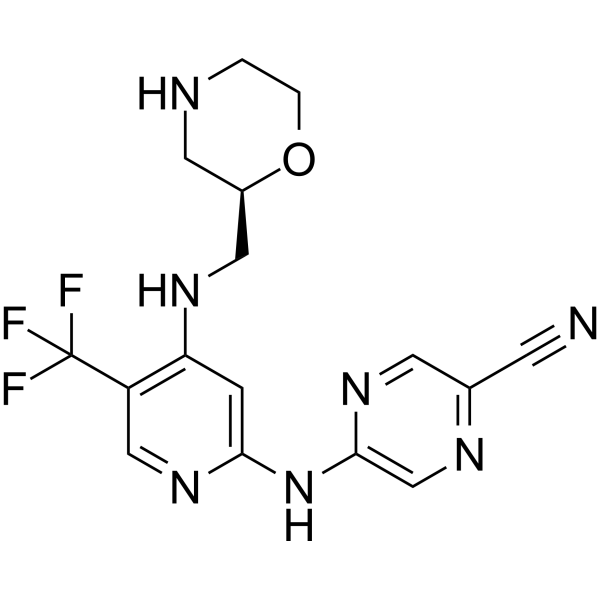
HY-18958
CCT245737CHK1 Inhibitor -
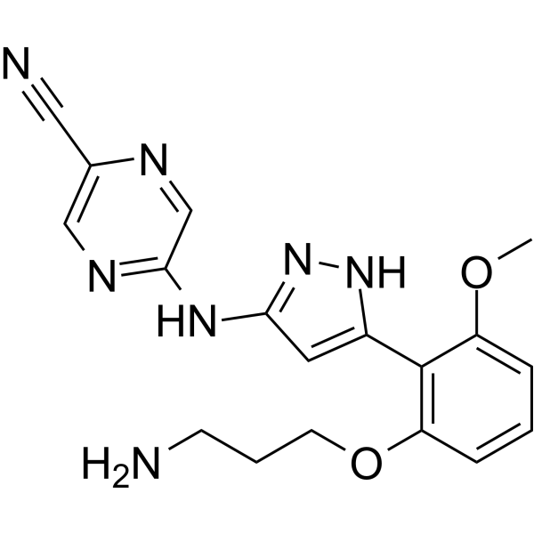
HY-18174
PrexasertibCHK1 Inhibitor -
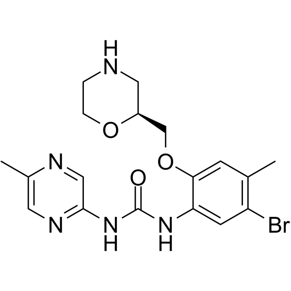
HY-14720
RabusertibCHK1 Inhibitor -
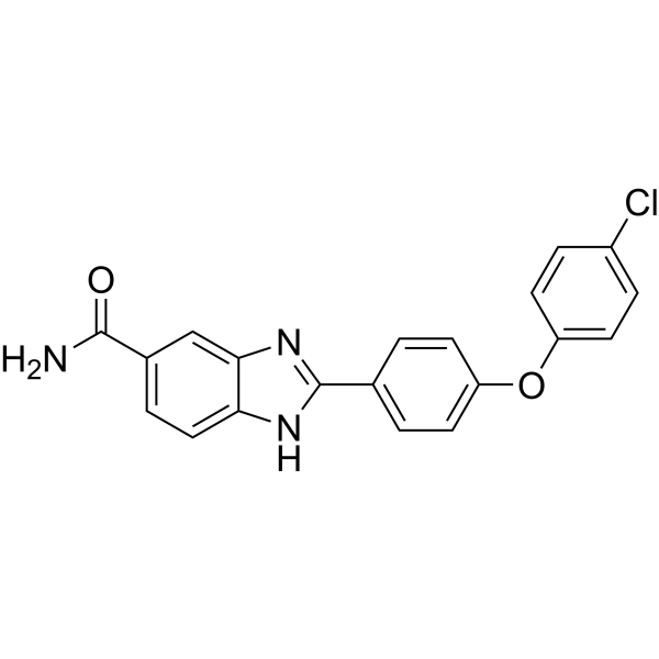
HY-13946
BML-277 -
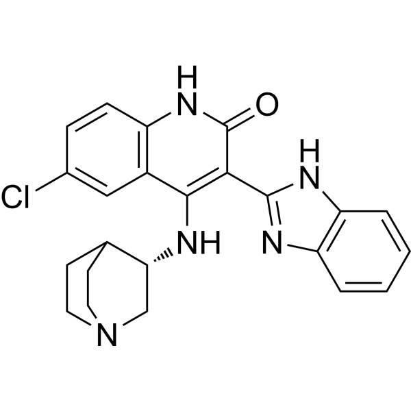
HY-13263
CHIR-124Checkpoint Kinase (Chk) Inhibitor -
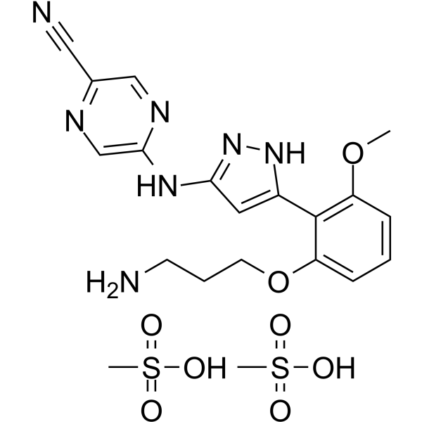
HY-18174E
Prexasertib dimesylateCHK1 Inhibitor -
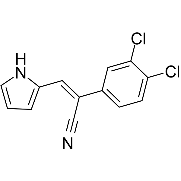
HY-117102
ANI-7AhR Activator -
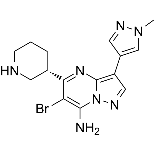
HY-15532B
SCH900776 (S-isomer)CHK1 Serine/Threonine Kinase Inhibitor -
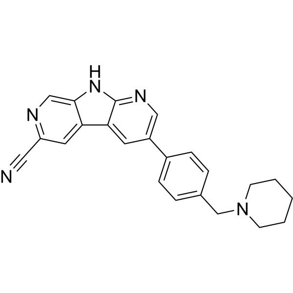
HY-15883
GNE-900ChK1 Inhibitor
Publications Citing Use of MedChemExpress (MCE) Prexasertib mesylate
More- Nat Commun. 2019 Aug 2;10(1):3485. [Abstract]
- Sci Adv. 2026 Jun 26;12(26):eadz3351. [Abstract]
- Cell Death Dis. 2026 Mar 26;17(1):375. [Abstract]
- Cancer Lett. 2026 Feb 4;642:218300.
- Cancer Lett. 2026 Apr 1:642:218300. [Abstract]
- Oncogene. 2026 Jun 10. [Abstract]
- Oncogene. 2022 Nov;41(46):5020-5031. [Abstract]
- Thorax. 2022 Mar;77(3):247-258. [Abstract]
- Cell Chem Biol. 2026 Jun 18;33(6):767-784.e7. [Abstract]
- Br J Cancer. 2021 Jul;125(1):101-111. [Abstract]
- Cell Rep. 2025 Apr 17;44(5):115605. [Abstract]
- Mol Cancer Ther. 2026 Jul 15:10.1158/1535-7163.MCT-26-0182.
- Mol Cancer Ther. 2025 Jun 4;24(6):920-930. [Abstract]
- Oncogenesis. 2025 Mar 1;14(1):4. [Abstract]
- Cells. 2024 Apr 19;13(8):710. [Abstract]
- Mol Cancer Res. 2019 Oct;17(10):2102-2114. [Abstract]
- Eur J Pharmacol. 2025 Sep 15:1003:177942. [Abstract]
- Cell Biol Toxicol. 2023 Jun;39(3):795-811. [Abstract]
- Int Immunopharmacol. 2026 Feb 15:171:116126. [Abstract]
- Int Immunopharmacol. 2025 Mar 26:150:114278. [Abstract]
- Sci Rep. 2025 Nov 27. [Abstract]
- Sci Rep. 2021 Feb 4;11(1):3176. [Abstract]
- Cancers (Basel). 2021 Aug 20;13(16):4200. [Abstract]
- Cancers (Basel). 2020 Aug 26;12(9):2426. [Abstract]
- Cancers (Basel). 2020 Jun 29;12(7):1726. [Abstract]
- Cell Signal. 2025 Jul:131:111709. [Abstract]
- Neurooncol Adv. 2024 Nov 19;6(1):vdae187. [Abstract]
- Discov Oncol. 2026 Jul 14. [Abstract]
- bioRxiv. 2025 Dec 25.
- bioRxiv. 2025 April 17.
- Research Square Preprint. 2024 Nov 06.
- bioRxiv. 2024 Nov 6:2024.11.04.621884. [Abstract]
- Universität Hamburg. 2022 Aug.
- Utrecht University. 2023 Feb.
- bioRxiv. January 04, 2022.
- Patent. US20210353605A1.
- bioRxiv. September 10, 2021.
- Research Square Preprint. 2021 May.
- Methods Mol Biol. 2018:1711:351-398. [Abstract]
 Customer Validation & Images

-
WB
-
Flow Cytometry
-
In Vivo Efficacy Study
-
Apoptosis Analysis
-
RT-PCR
All DNA/RNA Synthesis Isoforms
More

Biological Activity
Description
IC50 & Target
[1]|
Chk1 0.9 nM (Ki) |
Chk1 <1 nM (IC50) |
Chk2 8 nM (IC50) |
In Vitro
Prexasertib (LY2606368) mesylate inhibits MELK (IC50=38 nM), SIK (IC50=42 nM), BRSK2 (IC50=48 nM), ARK5 (IC50=64 nM). Prexasertib mesylate requires CDC25A and CDK2 to cause DNA damage[1].
Prexasertib mesylate (33, 100 nM; for 7 hours) results in DNA damage during S-phase in HeLa cells[1].
Prexasertib mesylate (8-250 nM; pre-treated for 15 minutes) inhibits CHK1 autophosphorylation (S296) and CHK2 autophosphorylation (S516) in HT-29 cells[1].
Prexasertib mesylate (4 nM; 24 hours) results in a large shift in cell-cycle populations from G1 and G2-M to S-phase with an accompanied induction of H2AX phosphorylation in U-2 OS cells[1].
Prexasertib mesylate (33 nM; for 12 hours) causes chromosomal fragmentation in HeLa cells. Prexasertib mesylate (100 nM; 0.5 to 9 hours) induces replication stress and depletes the pool of available RPA2 for binding to DNA[1].
MedChemExpress (MCE) has not independently confirmed the accuracy of these methods. They are for reference only. Further protocols information, click here.
-
Cell Line:HeLa cells
-
Concentration:33, 100 nM
-
Incubation Time:7 hours
-
Result:Had an IC50 of 37 nM and resulted in the G2-M population received DNA damage during S-phase but continued to progress through the cell cycle into an early mitosis.
-
Cell Line:HT-29 cells
-
Concentration:8, 16, 31, 63, 125, 250 nM
-
Incubation Time:Pre-treated for 15 minutes
-
Result:Inhibited CHK1 autophosphorylation (S296) and CHK2 autophosphorylation (S516) (IC50 of less than 31 nM) in HT-29 cells.
In Vivo
Prexasertib mesylate (15 mg/kg; SC) causes CHK1 inhibition in the blood and the phosphorylation of both H2AX (S139) and RPA2 (S4/S8)[1].
MedChemExpress (MCE) has not independently confirmed the accuracy of these methods. They are for reference only.
-
Animal Model:Female CD-1 nu-/nu- mice (26-28 g) with Calu-6 cells[1]
-
Dosage:1, 3.3, or 10 mg/kg
-
Administration:SC; twice daily for 3 days, rest 4 days; for three cycles
-
Result:Caused statistically significant tumor growth inhibition (up to 72.3%).


Chemical Information
-
CAS No. 1234015-55-4
-
Molecular Weight 461.49
-
Formula C19H23N7O5S
-
SMILES
N#CC1=NC=C(NC2=NNC(C3=C(OC)C=CC=C3OCCCN)=C2)N=C1.CS(=O)(O)=O
-
Synonyms
LY2606368 mesylate
-
Shipping
Room temperature in continental US; may vary elsewhere.
-
Storage
Please store the product under the recommended conditions in the Certificate of Analysis.


Publications (39)
-
Journal Impact Factor
-
Most Recent
-
Nat Commun
MYC paralog-dependent apoptotic priming orchestrates a spectrum of vulnerabilities in small cell lung cancer. [Abstract]2019 Aug 2;10(1):3485. PMID: 31375684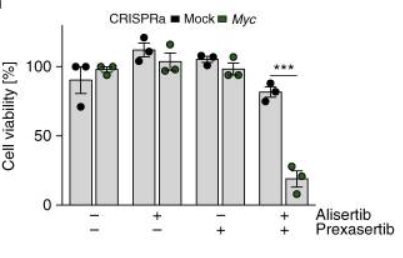
Prexasertib mesylate purchased from MedChemExpress. Usage Cited in: Nat Commun. 2019 Aug 2;10(1):3485. [Abstract]
Viability of mock control and Myc-activated CRISPRa cells upon treatment with 40 nM prexasertib for 96 h (n = 3).
-
Sci Adv
Patient-derived organoids across cancers reveal conserved tumor heterogeneity and actionable therapeutic vulnerabilities. [Abstract]2026 Jun 26;12(26):eadz3351. PMID: 42361179 -
Cell Death Dis
2026 Mar 26;17(1):375. PMID: 41888112 -
-
Cancer Lett
2026 Apr 1:642:218300. PMID: 41651400 -
Oncogene
DNA replication stress and translational repression converge to drive CDK1- and caspase-dependent apoptosis in Ewing sarcoma. [Abstract]2026 Jun 10. PMID: 42270776 -
Oncogene
Distinct roles of treatment schemes and BRCA2 on the restoration of homologous recombination DNA repair and PARP inhibitor resistance in ovarian cancer. [Abstract]2022 Nov;41(46):5020-5031. PMID: 36224341 -
Thorax
Potential for inhibition of checkpoint kinases 1/2 in pulmonary fibrosis and secondary pulmonary hypertension. [Abstract]2022 Mar;77(3):247-258. PMID: 34226205 -
Cell Chem Biol
The cytotoxic effect of prexasertib is a consequence of dual inhibition on both CHK1 and AMPK. [Abstract]2026 Jun 18;33(6):767-784.e7. PMID: 42061410 -
Br J Cancer
Interferon regulatory factor 1 (IRF-1) downregulates Checkpoint kinase 1 (CHK1) through miR-195 to upregulate apoptosis and PD-L1 expression in Hepatocellular carcinoma (HCC) cells. [Abstract]2021 Jul;125(1):101-111. PMID: 33772151
Prexasertib mesylate purchased from MedChemExpress. Usage Cited in: Br J Cancer. 2021 Jul;125(1):101-111. [Abstract]
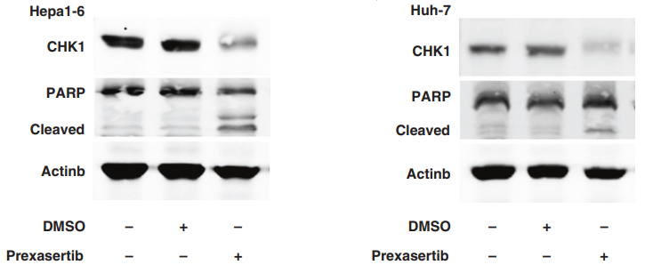
Full length and cleaved PARP protein expressions are determined by western blot in Hepa1-6 cells and Huh-7 cells induced by prexasertib with dose of 1 µM and 5 nM for 24 h, respectively.
Prexasertib mesylate purchased from MedChemExpress. Usage Cited in: Br J Cancer. 2021 Jul;125(1):101-111. [Abstract]
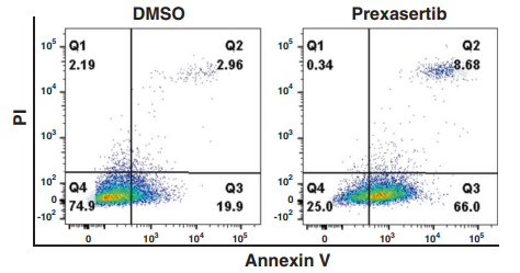
Representative image of FACS analysis of late apoptotic Hepa1-6 cell rate treated by DMSO or prexasertib with dose of 1 µM for 24 h are shown.
Prexasertib mesylate purchased from MedChemExpress. Usage Cited in: Br J Cancer. 2021 Jul;125(1):101-111. [Abstract]
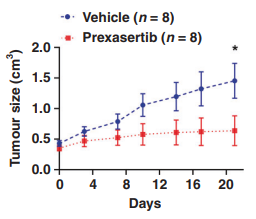
Tumour growth curves (Mean ± SEM) of murine HCC model treated by vehicle (n = 8) or prexasertib (10mg/kg, subcutaneous injection, 2 of 7 days, n = 8) are shown.
Prexasertib mesylate purchased from MedChemExpress. Usage Cited in: Br J Cancer. 2021 Jul;125(1):101-111. [Abstract]
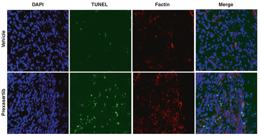
Prexasertib (10mg/kg, subcutaneous injection, 2 of 7 days). Representative images of TUNEL assay for cellular apoptosis (green staining) in tumours are shown.
Prexasertib mesylate purchased from MedChemExpress. Usage Cited in: Br J Cancer. 2021 Jul;125(1):101-111. [Abstract]
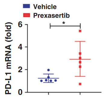
Prexasertib (10mg/kg, subcutaneous injection, 2 of 7 days, n = 8). PD-L1 mRNA expression is determined by qPCR in tumours treated by prexasertib or vehicle.
-
Cell Rep
The low-dose CHK1 inhibitor prexasertib triggers VDAC1 dephosphorylation to activate mtDNA-STING signaling and synergize immunotherapy. [Abstract]2025 Apr 17;44(5):115605. PMID: 40249707 -
-
Mol Cancer Ther
Targeting Monounsaturated Fatty Acid Metabolism for Radiosensitization of KRAS Mutant 3D Lung Cancer Models. [Abstract]2025 Jun 4;24(6):920-930. PMID: 39834305 -
Oncogenesis
STAG2 expression imparts distinct therapeutic vulnerabilities in muscle-invasive bladder cancer cells. [Abstract]2025 Mar 1;14(1):4. PMID: 40025053 -
Cells
Genomic Engineering of Oral Keratinocytes to Establish In Vitro Oral Potentially Malignant Disease Models as a Platform for Treatment Investigation. [Abstract]2024 Apr 19;13(8):710. PMID: 38667326 -
Mol Cancer Res
Pharmacologic Ascorbate Primes Pancreatic Cancer Cells for Death by Rewiring Cellular Energetics and Inducing DNA Damage. [Abstract]2019 Oct;17(10):2102-2114. PMID: 31337671 -
Eur J Pharmacol
VOPP1, a determinant of the sensitivity of non-small cell lung cancer cells to NAE inhibitors. [Abstract]2025 Sep 15:1003:177942. PMID: 40651787 -
Cell Biol Toxicol
Exploring the ATR-CHK1 pathway in the response of doxorubicin-induced DNA damages in acute lymphoblastic leukemia cells. [Abstract]2023 Jun;39(3):795-811. PMID: 34519926 -
Int Immunopharmacol
CHK1 inhibition by prexasertib sensitizes cisplatin-resistant malignant tumor cells via checkpoint abrogation and STAT1-driven PD-L1 upregulation. [Abstract]2026 Feb 15:171:116126. PMID: 41477994 -
Int Immunopharmacol
Prexasertib exerts a synergistic effect on the antitumor activity of Lenvatinib through ALOX15-mediated ferroptosis in hepatocellular carcinoma. [Abstract]2025 Mar 26:150:114278. PMID: 39954659 -
Sci Rep
Synergistic inhibition of CHK1 and MUS81 to combat replication stress resistance in high-risk neuroblastoma. [Abstract]2025 Nov 27. PMID: 41310217 -
Sci Rep
The mitotic checkpoint is a targetable vulnerability of carboplatin-resistant triple negative breast cancers. [Abstract]2021 Feb 4;11(1):3176. PMID: 33542435 -
Cancers (Basel)
Novel Insights into the Molecular Regulation of Ribonucleotide Reductase in Adrenocortical Carcinoma Treatment. [Abstract]2021 Aug 20;13(16):4200. PMID: 34439352 -
Cancers (Basel)
Systems Biology Approach Identifies Prognostic Signatures of Poor Overall Survival and Guides the Prioritization of Novel BET-CHK1 Combination Therapy for Osteosarcoma. [Abstract]2020 Aug 26;12(9):2426. PMID: 32859084 -
Cancers (Basel)
Cotargeting CHK1 and PI3K Synergistically Suppresses Tumor Growth of Oral Cavity Squamous Cell Carcinoma in Patient-Derived Xenografts. [Abstract]2020 Jun 29;12(7):1726. PMID: 32610557 -
Cell Signal
2025 Jul:131:111709. PMID: 40037423 -
Neurooncol Adv
Ion channel modulator DPI-201-106 significantly enhances antitumor activity of DNA damage response inhibitors in glioblastoma. [Abstract]2024 Nov 19;6(1):vdae187. PMID: 39659830 -
Discov Oncol
Chk1 inhibition emerges as the most effective partner for PARP inhibition in BRCA-proficient pancreatic cancer. [Abstract]2026 Jul 14. PMID: 42446852 -
-
-
-
bioRxiv
PAIRWISE: Deep Learning-based Prediction of Effective Personalized Drug Combinations in Cancer. [Abstract]2024 Nov 6:2024.11.04.621884. PMID: 39574568 -
-
-
-
-
-
-
Methods Mol Biol
2018:1711:351-398. PMID: 29344898

Protocols
-
Apoptosis
Apoptosis, also called programmed cell death, is generally characterized by distinct morphological characteristics.
-
TUNEL staining for apoptotic DNA fragmentation
TUNEL staining detects DNA strand breaks by using terminal deoxynucleotidyl transferase to add labeled nucleotides to exposed 3′-OH DNA termini, generating either microscopic staining in fixed cells or tissue sections, or fluorescence/cytometric signal in cell suspensions. TUNEL positivity reflects DNA fragmentation but should not be interpreted alone as definitive apoptosis, because TUNEL can also label necrotic, autolytic, mechanically damaged, or DNA-repair-associated DNA breaks.
-
Cell Cytotoxicity Assay
Cytotoxicity assays are usually based on the assessment of cell membrane damage, which can also be indirectly detected by measuring cell viability. Detection methods include MTT assay, CKK-8 assay, LDH assay and ATP assay, etc.
-
Annexin V plus membrane-impermeant dye apoptosis staining
Annexin V-based apoptosis assays rely on the detection of phosphatidylserine (PS) externalization from the inner leaflet of the plasma membrane to the outer leaflet, an early biochemical hallmark of apoptosis. Fluorescently labeled Annexin V binds PS in a calcium-dependent manner, enabling identification of early apoptotic cells by flow cytometry or fluorescence microscopy. When combined with a membrane-impermeant DNA-binding dye (e. g. , propidium iodide), this approach allows discrimination between viable (Annexin V−/dye−), early apoptotic (Annexin V+/dye−), and late apoptotic or necrotic (Annexin V+/dye+) cell populations by assessing membrane integrity and PS exposure.
-
Apoptosis Solutions
Apoptosis is a regulated, generally non-lytic cell-death pathway that removes unwanted, damaged, infected, or abnormal cells through coordinated morphological changes, caspase activation, DNA fragmentation, and membrane remodeling. The intrinsic apoptosis pathway is controlled mainly by mitochondrial outer membrane permeabilization, BCL-2 family proteins, cytochrome c release, apoptosome formation, caspase-9 activation, and downstream executioner caspase-3/7 activation. The extrinsic apoptosis pathway is initiated by death receptors such as Fas, TNFR, and TRAIL receptors, which recruit adaptor proteins and activate caspase-8 before engaging executioner caspases or mitochondrial amplification through BID cleavage. Apoptosis is linked to many phenotypes, including cancer cell killing, tissue homeostasis, immune regulation, neurodegeneration, infection response, and treatment-induced cytotoxicity; unresolved questions include how apoptosis interacts with necroptosis, pyroptosis, ferroptos


Purity & Documentation
References
[1]. King C, et al. LY2606368 Causes Replication Catastrophe and Antitumor Effects through CHK1-Dependent Mechanisms. Mol Cancer Ther. 2015 Sep;14(9):2004-1. [Content Brief]
[2]. Yin Y, et al. Chk1 inhibition potentiates the therapeutic efficacy of PARP inhibitor BMN673 in gastric cancer. Am J Cancer Res. 2017 Mar 1;7(3):473-483. [Content Brief]

Calculators
Concentration (start) × Volume (start) = Concentration (final) × Volume (final)


Prexasertib mesylate Related Classifications
HY-18174C Related Classifications
 Powered by Bioz