



SK-3-91

Based on 1 Customer Validation
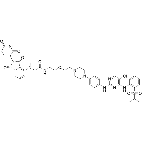
SK-3-91 is a multi-kinase PROTAC degrader that induces the degradation of the maximum number of unique kinases (over 125 distinct kinases) simultaneously. SK-3-91 induces protein degradation via the ubiquitin biotinylation (E-STUB) pathway, such as inducing the degradation of YTHDF2. SK-3-91 also inhibits cell proliferation and induces cell morphological changes. SK-3-91 can be used in research related to acute lymphoblastic leukemia, multiple myeloma, neuroblastoma, ovarian cancer, mantle cell lymphoma, and gastric cancer.
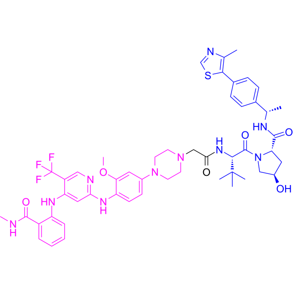
(Pink: YTHDF2 ligand (HY-169396); Blue: Cereblon ligand (HY-131717); Black: linker (HY-140819)).
For research use only. We do not sell to patients.
- Purity : 98.33%
- CAS No.: 2769753-23-1
- Formula: C42H47ClN10O8S
- Molecular Weight:887.40
-
Storage:
-20°C, protect from light
* In solvent : -80°C, 6 months; -20°C, 1 month (protect from light)

To place orders, for customer services and technical support, please contact: MedChemExpress USA
Tel: 609-228-6898 E-mail: [email protected] [email protected]


-
 Biological Activity
-
Chemical Information
-
Solvent & Solubility
- Protocols
- Purity & Documentation
- References
-
 Help & FAQs


All PROTACs Isoforms
More

Biological Activity
Description
IC50 & Target
[1]|
YTHDF2 |
Cereblon |
In Vitro
SK-3-91 (1-50 μM; 20 h) induces dose-dependent changes in cell morphology in U2OS cells, shows high biological similarity to aurora kinase inhibitors at 1 μM, and exhibits lower cytotoxicity than corresponding small-molecule kinase inhibitors at concentrations ≤10 μM[1].
SK-3-91 (400 nM; 72 h) inhibits the proliferation of K562 cells by 88.2%[1].
Treatment of MOLT-4 cells with SK-3-91 (1 μM; 1-8 h) induces no significant transcriptional changes within 4 h, but causes complete transcriptional collapse at 8 h[2].
MedChemExpress (MCE) has not independently confirmed the accuracy of these methods. They are for reference only.
-
Cell Line:K562 human leukemia cells
-
Concentration:0.12, 0.37, 1.1, 3.3, 10 μM
-
Incubation Time:24 h
-
Result:Induced degradation of the RNA-binding protein YTHDF2 in K562 cells.
Showed maximal degradation at concentrations between 0.37 μM and 1.1 μM.
Exhibited a hook effect (reduced degradation) at the higher concentration of 1.1 μM.
-
Cell Line:K562 human leukemia cells
-
Concentration:400 nM
-
Incubation Time:72 h
-
Result:Inhibited K562 cell proliferation by 88.2% relative to DMSO control.
- BrdU Incorporation Assay
- Somatic Cell Culture
- CFSE Dye Dilution Proliferation Assay
- Dye-dilution cell tracking and proliferation staining
- CCK-8/WST-8 Cell Proliferation Assay
- Protocol for Cell Cycle
- Cell Counting-Based Growth Curve Assay
- Colony Formation (Clonogenic) Assay
- Cell Viability Determination by MTT Colorimetric Assay
- EdU Incorporation Assay (Click Chemistry-Based DNA Synthesis Measurement)
- Ki-67 Immunostaining Proliferation Assay
- PCNA Immunodetection Proliferation Assay
- Protocol for Cell Counting and Cell Density Analysis
- MTT Cell Proliferation Assay


Chemical Information
-
CAS No. 2769753-23-1
-
Appearance Solid
-
Molecular Weight 887.40
-
Formula C42H47ClN10O8S
-
Color Light yellow to yellow
-
SMILES
O=C(N1C2C(NC(CC2)=O)=O)C3=C(NCC(NCCOCCN4CCN(C5=CC=C(NC6=NC(NC7=C(S(C(C)C)(=O)=O)C=CC=C7)=C(Cl)C=N6)C=C5)CC4)=O)C=CC=C3C1=O
-
Shipping
Room temperature in continental US; may vary elsewhere.
-
Storage
-20°C, protect from light
* In solvent : -80°C, 6 months; -20°C, 1 month (protect from light)


Solvent & Solubility
In Vitro:
DMSO : 200 mg/mL (225.38 mM; Need ultrasonic; Hygroscopic DMSO has a significant impact on the solubility of product, please use newly opened DMSO)
H2O : < 0.1 mg/mL (insoluble)
Please refer to the solubility information to select the appropriate solvent. Once prepared, please aliquot and store the solution to prevent product inactivation from repeated freeze-thaw cycles.
Storage method and period of stock solution: -80°C, 6 months; -20°C, 1 month (protect from light). When stored at -80°C, please use it within 6 months. When stored at -20°C, please use it within 1 month.
Please refer to the solubility information to select the appropriate solvent. Once prepared, please aliquot and store the solution to prevent product inactivation from repeated freeze-thaw cycles.
Storage method and period of stock solution: -80°C, 6 months; -20°C, 1 month (protect from light). When stored at -80°C, please use it within 6 months. When stored at -20°C, please use it within 1 month.
Concentration (start) × Volume (start) = Concentration (final) × Volume (final)
In Vivo:
Select the appropriate dissolution method based on your experimental animal and administration route.
- For the following dissolution methods, please ensure to first prepare a clear stock solution using an In Vitro approach and then sequentially add co-solvents:
- To ensure reliable experimental results, the clarified stock solution can be appropriately stored based on storage conditions. As for the working solution for In Vivo experiments, it is recommended to prepare freshly and use it on the same day.
- The percentages shown for the solvents indicate their volumetric ratio in the final prepared solution. If precipitation or phase separation occurs during preparation, heat and/or sonication can be used to aid dissolution.
Add each solvent one by one: 10% DMSO 40% PEG300 5% Tween-80 45% Saline
Solubility: 5 mg/mL (5.63 mM); Suspended solution; Need ultrasonic
This protocol yields a suspended solution of 5 mg/mL. Suspended solution can be used for oral and intraperitoneal injection.
Taking 1 mL working solution as an example, add 100 μL DMSO stock solution (50.0 mg/mL) to 400 μL PEG300, and mix evenly; then add 50 μL Tween-80 and mix evenly; then add 450 μL Saline to adjust the volume to 1 mL.
Preparation of Saline: Dissolve 0.9 g sodium chloride in ddH₂O and dilute to 100 mL to obtain a clear Saline solution.
Add each solvent one by one: 10% DMSO 90% (20% SBE-β-CD in Saline)
Solubility: 5 mg/mL (5.63 mM); Suspended solution; Need ultrasonic
This protocol yields a suspended solution of 5 mg/mL. Suspended solution can be used for oral and intraperitoneal injection.
Taking 1 mL working solution as an example, add 100 μL DMSO stock solution (50.0 mg/mL) to 900 μL 20% SBE-β-CD in Saline, and mix evenly.
Preparation of 20% SBE-β-CD in Saline (4°C, storage for one week): 2 g SBE-β-CD powder is dissolved in 10 mL Saline, completely dissolve until clear.
In Vivo Dissolution Calculator
Please enter the basic information of animal experiments:
-
-
-
-
Recommended: Prepare an additional quantity of animals to account for potential losses during experiments.
Please enter your animal formula composition:
-
%DMSO +
Recommended: Keep the proportion of DMSO in working solution below 2% if your animal is weak.
-
%+
-
+%Tween-80 + +
-
%Saline +
The co-solvents required include: DMSO, . All of co-solvents are available by MedChemExpress (MCE). , Tween 80. All of co-solvents are available by MedChemExpress (MCE).
Working solution concentration: 0.22 mg/mL
Method for preparing stock solution: mg drug dissolved in μL DMSO. Stock solution concentration: mg/mL. * In solvent : -80°C, 6 months; -20°C, 1 month (protect from light)
1. Take μL DMSO stock solution;
2. Add μL .
μL , mix evenly;
3. Then add μL Tween 80, mix evenly;
4. Then add μL
Please ensure that the stock solution in the first step is dissolved to a clear state, and add co-solvents in sequence. You can use ultrasonic heating (ultrasonic cleaner, recommended frequency 20-40 kHz), vortexing, etc. to assist dissolution.


Purity & Documentation
-
Data Sheet (273 KB)
-
SDS (252 KB)
- English - EN (252 KB)
- Français - FR (252 KB)
- Deutsch - DE (252 KB)
- Norwegian - NO (252 KB)
- Español - ES (252 KB)
- Swedish - SV (252 KB)
- Italian - IT (252 KB)
- Korean - KR (252 KB)
- Portuguese - PT (252 KB)
-
Handling Instructions (2659 KB)
References
[1]. Goebel GL, et al. Profiling Cellular Morphological Changes Induced by Dual-Targeting PROTACs of Aurora Kinase and RNA-Binding Protein YTHDF2. Chembiochem : a European journal of chemical biology. 2024 Oct 01;25(19):e202400183. [Content Brief]
[2]. Donovan KA, et al. Mapping the Degradable Kinome Provides a Resource for Expedited Degrader Development. Cell. 2020 Dec 10;183(6):1714-1731.e10. [Content Brief]
[3]. Huang HT, et al. Ubiquitin-specific proximity labeling for the identification of E3 ligase substrates. Nature chemical biology. 2024 Sep;20(9):1227-1236. [Content Brief]


Complete Stock Solution Preparation Table
Please refer to the solubility information to select the appropriate solvent. Once prepared, please aliquot and store the solution to prevent product inactivation from repeated freeze-thaw cycles.
Storage method and period of stock solution: -80°C, 6 months; -20°C, 1 month (protect from light). When stored at -80°C, please use it within 6 months. When stored at -20°C, please use it within 1 month.
| Optional Solvent | Concentration Solvent Mass | 1 mg | 5 mg | 10 mg | 25 mg |
|---|---|---|---|---|---|
| DMSO | 1 mM | 1.1269 mL | 5.6344 mL | 11.2689 mL | 28.1722 mL |
| 5 mM | 0.2254 mL | 1.1269 mL | 2.2538 mL | 5.6344 mL | |
| 10 mM | 0.1127 mL | 0.5634 mL | 1.1269 mL | 2.8172 mL | |
| 15 mM | 0.0751 mL | 0.3756 mL | 0.7513 mL | 1.8781 mL | |
| 20 mM | 0.0563 mL | 0.2817 mL | 0.5634 mL | 1.4086 mL | |
| 25 mM | 0.0451 mL | 0.2254 mL | 0.4508 mL | 1.1269 mL | |
| 30 mM | 0.0376 mL | 0.1878 mL | 0.3756 mL | 0.9391 mL | |
| 40 mM | 0.0282 mL | 0.1409 mL | 0.2817 mL | 0.7043 mL | |
| 50 mM | 0.0225 mL | 0.1127 mL | 0.2254 mL | 0.5634 mL | |
| 60 mM | 0.0188 mL | 0.0939 mL | 0.1878 mL | 0.4695 mL | |
| 80 mM | 0.0141 mL | 0.0704 mL | 0.1409 mL | 0.3522 mL | |
| 100 mM | 0.0113 mL | 0.0563 mL | 0.1127 mL | 0.2817 mL |


SK-3-91 Related Classifications
HY-137341 Related Classifications
 Powered by Bioz