



Urolithin M5
Based on 1 publication(s) in Google Scholar
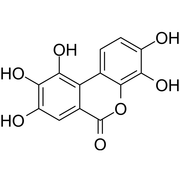
Urolithin M5 (Decarboxyellagic acid) is an orally active influenza virus neuraminidase inhibitor and neuroprotective agent, with IC50 values of 174.8 μM (HK68), 191.5 μM (pdm09), 243.2 μM (WSN) and 257.1 μM (PR8) against four influenza virus neuraminidases, respectively. Urolithin M5 inhibits viral neuraminidase activity, thereby blocking influenza virus replication (including oseltamivir (HY-13317)-resistant strains), protecting infected mammals from death and improving pulmonary edema. Urolithin M5 forms a hydrogen-bond stabilized complex with IGF1R, and binds to MAPK14, AKT1, NFKB1 and EGFR. Urolithin M5 reduces reactive oxygen species production, inhibits neuronal apoptosis, restores mitochondrial transmembrane potential, and promotes neurite outgrowth of damaged neuronal cells. Urolithin M5 can be used in research related to influenza virus infection and Alzheimer's disease.
Nur für Forschungszwecke. Wir verkaufen nicht an Patienten.
- Reinheit : 97.31%
- CAS. Nr.: 91485-02-8
- Formel: C13H8O7
- Molecular Weight:276.20
-
Speicherung:Powder -20°C, 3 years ; In solvent -80°C, 6 months , -20°C, 1 month

To place orders, for customer services and technical support, please contact: MedChemExpress USA
Tel: 609-228-6898 E-mail: [email protected] [email protected]


-
 Biologische Aktivität
-
Chemical Information
-
Lösungsmittel & Löslichkeit
- Protokoll
- Reinheit & Dokumentation
- Verweise
-
 Help & FAQs


-
HY-133178
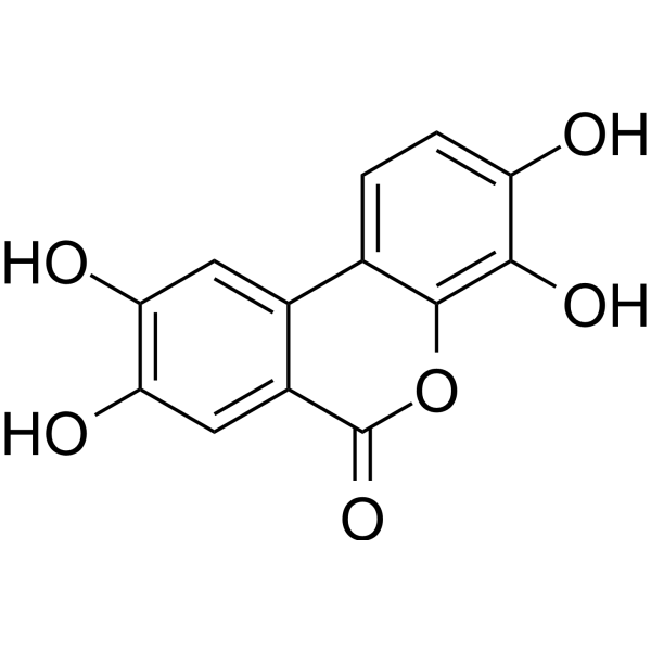
Urolithin DEphA Receptor Antagonist -
HY-126307
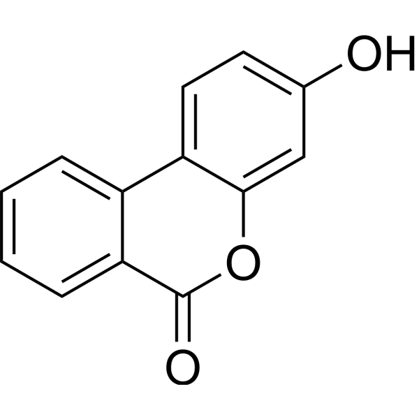
Urolithin BNF-κB Inhibitor -

HY-13259
MG-132Proteasome Inhibitor -
HY-100599
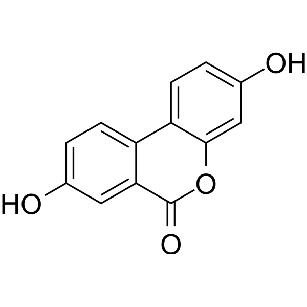
Urolithin AMitophagy Activator -
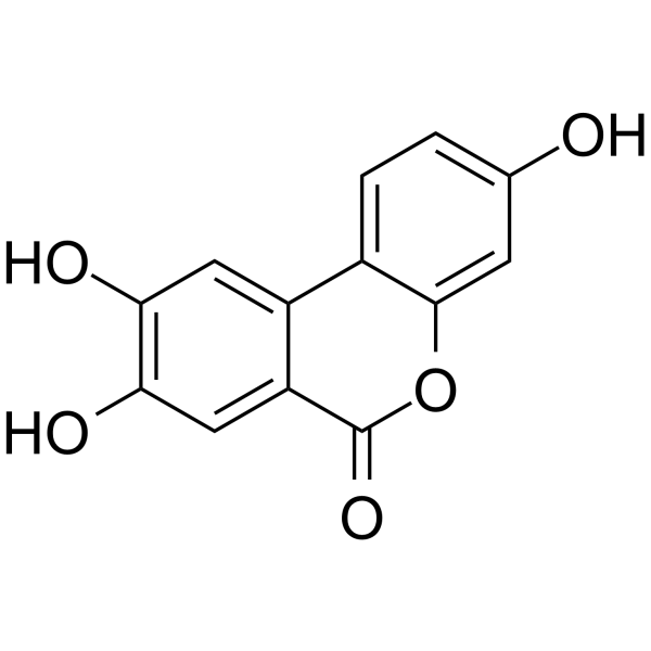
HY-135897
Urolithin CInsulin Secretion Activator -
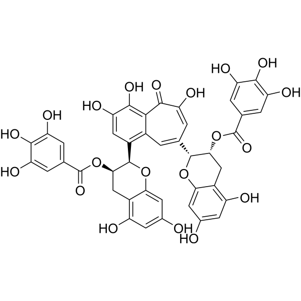
HY-N1992
Theaflavin 3,3'-digallateVirus Protease Inhibitor -
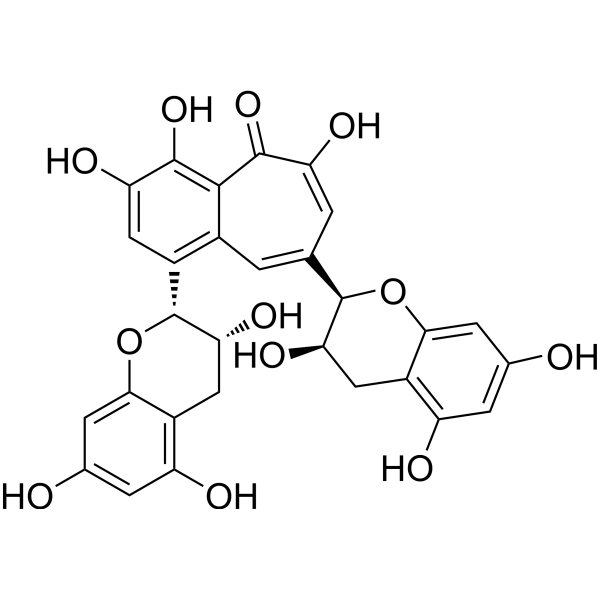
HY-N0243
TheaflavinEndogenous Metabolite -

HY-W746164
Urolithin E
Publications Citing Use of MedChemExpress (MCE) Urolithin M5
MoreAlle EGFR Isoform-spezifische Produkte anzeigen
More

Biologische Aktivität
Beschreibung
In Vitro
Urolithin M5 (two-fold dilution series; 48 h) exhibits cytotoxicity against MDCK cells with a CC50 of 227.4 μM[1].
Urolithin M5 (5-500 μM; 30 min virus pre-incubation, 30 min erythrocyte incubation) does not inhibit hemagglutination induced by influenza A/WSN/33(H1N1) virus in chicken erythrocytes[1].
MedChemExpress (MCE) has not independently confirmed the accuracy of these methods. They are for reference only.
-
Cell Line:Madin-Darby canine kidney (MDCK) cells
-
Concentration:10 μM, 100 μM, 1 mM
-
Incubation Time:48 h
-
Result:Exhibited a CC50 value of 227.4 μM against madin-Darby canine kidney (MDCK) cells.
In Vivo
MedChemExpress (MCE) has not independently confirmed the accuracy of these methods. They are for reference only.
-
Animal Model:BALB/c (female, 16-18 g, intranasally infected with influenza A virus strain PR8)[1]
-
Dosage:200 mg/kg/day
-
Administration:p.o.; once daily; 6 consecutive days
-
Result:Protected 50% of mice from death.
Showed rebound in body weight starting at day 9.Reduced lung index from 1.445 to 0.8875.\n
Reduced lung viral titer by 0.52 log.Decreased lung tissue levels of NF-κB, TNF-α, and IL-6.
Alleviated histopathological lung lesions including reduced bronchial injury and inflammatory cell infiltration.
- PC12 NGF-Induced Neuronal Differentiation Culture
- Research Protocol for Infectious Diseases
- Detection of Apoptosis
- TUNEL staining for apoptotic DNA fragmentation
- SH-SY5Y Neuronal Differentiation Culture
- Annexin V plus membrane-impermeant dye apoptosis staining
- ROS/oxidative-stress fluorescent staining
- SH-SY5Y neuronal-like differentiation
- Apoptosis Solutions
- Cell differentiation
- PC12 NGF-induced neuronal-like differentiation
- Alzheimer’s Disease Modeling


Chemical Information
-
CAS. Nr. 91485-02-8
-
Appearance Solid
-
Molecular Weight 276.20
-
Formel C13H8O7
-
Color White to yellow
-
SMILES
O=C1C2=CC(O)=C(O)C(O)=C2C3=CC=C(O)C(O)=C3O1
-
Synonyms
Decarboxyellagic acid
-
Structure Classification
-
Initial Source
-
Versand
Room temperature in continental US; may vary elsewhere.
-
Speicherung
Powder -20°C 3 years In solvent -80°C 6 months -20°C 1 month


Publications (1)
-
Journal Impact Factor
-
Most Recent
-
Vet Microbiol
The Chinese medicine monomer Schisandrin C inhibits PRRSV infection by regulating the OGT-PI3K/AKT/mTOR signaling pathway. [Abstract]2026 May:316:110992. PMID: 41865607


Lösungsmittel & Löslichkeit
In Vitro:
DMSO : ≥ 100 mg/mL (362.06 mM; Hygroscopic DMSO has a significant impact on the solubility of product, please use newly opened DMSO)
* "≥" means soluble, but saturation unknown.
Please refer to the solubility information to select the appropriate solvent. Once prepared, please aliquot and store the solution to prevent product inactivation from repeated freeze-thaw cycles.
Storage method and period of stock solution: -80°C, 6 months; -20°C, 1 month. When stored at -80°C, please use it within 6 months. When stored at -20°C, please use it within 1 month.
Please refer to the solubility information to select the appropriate solvent. Once prepared, please aliquot and store the solution to prevent product inactivation from repeated freeze-thaw cycles.
Storage method and period of stock solution: -80°C, 6 months; -20°C, 1 month. When stored at -80°C, please use it within 6 months. When stored at -20°C, please use it within 1 month.
Konzentration (Stammlösung) × Volumen (Stammlösung) = Konzentration (Ziellösung) × Volumen (Ziellösung)


Reinheit & Dokumentation
-
Data Sheet (280 KB)
-
SDS (251 KB)
- English - EN (251 KB)
- Français - FR (251 KB)
- Deutsch - DE (251 KB)
- Norwegian - NO (251 KB)
- Español - ES (251 KB)
- Swedish - SV (251 KB)
- Italian - IT (251 KB)
- Korean - KR (251 KB)
- Portuguese - PT (251 KB)
-
Handling Instructions (2659 KB)
Verweise
[1]. Xiao M, et al. Urolithin M5 from the Leaves of Canarium album (Lour.) DC. Inhibits Influenza Virus by Targeting Neuraminidase. Molecules. 2022 Sep 5;27(17):5724. [Content Brief]
[2]. Li HL, et al. Identification of ellagic acid and urolithins as natural inhibitors of Aβ25-35-induced neurotoxicity and the mechanism predication using network pharmacology analysis and molecular docking. Front Nutr. 2022;9:966276. Published 2022 Aug 2. [Content Brief]


Complete Stock Solution Preparation Table
Please refer to the solubility information to select the appropriate solvent. Once prepared, please aliquot and store the solution to prevent product inactivation from repeated freeze-thaw cycles.
Storage method and period of stock solution: -80°C, 6 months; -20°C, 1 month. When stored at -80°C, please use it within 6 months. When stored at -20°C, please use it within 1 month.
| Optional Solvent | Concentration Solvent Mass | 1 mg | 5 mg | 10 mg | 25 mg |
|---|---|---|---|---|---|
| DMSO | 1 mM | 3.6206 mL | 18.1028 mL | 36.2056 mL | 90.5141 mL |
| 5 mM | 0.7241 mL | 3.6206 mL | 7.2411 mL | 18.1028 mL | |
| 10 mM | 0.3621 mL | 1.8103 mL | 3.6206 mL | 9.0514 mL | |
| 15 mM | 0.2414 mL | 1.2069 mL | 2.4137 mL | 6.0343 mL | |
| 20 mM | 0.1810 mL | 0.9051 mL | 1.8103 mL | 4.5257 mL | |
| 25 mM | 0.1448 mL | 0.7241 mL | 1.4482 mL | 3.6206 mL | |
| 30 mM | 0.1207 mL | 0.6034 mL | 1.2069 mL | 3.0171 mL | |
| 40 mM | 0.0905 mL | 0.4526 mL | 0.9051 mL | 2.2629 mL | |
| 50 mM | 0.0724 mL | 0.3621 mL | 0.7241 mL | 1.8103 mL | |
| 60 mM | 0.0603 mL | 0.3017 mL | 0.6034 mL | 1.5086 mL | |
| 80 mM | 0.0453 mL | 0.2263 mL | 0.4526 mL | 1.1314 mL | |
| 100 mM | 0.0362 mL | 0.1810 mL | 0.3621 mL | 0.9051 mL |


Urolithin M5 Related Classifications
HY-N7922 Related Classifications
 Powered by Bioz
Keywords
- Urolithin M5
- 91485-02-8
- Decarboxyellagic acid
- Urolithin M 5
- Urolithin M-5
- Influenza Virus
- p38 MAPK
- EGFR
- Akt
- Reactive Oxygen Species (ROS)
- Apoptosis
- oseltamivir-resistant strains
- NFKB1
- MDCK cells
- *Canarium album* (Lour.) DC.
- influenza virus neuraminidase
- IGF1R
- AKT1
- influenza virus replication
- MAPK14
- Inhibitor
- inhibitor
- inhibit