



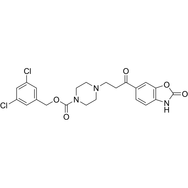
PF-8380
Based on 5 publication(s) in Google Scholar
PF-8380 is a potent autotaxin inhibitor with an IC50 of 2.8 nM in isolated enzyme assay and 101 nM in human whole blood.
For research use only. We do not sell to patients.
- Purity : 99.05%
- CAS No.: 1144035-53-9
- Formula: C22H21Cl2N3O5
- Molecular Weight:478.33
-
Storage:Powder -20°C, 3 years , 4°C, 2 years ; In solvent -80°C, 2 years , -20°C, 1 year

To place orders, for customer services and technical support, please contact: MedChemExpress USA
Tel: 609-228-6898 E-mail: [email protected] [email protected]


-
 Biological Activity
-
Chemical Information
-
Solvent & Solubility
- Purity & Documentation
- References
-
 Help & FAQs


-
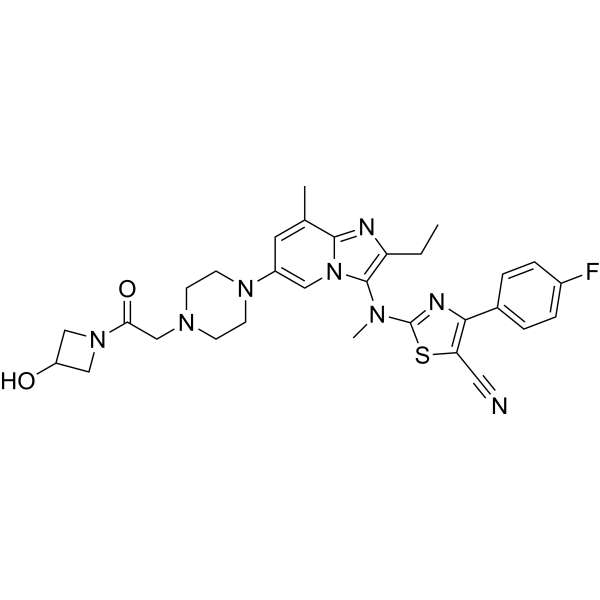
HY-101772
ZiritaxestatAutotaxin Inhibitor -
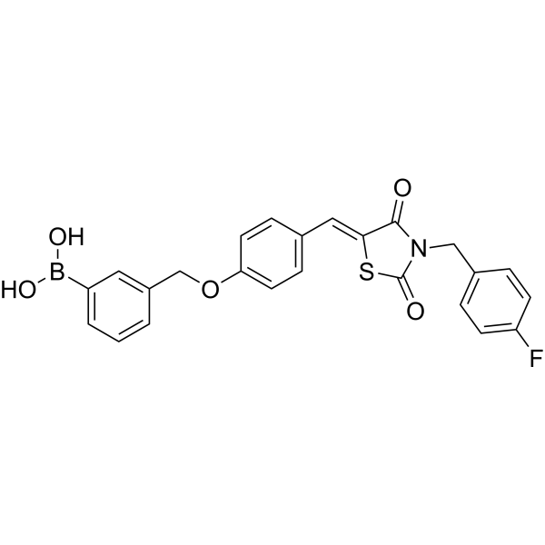
HY-19329
HA130Autotaxin Inhibitor -
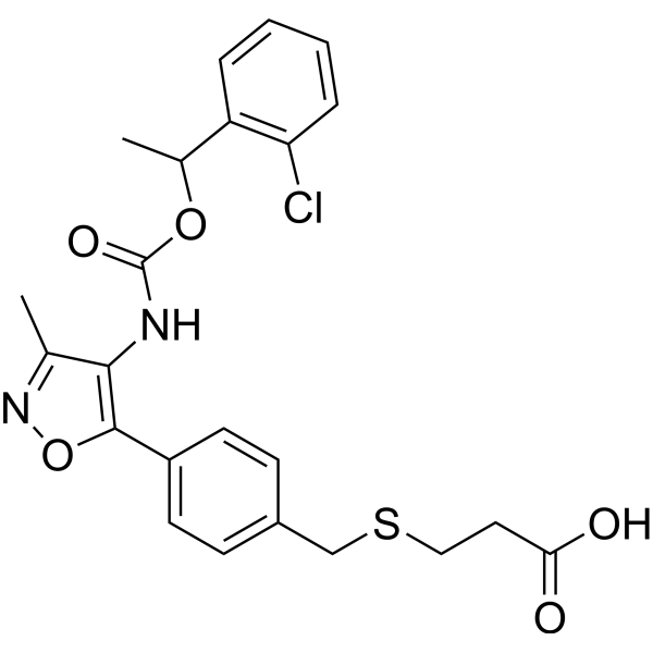
HY-13285
Ki16425LPA1/3 Antagonist -
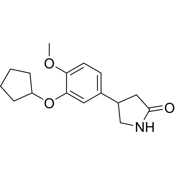
HY-16900
RolipramPDE4 Inhibitor -

HY-10431
SB-431542TGF-β Receptor Kinase Inhibitor -
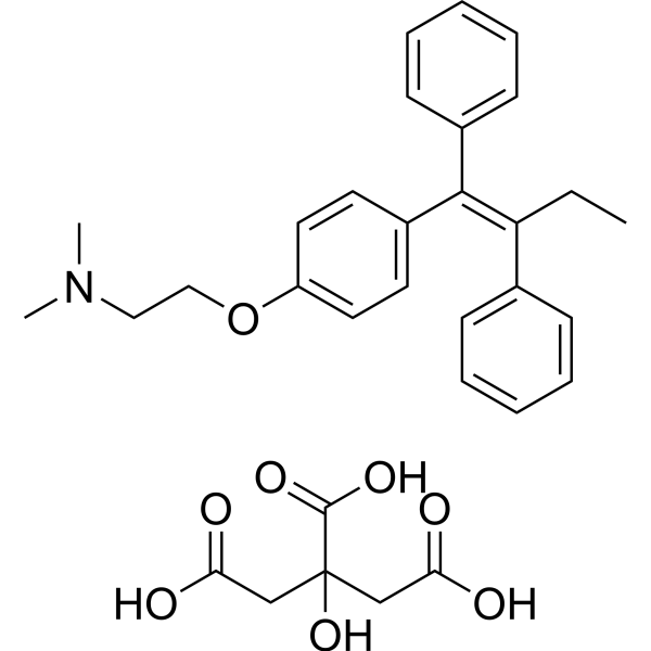
HY-13757
Tamoxifen CitrateEstrogen Receptor Modulator -
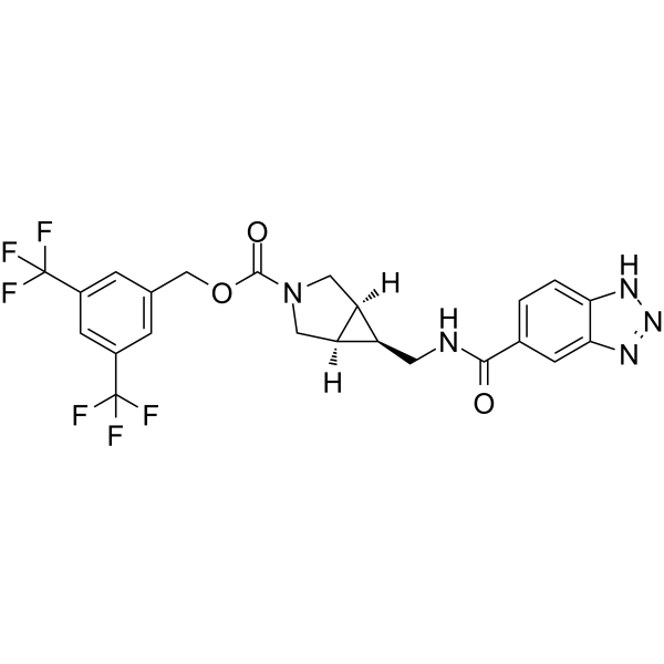
HY-124772
BI-2545ATX Inhibitor -
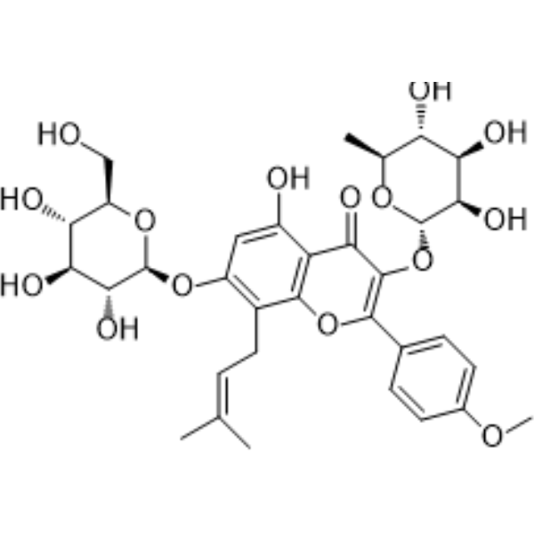
HY-N0014
IcariinPPAR Activator -

HY-13344R
PF-8380 (Standard)Autotaxin Inhibitor -
HY-16039
AM095LPA1 Antagonist -
HY-15455
RoflumilastPDE4 Inhibitor -
HY-B0312
DipyridamolePDE Inhibitor
Publications Citing Use of MedChemExpress (MCE) PF-8380
More Customer Validation & Images

-
WB


Biological Activity
Description
IC50 & Target
[1]|
Autotaxin 2.8 nM (IC50, In isolated enzyme assay) |
Cellular Effect
|
Cell Line

|
Type | Value | Description | References |
|---|---|---|---|---|
| A549 | IC50 |
>20 μM
Compound: PP-8380
|
Cytotoxicity against human A549 cells assessed as inhibition of cell growth incubated for 72 hrs by MTT assay
Cytotoxicity against human A549 cells assessed as inhibition of cell growth incubated for 72 hrs by MTT assay
|
[PMID: 35436669] |
| A549 | IC50 |
8.21 μM
Compound: PF-8380; 510k
|
Antiproliferative activity against human A549 cells measured after 72 hrs by MTT assay
Antiproliferative activity against human A549 cells measured after 72 hrs by MTT assay
|
[PMID: 32535330] |
| HEK293 | IC50 |
1.7 nM
Compound: PF-8380
|
Inhibition of recombinant human ATX beta expressed in HEK293 cells using LPC as substrate measured after 30 mins by LC-MS/MS analysis
Inhibition of recombinant human ATX beta expressed in HEK293 cells using LPC as substrate measured after 30 mins by LC-MS/MS analysis
|
[PMID: 28743508] |
| HEK293 | IC50 |
2.8 nM
Compound: 21
|
Inhibition of recombinant human ATX expressed in HEK293 cells using FS3 as substrate preincubated for 15 mins followed by substrate addition measured after 30 mins
Inhibition of recombinant human ATX expressed in HEK293 cells using FS3 as substrate preincubated for 15 mins followed by substrate addition measured after 30 mins
|
[PMID: 26745766] |
| HEK293 | IC50 |
2.8 nM
Compound: 6; PF-8380
|
Inhibition of human ATX expressed in HEK293 cells using FS-3 as substrate preincubated for 15 mins followed by substrate addition measured after 30 mins
Inhibition of human ATX expressed in HEK293 cells using FS-3 as substrate preincubated for 15 mins followed by substrate addition measured after 30 mins
|
[PMID: 27982588] |
| HEK293 | IC50 |
2.8 nM
Compound: PF-8380
|
Inhibition of recombinant ATX (unknown origin) expressed in HEK293 cells using FS-3 as substrate after 15 mins
Inhibition of recombinant ATX (unknown origin) expressed in HEK293 cells using FS-3 as substrate after 15 mins
|
[PMID: 27544588] |
| HEK293 | IC50 |
2.8 nM
Compound: PF-8380
|
Inhibition of recombinant human ATX beta expressed in HEK293 cells using FS-3 as substrate pretreated for 15 mins followed by substrate addition measured after 30 mins
Inhibition of recombinant human ATX beta expressed in HEK293 cells using FS-3 as substrate pretreated for 15 mins followed by substrate addition measured after 30 mins
|
[PMID: 28743508] |
| HEK293 | IC50 |
3 nM
Compound: 2
|
Inhibition of human ATX expressed in HEK293 cells using FS-3 as substrate after 15 mins
Inhibition of human ATX expressed in HEK293 cells using FS-3 as substrate after 15 mins
|
[PMID: 28165241] |
| Hep 3B2 | IC50 |
>20 μM
Compound: PP-8380
|
Cytotoxicity against human Hep3B cells assessed as inhibition of cell growth incubated for 72 hrs by MTT assay
Cytotoxicity against human Hep3B cells assessed as inhibition of cell growth incubated for 72 hrs by MTT assay
|
[PMID: 35436669] |
| MCF7 | IC50 |
>20 μM
Compound: PP-8380
|
Cytotoxicity against human MCF-7 cells assessed as inhibition of cell growth incubated for 72 hrs by MTT assay
Cytotoxicity against human MCF-7 cells assessed as inhibition of cell growth incubated for 72 hrs by MTT assay
|
[PMID: 35436669] |
| MCF7 | IC50 |
2.31 μM
Compound: PF-8380; 510k
|
Antiproliferative activity against human MCF7 cells measured after 72 hrs by MTT assay
Antiproliferative activity against human MCF7 cells measured after 72 hrs by MTT assay
|
[PMID: 32535330] |
| MDA-MB-231 | IC50 |
>20 μM
Compound: PP-8380
|
Cytotoxicity against human MDA-MB-231 cells assessed as inhibition of cell growth incubated for 72 hrs by MTT assay
Cytotoxicity against human MDA-MB-231 cells assessed as inhibition of cell growth incubated for 72 hrs by MTT assay
|
[PMID: 35436669] |
| MDA-MB-231 | IC50 |
4.17 μM
Compound: PF-8380; 510k
|
Antiproliferative activity against human MDA-MB-231 cells measured after 72 hrs by MTT assay
Antiproliferative activity against human MDA-MB-231 cells measured after 72 hrs by MTT assay
|
[PMID: 32535330] |
| NCI-H1581 | IC50 |
>20 μM
Compound: PP-8380
|
Cytotoxicity against human NCI-H1581 cells assessed as inhibition of cell growth incubated for 72 hrs by MTT assay
Cytotoxicity against human NCI-H1581 cells assessed as inhibition of cell growth incubated for 72 hrs by MTT assay
|
[PMID: 35436669] |
| NCI-H2228 | IC50 |
>20 μM
Compound: PP-8380
|
Cytotoxicity against human H2228 cells assessed as inhibition of cell growth incubated for 72 hrs by MTT assay
Cytotoxicity against human H2228 cells assessed as inhibition of cell growth incubated for 72 hrs by MTT assay
|
[PMID: 35436669] |
| NCI-H2228 | IC50 |
11.27 μM
Compound: PF-8380; 510k
|
Antiproliferative activity against human NCI-H2228 cells measured after 72 hrs by MTT assay
Antiproliferative activity against human NCI-H2228 cells measured after 72 hrs by MTT assay
|
[PMID: 32535330] |
| RAW264.7 | IC50 |
9.58 μM
Compound: PP-8380
|
Cytotoxicity against mouse RAW264.7 cells assessed as inhibition of cell growth incubated for 72 hrs by MTT assay
Cytotoxicity against mouse RAW264.7 cells assessed as inhibition of cell growth incubated for 72 hrs by MTT assay
|
[PMID: 35436669] |
In Vitro
PF-8380 also inhibits rat autotaxin with an IC50 of 1.16 nM with FS-3 substrate. Potency of PF-8380 is maintained when using enzyme produced from fetal fibroblasts used in combination with lysophosphatidyl choline (LPC) as a substrate. In human whole blood incubated with PF-8380 for 2 h, autotaxin is inhibited with an IC50 of 101 nM[1]. Autotaxin (ATX), an enzyme with lysophospholipase D (lysoPLD) activity, catalyzes the production of lysophosphatidic acid (LPA) from lysophosphatidylcholine (LPC). Pre-treatment of GL261 and U87-MG cells with 1 μM PF-8380 followed by 4 Gy irradiation results in decreased clonogenic survival, decreases migration (33% in GL261; P=0.002 and 17.9% in U87-MG; P=0.012), decreases invasion (35.6% in GL261; P=0.0037 and 31.8% in U87-MG; P=0.002), and attenuates radiation-induced Akt phosphorylation[2].
MedChemExpress (MCE) has not independently confirmed the accuracy of these methods. They are for reference only.
In Vivo
MedChemExpress (MCE) has not independently confirmed the accuracy of these methods. They are for reference only.


Chemical Information
-
CAS No. 1144035-53-9
-
Appearance Solid
-
Molecular Weight 478.33
-
Formula C22H21Cl2N3O5
-
Color White to light brown
-
SMILES
O=C(N1CCN(CC1)CCC(C2=CC(O3)=C(C=C2)NC3=O)=O)OCC4=CC(Cl)=CC(Cl)=C4
-
Shipping
Room temperature in continental US; may vary elsewhere.
-
Storage
Powder -20°C 3 years 4°C 2 years In solvent -80°C 2 years -20°C 1 year


Publications (5)
-
Journal Impact Factor
-
Most Recent
-
Autophagy
A defective lysophosphatidic acid-autophagy axis increases miscarriage risk by restricting decidual macrophage residence. [Abstract]2022 Oct;18(10):2459-2480. PMID: 35220880 -
J Clin Invest
Autocrine lysophosphatidic acid signaling activates β-catenin and promotes lung allograft fibrosis. [Abstract]2017 Apr 3;127(4):1517-1530. PMID: 28240604
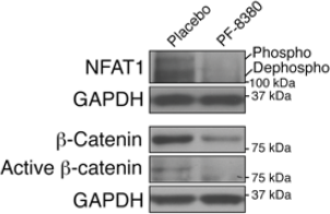
PF-8380 purchased from MedChemExpress. Usage Cited in: J Clin Invest. 2017 Apr 3;127(4):1517-1530. [Abstract]
Western blot analysis demonstrate decreased expression of dephosphorylated NFAT1 and total and active β-catenin proteins in allografts treated with PF-8380 (n = 4/group). All representative blots shown are from the same biological samples. Total and active β-catenin are blotted simultaneously on 2 parallel gels.
-
Cancer Lett
Combined inhibition of EZH2 and the autotaxin-LPA-LPA2 axis exerts synergistic antitumor effects on colon cancer cells. [Abstract]2023 Jul 10:566:216226. PMID: 37230222 -
J Mol Med (Berl)
Autotaxin stimulates LPA2 receptor in macrophages and exacerbates dextran sulfate sodium-induced acute colitis. [Abstract]2020 Dec;98(12):1781-1794. PMID: 33128578 -
Sci Rep
Loss of FOXF1 expression promotes human lung-resident mesenchymal stromal cell migration via ATX/LPA/LPA1 signaling axis. [Abstract]2020 Dec 4;10(1):21231. PMID: 33277571


Solvent & Solubility
In Vitro:
DMSO : 100 mg/mL (209.06 mM; Need ultrasonic; Hygroscopic DMSO has a significant impact on the solubility of product, please use newly opened DMSO)
Please refer to the solubility information to select the appropriate solvent. Once prepared, please aliquot and store the solution to prevent product inactivation from repeated freeze-thaw cycles.
Storage method and period of stock solution: -80°C, 2 years; -20°C, 1 year. When stored at -80°C, please use it within 2 years. When stored at -20°C, please use it within 1 year.
Please refer to the solubility information to select the appropriate solvent. Once prepared, please aliquot and store the solution to prevent product inactivation from repeated freeze-thaw cycles.
Storage method and period of stock solution: -80°C, 2 years; -20°C, 1 year. When stored at -80°C, please use it within 2 years. When stored at -20°C, please use it within 1 year.
Concentration (start) × Volume (start) = Concentration (final) × Volume (final)
In Vivo:
Select the appropriate dissolution method based on your experimental animal and administration route.
- For the following dissolution methods, please ensure to first prepare a clear stock solution using an In Vitro approach and then sequentially add co-solvents:
- To ensure reliable experimental results, the clarified stock solution can be appropriately stored based on storage conditions. As for the working solution for In Vivo experiments, it is recommended to prepare freshly and use it on the same day.
- The percentages shown for the solvents indicate their volumetric ratio in the final prepared solution. If precipitation or phase separation occurs during preparation, heat and/or sonication can be used to aid dissolution.
Add each solvent one by one: 10% DMSO 40% PEG300 5% Tween-80 45% Saline
Solubility: ≥ 0.67 mg/mL (1.40 mM); Clear solution
This protocol yields a clear solution of ≥ 0.67 mg/mL (saturation unknown).
Taking 1 mL working solution as an example, add 100 μL DMSO stock solution (6.7 mg/mL) to 400 μL PEG300, and mix evenly; then add 50 μL Tween-80 and mix evenly; then add 450 μL Saline to adjust the volume to 1 mL.
Preparation of Saline: Dissolve 0.9 g sodium chloride in ddH₂O and dilute to 100 mL to obtain a clear Saline solution.
Add each solvent one by one: 10% DMSO 90% (20% SBE-β-CD in Saline)
Solubility: ≥ 0.67 mg/mL (1.40 mM); Clear solution
This protocol yields a clear solution of ≥ 0.67 mg/mL (saturation unknown).
Taking 1 mL working solution as an example, add 100 μL DMSO stock solution (6.7 mg/mL) to 900 μL 20% SBE-β-CD in Saline, and mix evenly.
Preparation of 20% SBE-β-CD in Saline (4°C, storage for one week): 2 g SBE-β-CD powder is dissolved in 10 mL Saline, completely dissolve until clear.
For the following dissolution methods, please prepare the working solution directly:
It is recommended to prepare fresh solutions and use them promptly within a short period of time.
The percentages shown for the solvents indicate their volumetric ratio in the final prepared solution. If precipitation or phase separation occurs during preparation, heat and/or sonication can be used to aid dissolution.
Add each solvent one by one: 50% PEG300 50% Saline
Solubility: 10 mg/mL (20.91 mM); Suspended solution; Need ultrasonic
Add each solvent one by one: 0.5% CMC-Na/saline water
Solubility: 5 mg/mL (10.45 mM); Suspended solution; Need ultrasonic
Add each solvent one by one: 15% Solutol HS 15 10% Cremophor EL 35% PEG400 40% Water
Solubility: 10 mg/mL (20.91 mM); Suspended solution; Need ultrasonic
In Vivo Dissolution Calculator
Please enter the basic information of animal experiments:
-
-
-
-
Recommended: Prepare an additional quantity of animals to account for potential losses during experiments.
Please enter your animal formula composition:
-
%DMSO +
Recommended: Keep the proportion of DMSO in working solution below 2% if your animal is weak.
-
%+
-
+%Tween-80 + +
-
%Saline +
The co-solvents required include: DMSO, . All of co-solvents are available by MedChemExpress (MCE). , Tween 80. All of co-solvents are available by MedChemExpress (MCE).
Working solution concentration: 0.22 mg/mL
Method for preparing stock solution: mg drug dissolved in μL DMSO. Stock solution concentration: mg/mL.
1. Take μL DMSO stock solution;
2. Add μL .
μL , mix evenly;
3. Then add μL Tween 80, mix evenly;
4. Then add μL
Please ensure that the stock solution in the first step is dissolved to a clear state, and add co-solvents in sequence. You can use ultrasonic heating (ultrasonic cleaner, recommended frequency 20-40 kHz), vortexing, etc. to assist dissolution.


Purity & Documentation
-
Data Sheet (279 KB)
-
SDS (393 KB)
- English - EN (393 KB)
- Français - FR (393 KB)
- Deutsch - DE (393 KB)
- Norwegian - NO (393 KB)
- Español - ES (393 KB)
- Swedish - SV (393 KB)
- Italian - IT (393 KB)
- Korean - KR (393 KB)
- Portuguese - PT (393 KB)
-
Handling Instructions (2659 KB)
References
[1]. Gierse J, et al. A novel autotaxin inhibitor reduces lysophosphatidic acid levels in plasma and the site of inflammation. J Pharmacol Exp Ther. 2010 Jul;334(1):310-7. [Content Brief]
[2]. Bhave SR, et al. Autotaxin Inhibition with PF-8380 Enhances the Radiosensitivity of Human and Murine Glioblastoma Cell Lines. Front Oncol. 2013 Sep 17;3:236. [Content Brief]
[3]. Cao P, et al. Autocrine lysophosphatidic acid signaling activates β-catenin and promotes lung allograft fibrosis. J Clin Invest. 2017 Apr 3;127(4):1517-1530. [Content Brief]


Complete Stock Solution Preparation Table
Please refer to the solubility information to select the appropriate solvent. Once prepared, please aliquot and store the solution to prevent product inactivation from repeated freeze-thaw cycles.
Storage method and period of stock solution: -80°C, 2 years; -20°C, 1 year. When stored at -80°C, please use it within 2 years. When stored at -20°C, please use it within 1 year.
| Optional Solvent | Concentration Solvent Mass | 1 mg | 5 mg | 10 mg | 25 mg |
|---|---|---|---|---|---|
| DMSO | 1 mM | 2.0906 mL | 10.4530 mL | 20.9061 mL | 52.2652 mL |
| 5 mM | 0.4181 mL | 2.0906 mL | 4.1812 mL | 10.4530 mL | |
| 10 mM | 0.2091 mL | 1.0453 mL | 2.0906 mL | 5.2265 mL | |
| 15 mM | 0.1394 mL | 0.6969 mL | 1.3937 mL | 3.4843 mL | |
| 20 mM | 0.1045 mL | 0.5227 mL | 1.0453 mL | 2.6133 mL | |
| 25 mM | 0.0836 mL | 0.4181 mL | 0.8362 mL | 2.0906 mL | |
| 30 mM | 0.0697 mL | 0.3484 mL | 0.6969 mL | 1.7422 mL | |
| 40 mM | 0.0523 mL | 0.2613 mL | 0.5227 mL | 1.3066 mL | |
| 50 mM | 0.0418 mL | 0.2091 mL | 0.4181 mL | 1.0453 mL | |
| 60 mM | 0.0348 mL | 0.1742 mL | 0.3484 mL | 0.8711 mL | |
| 80 mM | 0.0261 mL | 0.1307 mL | 0.2613 mL | 0.6533 mL | |
| 100 mM | 0.0209 mL | 0.1045 mL | 0.2091 mL | 0.5227 mL |


PF-8380 Related Classifications
HY-13344 Related Classifications
 Powered by Bioz