



EYA2 Antibody (YA2905)
(Synonyms: EAB1)
Based on 1 Customer Validation
EYA2 Antibody (YA2905) is a Rabbit-derived and non-conjugated IgG monoclonal antibody, targeting to EYA2.
-
Host:
Rabbit
-
Isotype:
IgG
-
Application:
WB, IP
-
Reactivity :
Human
-
Formulation:
Supplied in 50mM Tris-Glycine(pH 7.4), 0.15M NaCl, 40% Glycerol, 0.01% Sodium azide and 0.05% BSA
-
Conjugation:
Non-conjugated


To place orders, for customer services and technical support, please contact: MedChemExpress USA
Tel: 609-228-6898 E-mail: [email protected] [email protected]





Applications
| Application |
WB

WB: Western Blot
|
IP
IP: Immunoprecipitation
|
|---|---|---|
| Dilution Ratio | 1:500-1:1000 | 1:20 |


Product Details
EYA2 Antibody (YA2905) is a Rabbit-derived and non-conjugated IgG monoclonal antibody, targeting to EYA2.
-
Host Rabbit

-
Clonality Recombinant,Monoclonal

-
Species ReactivityHuman
-
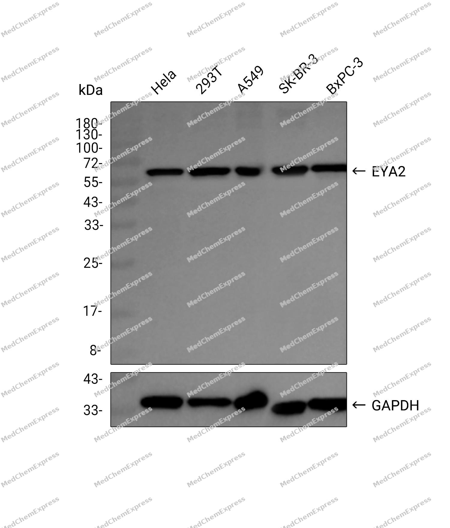
Observed Molecular WeightObserved band size: 59 kDa
Note: Due to possible protein modifications or aggregation, the molecular weight should be confirmed by actual measurement, and the predicted value is for reference only.

-
Calculated Molecular Weight Predicted band size: 59 kDa

A synthetic peptide of human EYA2
Endogenous
Affinity Purified
Non-conjugated
Unmodified
IgG


Product Properties
-
Appearance
Solution
-
Formulation
Supplied in 50mM Tris-Glycine(pH 7.4), 0.15M NaCl, 40% Glycerol, 0.01% Sodium azide and 0.05% BSA
-
Concentration
Batch-dependent, Please check the COA for the concentration of each lot. Check Lot Concentration
-
Storage & Stability
Stored at -20°C for 1 year. Avoid repeated freeze / thaw cycles.
-
Shipping
Shipping with blue ice.


Verification Images
-
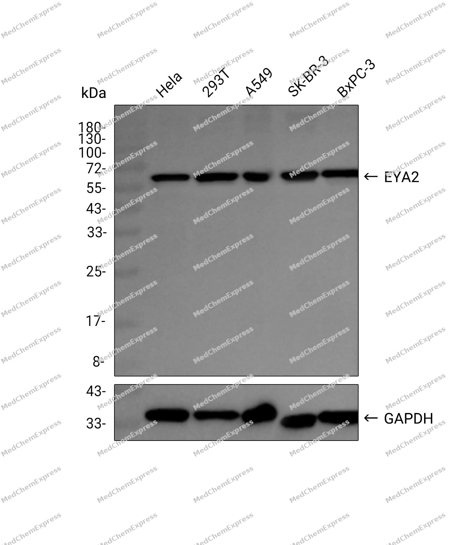 Western blot analysis was performed on extracts from Hela (lane 1, 15 μg), 293T (lane 2, 15 μg), A549 (lane 3, 15 μg), SK-BR-3 (lane 4, 15 μg), and BxPC-3 (lane 5, 15 μg) using EYA2 Rabbit mAb.Proteins were transferred to a PVDF membrane and blocked with 5% non-fat milk in TBST at 4°C overnight.The primary antibody (1:1000 dilution) and the loading control antibody (GAPDH, HY-P80137, 1:20000 dilution) were incubated in 5% non-fat milk in TBST for 1 hour at 37°C.Goat Anti-Rabbit IgG-HRP Secondary Antibody (1:20000 dilution) was then applied for 40 minutes at 37°C.


Background
-
Function
EYA2 functions both as protein phosphatase and as transcriptional coactivator for SIX1, and probably also for SIX2, SIX4 and SIX5. Tyrosine phosphatase that dephosphorylates 'Tyr-142' of histone H2AX (H2AXY142ph) and promotes efficient DNA repair via the recruitment of DNA repair complexes containing MDC1. 'Tyr-142' phosphorylation of histone H2AX plays a central role in DNA repair and acts as a mark that distinguishes between apoptotic and repair responses to genotoxic stress. Its function as histone phosphatase may contribute to its function in transcription regulation during organogenesis. Plays an important role in hypaxial muscle development together with SIX1 and DACH2; in this it is functionally redundant with EYA1[1][2][3].
-
Subcellular Localization
Cytoplasm; Nucleus
-
Expression
Tissue_specificity:Highest expression in muscle with lower levels in kidney, placenta, pancreas, brain and heart -
Isoforms & Post-Translational Modification
O00167 has 3 isomers: O00167-1: 59232 Da (predicted); O00167-2: 56688 Da (predicted); O00167-3: 50533 Da (predicted).
-
Subunit
Interacts with DACH2 and SIX1, and probably with SIX2, SIX4 and SIX5. Interacts with CAPN8 (By similarity). Interacts with GNAZ and GNAI2; this precludes interaction with SIX1
-
SwissProt ID
-
Synonyms
EAB1
-
Research Field
Epigenetics and Nuclear Signaling


Documentation
-
Data Sheet (260 KB)
-
SDS (251 KB)
- English - EN (251 KB)
- Français - FR (251 KB)
- Deutsch - DE (251 KB)
- Norwegian - NO (251 KB)
- Español - ES (251 KB)
- Swedish - SV (251 KB)
- Italian - IT (251 KB)
- Korean - KR (251 KB)
- Portuguese - PT (251 KB)
-
User Guide for Antibodies (1077 KB)
References
[1]. Fougerousse F, et al. Six and Eya expression during human somitogenesis and MyoD gene family activation. J Muscle Res Cell Motil. 2002;23(3):255-64. [Content Brief]
[2]. Patrick AN, et al. Structure-function analyses of the human SIX1-EYA2 complex reveal insights into metastasis and BOR syndrome. Nat Struct Mol Biol. 2013 Apr;20(4):447-53. [Content Brief]
[3]. Krishnan N, et al. Dephosphorylation of the C-terminal tyrosyl residue of the DNA damage-related histone H2A.X is mediated by the protein phosphatase eyes absent. J Biol Chem. 2009 Jun 12;284(24):16066-16070. [Content Brief]

 Powered by Bioz
