



Fbxw7 Antibody (YA1270)
(Synonyms: FBXW7; FBW7 ; FBX30; SEL10 ; F-box/WD repeat-containing protein 7; F-box protein FBX30; SEL-10; hCdc4)
Based on 1 Customer Validation
Fbxw7 Antibody (YA1270) is a Rabbit-derived and non-conjugated IgG monoclonal antibody, targeting to Fbxw7.
-
Host:
Rabbit
-
Isotype:
IgG
-
Application:
IHC-P
-
Reactivity :
Human
-
Formulation:
Supplied in 50mM Tris-Glycine(pH 7.4), 0.15M NaCl, 40%Glycerol, 0.01% sodium azide and 0.05% BSA.
-
Conjugation:
Non-conjugated


To place orders, for customer services and technical support, please contact: MedChemExpress USA
Tel: 609-228-6898 E-mail: [email protected] [email protected]





Applications
| Application |
IHC-P

IHC-P: Immunohistochemistry-Paraffin
|
|---|---|
| Dilution Ratio | 1:100-1:200 |


Product Details
Fbxw7 Antibody (YA1270) is a Rabbit-derived and non-conjugated IgG monoclonal antibody, targeting to Fbxw7.
-
Host Rabbit

-
Clonality Recombinant,Monoclonal

-
Species ReactivityHuman
-
Observed Molecular WeightObserved band size: 69 kDa
Note: Due to possible protein modifications or aggregation, the molecular weight should be confirmed by actual measurement, and the predicted value is for reference only.

-
Calculated Molecular Weight Predicted band size: 80 kDa

A synthesized peptide derived from human Fbxw7
Endogenous
Affinity Purified
Non-conjugated
Unmodified
IgG


Product Properties
-
Appearance
Liquid
-
Formulation
Supplied in 50mM Tris-Glycine(pH 7.4), 0.15M NaCl, 40%Glycerol, 0.01% sodium azide and 0.05% BSA.
-
Concentration
Batch-dependent, Please check the COA for the concentration of each lot. Check Lot Concentration
-
Storage & Stability
Stored at -20°C for 1 year. Avoid repeated freeze / thaw cycles.
-
Shipping
Shipping with blue ice.


Verification Images
-
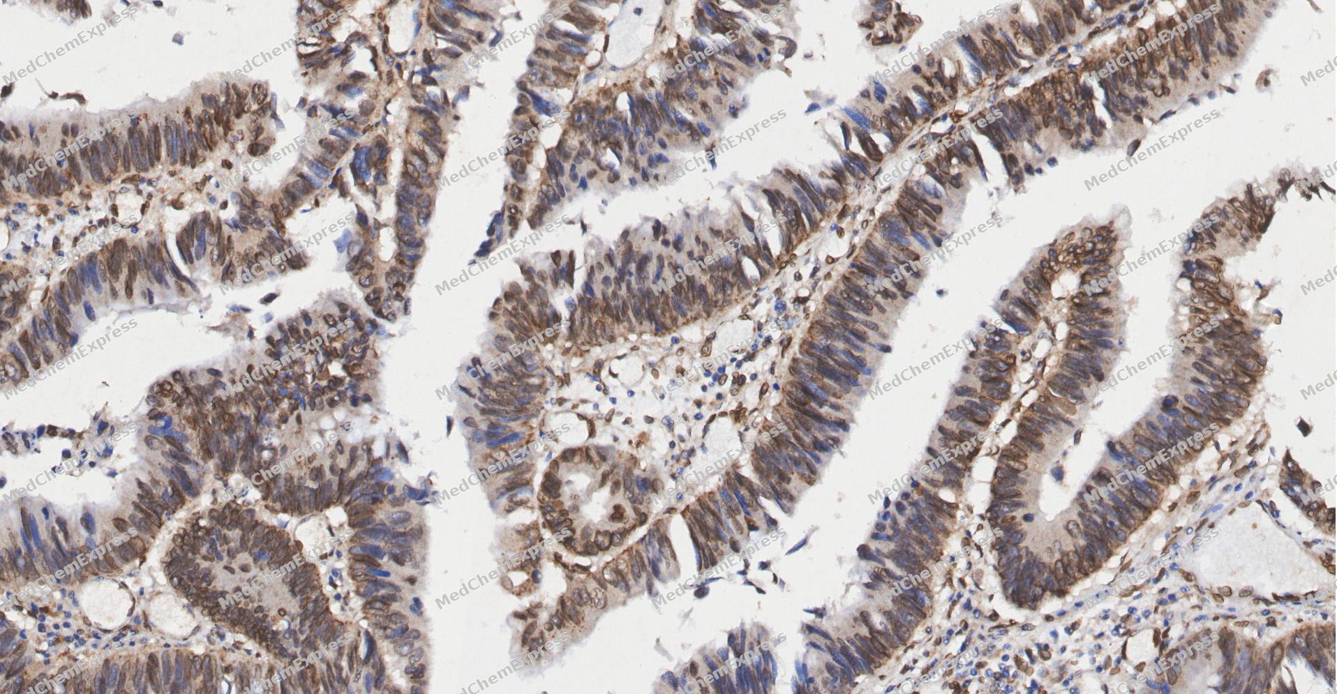 Immunohistochemical analysis of paraffin-embedded human Colon cancer tissue using Fbxw7 antibody was performed. The section was pretreated using high-temperature and high-pressure mediated EDTA antigen retrieval buffer (pH 9.0), for 5 minutes. The tissues were incubated with primary antibody (HY-P81525, 1:100 dilution) at room temperature for 60 minutes. Detection was performed using an HRP conjugated compact polymer system. DAB was used as the chromogen. The tissues were counterstained with hematoxylin and mounted with neutral balsam mounting medium.
 -
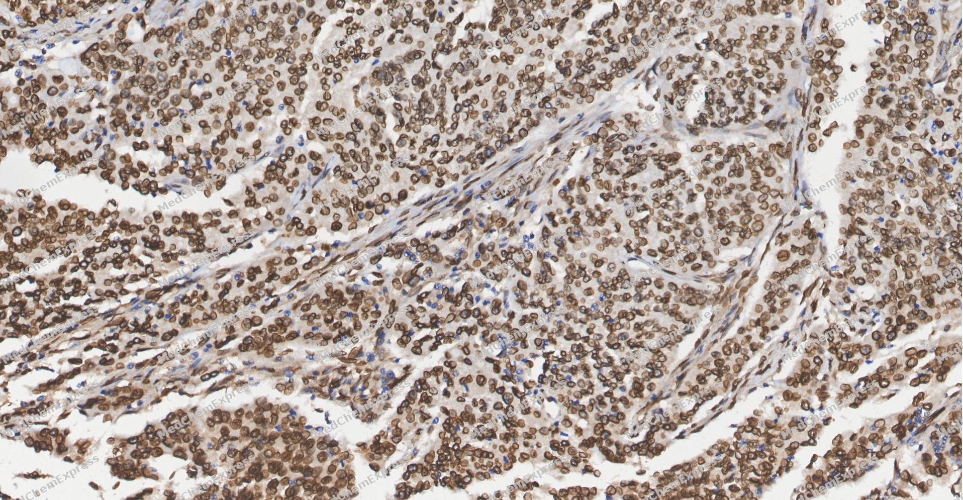 Immunohistochemical analysis of paraffin-embedded human Gastric Cancer tissue using Fbxw7 antibody was performed. The section was pretreated using high-temperature and high-pressure mediated EDTA antigen retrieval buffer (pH 9.0), for 5 minutes. The tissues were incubated with primary antibody (HY-P81525, 1:100 dilution) at room temperature for 60 minutes. Detection was performed using an HRP conjugated compact polymer system. DAB was used as the chromogen. The tissues were counterstained with hematoxylin and mounted with neutral balsam mounting medium.
 -
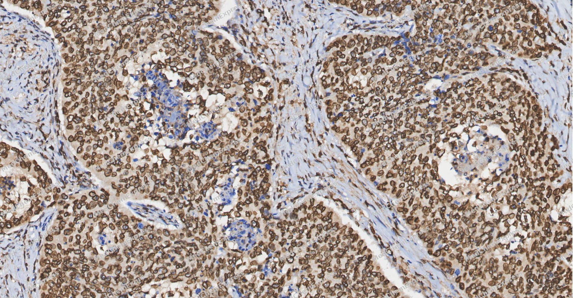 Immunohistochemical analysis of paraffin-embedded human Esophageal Carcinoma tissue using Fbxw7 antibody was performed. The section was pretreated using high-temperature and high-pressure mediated EDTA antigen retrieval buffer (pH 9.0), for 5 minutes. The tissues were incubated with primary antibody (HY-P81525, 1:100 dilution) at room temperature for 60 minutes. Detection was performed using an HRP conjugated compact polymer system. DAB was used as the chromogen. The tissues were counterstained with hematoxylin and mounted with neutral balsam mounting medium.
 -
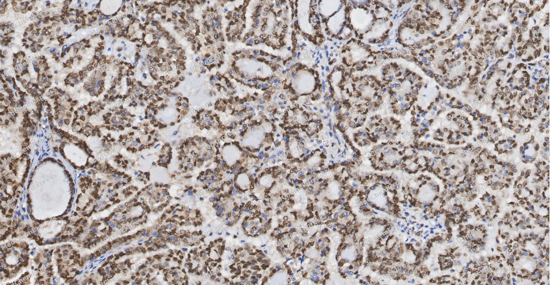 Immunohistochemical analysis of paraffin-embedded human ovarian carcinoma tissue using Fbxw7 antibody was performed. The section was pretreated using high-temperature and high-pressure mediated EDTA antigen retrieval buffer (pH 9.0), for 5 minutes. The tissues were incubated with primary antibody (HY-P81525, 1:100 dilution) at room temperature for 60 minutes. Detection was performed using an HRP conjugated compact polymer system. DAB was used as the chromogen. The tissues were counterstained with hematoxylin and mounted with neutral balsam mounting medium.
 -
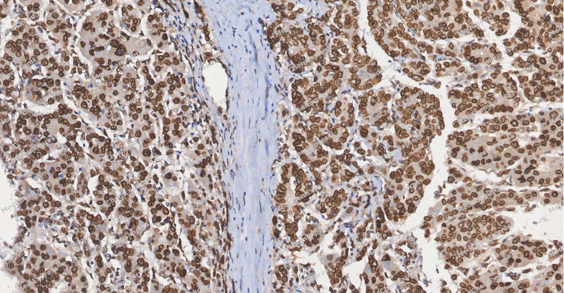 Immunohistochemical analysis of paraffin-embedded human Liver cancer tissue using Fbxw7 antibody was performed. The section was pretreated using high-temperature and high-pressure mediated EDTA antigen retrieval buffer (pH 9.0), for 5 minutes. The tissues were incubated with primary antibody (HY-P81525, 1:100 dilution) at room temperature for 60 minutes. Detection was performed using an HRP conjugated compact polymer system. DAB was used as the chromogen. The tissues were counterstained with hematoxylin and mounted with neutral balsam mounting medium.
 -
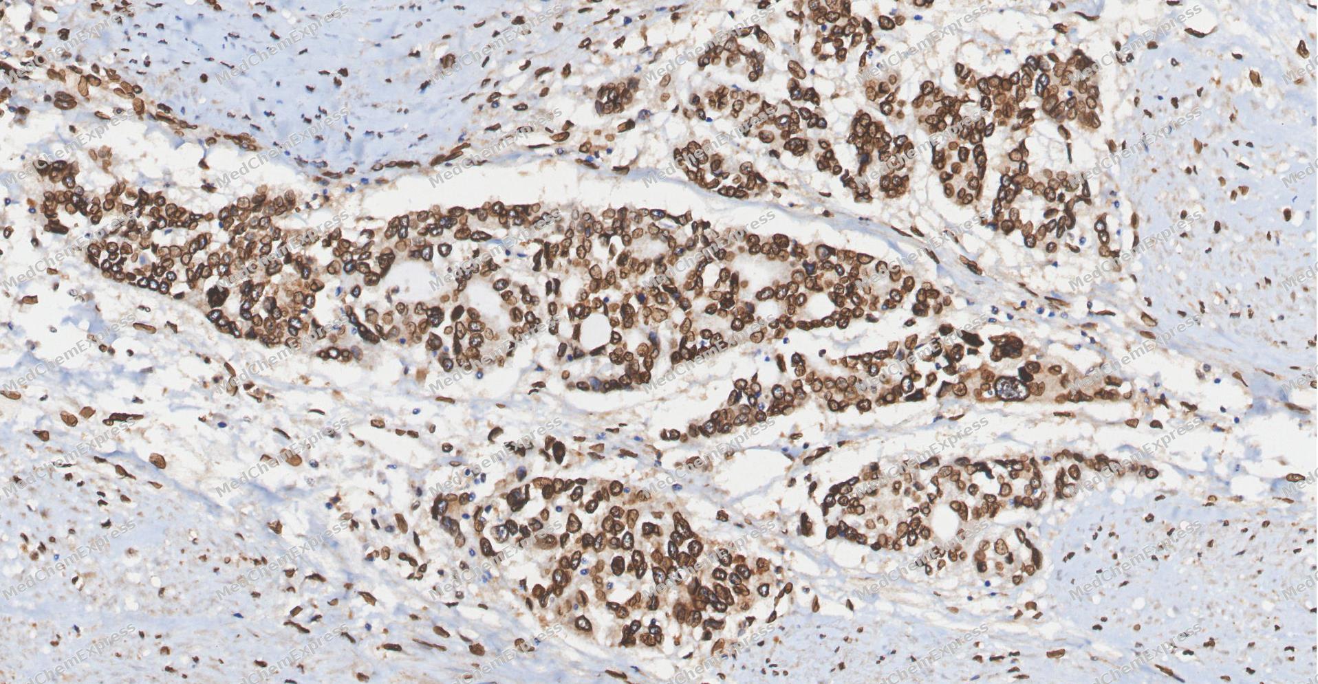 Immunohistochemical analysis of paraffin-embedded human bladder cancer tissue using Fbxw7 antibody was performed. The section was pretreated using high-temperature and high-pressure mediated EDTA antigen retrieval buffer (pH 9.0), for 5 minutes. The tissues were incubated with primary antibody (HY-P81525, 1:100 dilution) at room temperature for 60 minutes. Detection was performed using an HRP conjugated compact polymer system. DAB was used as the chromogen. The tissues were counterstained with hematoxylin and mounted with neutral balsam mounting medium.


Background
-
Function
Fbxw7 is a Substrate recognition component of a SCF (SKP1-CUL1-F-box protein) E3 ubiquitin-protein ligase complex which mediates the ubiquitination and subsequent proteasomal degradation of target proteins. Recognizes and binds phosphorylated sites/phosphodegrons within target proteins and thereafter brings them to the SCF complex for ubiquitination. Identified substrates include cyclin-E (CCNE1 or CCNE2), DISC1, JUN, MYC, NOTCH1 released notch intracellular domain (NICD), NFE2L1, NOTCH2, MCL1, MLST8, RICTOR, and probably PSEN1. Acts as a negative regulator of JNK signaling by binding to phosphorylated JUN and promoting its ubiquitination and subsequent degradation. Involved in bone homeostasis and negative regulation of osteoclast differentiation. Regulates the amplitude of the cyclic expression of hepatic core clock genes and genes involved in lipid and glucose metabolism via ubiquitination and proteasomal degradation of their transcriptional repressor NR1D1; CDK1-dependent phosphorylation of NR1D1 is necessary for SCF(FBXW7)-mediated ubiquitination. Also able to promote 'Lys-63'-linked ubiquitination in response to DNA damage. The SCF(FBXW7) complex facilitates double-strand break repair following phosphorylation by ATM: phosphorylation promotes localization to sites of double-strand breaks and 'Lys-63'-linked ubiquitination of phosphorylated XRCC4, enhancing DNA non-homologous end joining[1][2][3][4][5][6][7][8][9][10][11][12][13][14][15][16][17][18][19][20][21].
-
Subcellular Localization
Nucleus, nucleoplasm; Chromosome; Cytoplasm; Nucleus, nucleolus
-
Expression
Tissue_specificity:Widely expressed; Expressed in brain -
Isoforms & Post-Translational Modification
Q969H0 has 3 isomers: Q969H0-1: 79663 Da (predicted); Q969H0-2: 70324 Da (predicted); Q969H0-4: 66120 Da (predicted).
Phosphorylation at Thr-205 promotes interaction with PIN1, leading to disrupt FBXW7 dimerization and promoting FBXW7 autoubiquitination and degradation (PubMed:22608923). Phosphorylated by ATM at Ser-26 in response to DNA damage, promoting recruitment to DNA damage sites and 'Lys-63'-linked ubiquitination of phosphorylated XRCC4 (PubMed:26774286);Ubiquitinated: autoubiquitinates following phosphorylation at Thr-205 and subsequent interaction with PIN1. Ubiquitination leads to its proteasomal degradation (PubMed:22608923) -
Subunit
Homodimer; homodimerization plays a role in substrate binding and/or ubiquitination and degradation (PubMed:17434132, PubMed:22608923, PubMed:28007894). Component of the SCF(FBXW7) complex consisting of CUL1, RBX1, SKP1 and FBXW7 (PubMed:11565034, PubMed:15103331, PubMed:22748924, PubMed:26774286, PubMed:26976582, PubMed:28007894, PubMed:28727686, PubMed:34741373). Interacts (via F-box domain) with SKP1 (PubMed:11585921, PubMed:17434132, PubMed:26976582, PubMed:28007894, PubMed:28727686). Interacts (via F-box domain) with pseudophosphatase STYX; the interaction is direct and prevents FBXW7 interaction with SKP1 (PubMed:28007894). Interacts with cyclin-E (CCNE1 or CCNE2) (PubMed:11565034, PubMed:17434132). Interacts with PSEN1 (PubMed:12354302). Forms a trimeric complex with NOTCH1 and SGK1 (PubMed:21147854). Interacts with NOTCH1 intracellular domain/NICD and NOTCH4 intracellular domain/NICD (PubMed:11585921). Interacts with NOTCH2 intracellular domain (N2ICD) (PubMed:29149593). Interacts with MYC (when phosphorylated) (PubMed:17873522, PubMed:25775507, PubMed:28007894). Interacts with USP28, counteracting ubiquitination of MYC (PubMed:17873522). Interacts with JUN (PubMed:14739463, PubMed:22608923). Found in a complex with JUN and PRR7 (PubMed:27458189). Interacts with JUN and PRR7; the interaction inhibits ubiquitination-mediated JUN degradation, promoting its phosphorylation and transcriptional activity (PubMed:27458189). Interacts (when phosphorylated at Thr-205) with PIN1, disrupting FBXW7 dimerization and promoting FBXW7 autoubiquitination and degradation (PubMed:22608923). Interacts with UBE2QL1 (PubMed:24000165). Interacts with FAM83D; promotes FBXW7 degradation (PubMed:24344117). Interacts with MYCN; FBXW7 competes with AURKA for binding to unphosphorylated MYCN but not for binding to phosphorylated MYCN (PubMed:27837025). Interacts with STOML1 (PubMed:23082202). Interacts with NFE2L1 (By similarity). Interacts with USP36, counteracting ubiquitination of MYC (PubMed:25775507). Interacts with NR1D1 (PubMed:27238018). Interacts with RICTOR; mediates RICTOR ubiquitination and degradation (PubMed:25897075). Interacts with USP38, counteracting ubiquitination of MYC (PubMed:34102342)
-
SwissProt ID
-
Synonyms
FBXW7; FBW7 ; FBX30; SEL10 ; F-box/WD repeat-containing protein 7; F-box protein FBX30; SEL-10; hCdc4
-
Research Field
Cell Biology


Documentation
-
Data Sheet (235 KB)
-
SDS (252 KB)
- English - EN (252 KB)
- Français - FR (252 KB)
- Deutsch - DE (252 KB)
- Norwegian - NO (252 KB)
- Español - ES (252 KB)
- Swedish - SV (252 KB)
- Italian - IT (252 KB)
- Korean - KR (252 KB)
- Portuguese - PT (252 KB)
-
User Guide for Antibodies (1077 KB)

 Powered by Bioz
