



HSD11B1 Antibody (YA3376)
(Synonyms: 11-DH; 11-beta-HSD1; HSD11B1; HSD11; HSD11L; 11 beta HSD1)
Based on 1 Customer Validation
HSD11B1 Antibody (YA3376) is a Rabbit-derived and non-conjugated IgG monoclonal antibody, targeting to HSD11B1.
-
Host:
Rabbit
-
Isotype:
IgG
-
Application:
WB, IHC-P
-
Reactivity :
Human, Mouse, Rat
-
Formulation:
Supplied in 50mM Tris-Glycine(pH 7.4), 0.15M NaCl, 40%Glycerol, 0.01% sodium azide and 0.05% BSA.
-
Conjugation:
Non-conjugated


To place orders, for customer services and technical support, please contact: MedChemExpress USA
Tel: 609-228-6898 E-mail: [email protected] [email protected]





Applications
| Application |
WB

WB: Western Blot
|
IHC-P
IHC-P: Immunohistochemistry-Paraffin
|
|---|---|---|
| Dilution Ratio | 1:500-1:1000 | 1:50-1:100 |


Product Details
HSD11B1 Antibody (YA3376) is a Rabbit-derived and non-conjugated IgG monoclonal antibody, targeting to HSD11B1.
-
Host Rabbit

-
Clonality Recombinant,Monoclonal

-
Species ReactivityHuman, Mouse, Rat
-
Observed Molecular WeightObserved band size: 32 kDa
Note: Due to possible protein modifications or aggregation, the molecular weight should be confirmed by actual measurement, and the predicted value is for reference only.

-
Calculated Molecular Weight Predicted band size: 32 kDa

A synthesized peptide derived from human HSD11B1 aa1-57/292.
Endogenous
Affinity Chromatography
Non-conjugated
Unmodified
IgG


Product Properties
-
Appearance
Liquid
-
Formulation
Supplied in 50mM Tris-Glycine(pH 7.4), 0.15M NaCl, 40%Glycerol, 0.01% sodium azide and 0.05% BSA.
-
Concentration
Batch-dependent, Please check the COA for the concentration of each lot. Check Lot Concentration
-
Storage & Stability
Stored at -20°C for 1 year. Avoid repeated freeze / thaw cycles.
-
Shipping
Shipping with blue ice.


Verification Images
-
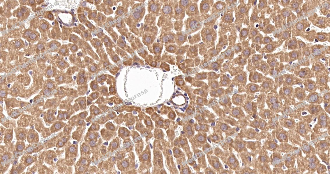 Immunohistochemical analysis of paraffin-embedded mouse liver tissue using HSD11B1 Antibody. The section was pre-treated using heat mediated antigen retrieval with sodium citrate buffer (pH 6.0) for 8 minutes. The tissues were blocked in QuickBlock for 20 minutes at room temperature, washed with ddH2O and PBS, and then probed with the primary antibody at 1/100 dilution in 4℃ overnight. The detection was performed using an HRP conjugated compact polymer system. DAB was used as the chromogen. Tissues were counterstained with hematoxylin and mounted with DPX.
 -
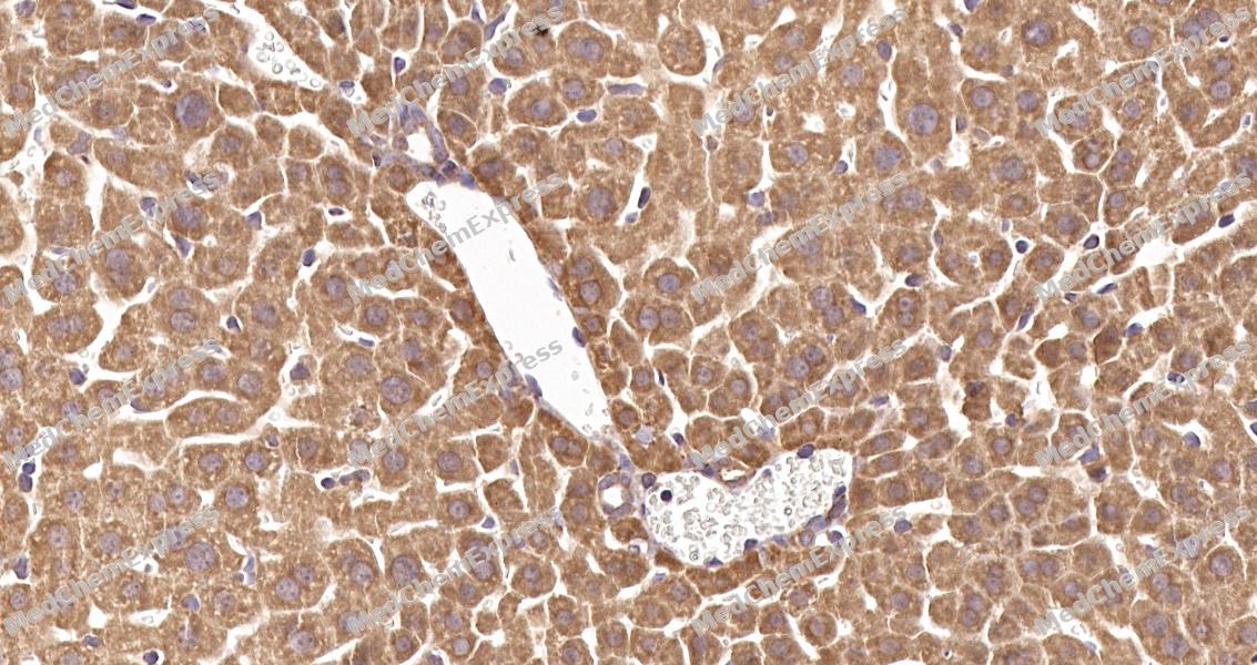 Immunohistochemical analysis of paraffin-embedded mouse liver tissue using HSD11B1 Antibody. The section was pre-treated using heat mediated antigen retrieval with sodium citrate buffer (pH 6.0) for 8 minutes. The tissues were blocked in QuickBlock for 20 minutes at room temperature, washed with ddH2O and PBS, and then probed with the primary antibody at 1/100 dilution in 4℃ overnight. The detection was performed using an HRP conjugated compact polymer system. DAB was used as the chromogen. Tissues were counterstained with hematoxylin and mounted with DPX.


Background
-
Function
HSD11B1 controls the reversible conversion of biologically active glucocorticoids such as cortisone to cortisol, and 11-dehydrocorticosterone to corticosterone in the presence of NADP(H). Participates in the corticosteroid receptor-mediated anti-inflammatory response, as well as metabolic and homeostatic processes. Plays a role in the secretion of aqueous humor in the eye, maintaining a normotensive, intraocular environment. Bidirectional in vitro, predominantly functions as a reductase in vivo, thereby increasing the concentration of active glucocorticoids. It has broad substrate specificity, besides glucocorticoids, it accepts other steroid and sterol substrates. Interconverts 7-oxo- and 7-hydroxy-neurosteroids such as 7-oxopregnenolone and 7beta-hydroxypregnenolone, 7-oxodehydroepiandrosterone (3beta-hydroxy-5-androstene-7,17-dione) and 7beta-hydroxydehydroepiandrosterone (3beta,7beta-dihydroxyandrost-5-en-17-one), among others. Catalyzes the stereo-specific conversion of the major dietary oxysterol, 7-ketocholesterol (7-oxocholesterol), into the more polar 7-beta-hydroxycholesterol metabolite. 7-oxocholesterol is one of the most important oxysterols, it participates in several events such as induction of apoptosis, accumulation in atherosclerotic lesions, lipid peroxidation, and induction of foam cell formation. Mediates the 7-oxo reduction of 7-oxolithocholate mainly to chenodeoxycholate, and to a lesser extent to ursodeoxycholate, both in its free form and when conjugated to glycine or taurine, providing a link between glucocorticoid activation and bile acid metabolism. Catalyzes the synthesis of 7-beta-25-dihydroxycholesterol from 7-oxo-25-hydroxycholesterol in vitro, which acts as a ligand for the G-protein-coupled receptor (GPCR) Epstein-Barr virus-induced gene 2 (EBI2) and may thereby regulate immune cell migration[1][2][3][4][5][6][7][8][9][10][11][12].
-
Subcellular Localization
Endoplasmic reticulum membrane; Single-pass type II membrane protein
-
Expression
Tissue_specificity:Widely expressed, highest expression in liver, lower in testis, ovary, lung, foreskin fibroblasts, and much lower in kidney (PubMed:1885595) . Expressed in liver (at protein level) (PubMed:21453287) . Expressed in the basal cells of the corneal epithelium and in the ciliary nonpigmented epithelium (both at mRNA and at protein level) (PubMed:11481269) -
Subunit
Homodimer
-
SwissProt ID
-
Synonyms
11-DH; 11-beta-HSD1; HSD11B1; HSD11; HSD11L; 11 beta HSD1
-
Research Field
Signal Transduction


Documentation
-
Data Sheet (234 KB)
-
SDS (251 KB)
- English - EN (251 KB)
- Français - FR (251 KB)
- Deutsch - DE (251 KB)
- Norwegian - NO (251 KB)
- Español - ES (251 KB)
- Swedish - SV (251 KB)
- Italian - IT (251 KB)
- Korean - KR (251 KB)
- Portuguese - PT (251 KB)
-
User Guide for Antibodies (1077 KB)

 Powered by Bioz
