



Ihh Antibody (YA3011)
(Synonyms: BDA1; HHG2)Based on 1 Customer Validation
Ihh Antibody (YA3011) is a Rabbit-derived and non-conjugated IgG monoclonal antibody, targeting to Ihh.
-
Host:
Rabbit
-
Isotype:
IgG
-
Application:
WB, IHC-P
-
Reactivity :
Human, Mouse
-
Formulation:
Supplied in 50mM Tris-Glycine(pH 7.4), 0.15M NaCl, 40% Glycerol, 0.01% Sodium azide and 0.05% BSA
-
Conjugation:
Non-conjugated


To place orders, for customer services and technical support, please contact: MedChemExpress USA
Tel: 609-228-6898 E-mail: [email protected] [email protected]





Applications
| Application |
WB

WB: Western Blot
|
IHC-P
IHC-P: Immunohistochemistry-Paraffin
|
|---|---|---|
| Dilution Ratio | 1:500-1:1000 | 1:50-1:100 |


Product Details
Ihh Antibody (YA3011) is a Rabbit-derived and non-conjugated IgG monoclonal antibody, targeting to Ihh.
-
Host Rabbit

-
Clonality Recombinant,Monoclonal

-
Species ReactivityHuman, Mouse
-
Observed Molecular WeightObserved band size: 45 kDa
Note: Due to possible protein modifications or aggregation, the molecular weight should be confirmed by actual measurement, and the predicted value is for reference only.

-
Calculated Molecular Weight Predicted band size: 45 kDa

A synthetic peptide of human Ihh
Endogenous
Affinity Purified
Non-conjugated
Unmodified
IgG


Product Properties
-
Appearance
Solution
-
Formulation
Supplied in 50mM Tris-Glycine(pH 7.4), 0.15M NaCl, 40% Glycerol, 0.01% Sodium azide and 0.05% BSA
-
Concentration
Batch-dependent, Please check the COA for the concentration of each lot. Check Lot Concentration
-
Storage & Stability
Stored at -20°C for 1 year. Avoid repeated freeze / thaw cycles.
-
Shipping
Shipping with blue ice.


Verification Images
-
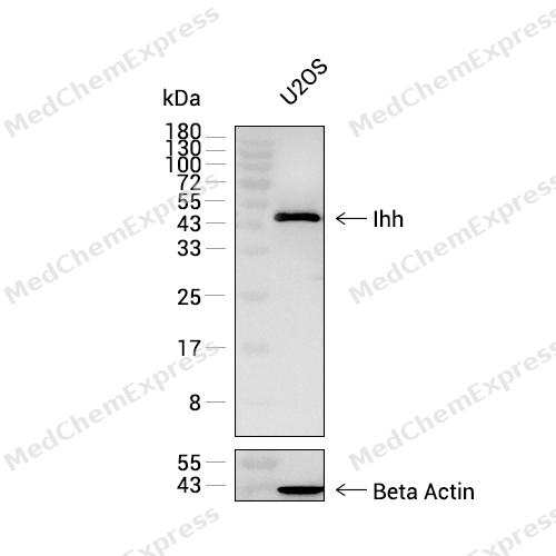 Western blot analysis was performed on extracts from U2OS (lane 1, 20 μg) using Ihh Rabbit mAb.Proteins were transferred to a PVDF membrane and blocked with 5% non - fat milk in TBST at 4°C overnight.The primary antibody (1:1000 dilution) and the loading control antibody (beta-Actin, HY-P83730, 1:5000 dilution) were incubated in 5% non-fat milk in TBST for 1 hour at 37°C.Goat Anti - Rabbit IgG - HRP Secondary Antibody (1:20000 dilution) was then applied for 40 minutes at 37°C.
 -
 Immunohistochemical analysis of paraffin-embedded human pancreas using Ihh antibody. The section was pre-treated using heat mediated antigen retrieval with EDTA (pH 9.0) for 14 minutes. The tissues were blocked in 5% BSA for 30 minutes at room temperature, washed with TBST, and then probed with the primary antibody (HY-P83266, 1/100) for 30 minutes at room temperature. The detection was performed using Polymer HRP-conjugated Goat Anti-Mouse/Rabbit lgG(H&L) secondary antibody (HY-P83652). DAB was used as the chromogen. Tissues were counterstained with hematoxylin and mounted with DPX.
 -
 Immunohistochemical analysis of paraffin-embedded human lung cancer using Ihh antibody. The section was pre-treated using heat mediated antigen retrieval with EDTA (pH 9.0) for 14 minutes. The tissues were blocked in 5% BSA for 30 minutes at room temperature, washed with TBST, and then probed with the primary antibody (HY-P83266, 1/100) for 30 minutes at room temperature. The detection was performed using Polymer HRP-conjugated Goat Anti-Mouse/Rabbit lgG(H&L) secondary antibody (HY-P83652). DAB was used as the chromogen. Tissues were counterstained with hematoxylin and mounted with DPX.
 -
 Immunohistochemical analysis of paraffin-embedded human ovarian cancer using Ihh antibody. The section was pre-treated using heat mediated antigen retrieval with EDTA (pH 9.0) for 14 minutes. The tissues were blocked in 5% BSA for 30 minutes at room temperature, washed with TBST, and then probed with the primary antibody (HY-P83266, 1/100) for 30 minutes at room temperature. The detection was performed using Polymer HRP-conjugated Goat Anti-Mouse/Rabbit lgG(H&L) secondary antibody (HY-P83652). DAB was used as the chromogen. Tissues were counterstained with hematoxylin and mounted with DPX.
 -
 Immunohistochemical analysis of paraffin-embedded human prostate cancer using Ihh antibody. The section was pre-treated using heat mediated antigen retrieval with EDTA (pH 9.0) for 14 minutes. The tissues were blocked in 5% BSA for 30 minutes at room temperature, washed with TBST, and then probed with the primary antibody (HY-P83266, 1/100) for 30 minutes at room temperature. The detection was performed using Polymer HRP-conjugated Goat Anti-Mouse/Rabbit lgG(H&L) secondary antibody (HY-P83652). DAB was used as the chromogen. Tissues were counterstained with hematoxylin and mounted with DPX.
 -
 Immunohistochemical analysis of paraffin-embedded human liver using Ihh antibody. The section was pre-treated using heat mediated antigen retrieval with EDTA (pH 9.0) for 14 minutes. The tissues were blocked in 5% BSA for 30 minutes at room temperature, washed with TBST, and then probed with the primary antibody (HY-P83266, 1/100) for 30 minutes at room temperature. The detection was performed using Polymer HRP-conjugated Goat Anti-Mouse/Rabbit lgG(H&L) secondary antibody (HY-P83652). DAB was used as the chromogen. Tissues were counterstained with hematoxylin and mounted with DPX.
 -
 Immunohistochemical analysis of paraffin-embedded human bladder cacner using Ihh antibody. The section was pre-treated using heat mediated antigen retrieval with EDTA (pH 9.0) for 14 minutes. The tissues were blocked in 5% BSA for 30 minutes at room temperature, washed with TBST, and then probed with the primary antibody (HY-P83266, 1/100) for 30 minutes at room temperature. The detection was performed using Polymer HRP-conjugated Goat Anti-Mouse/Rabbit lgG(H&L) secondary antibody (HY-P83652). DAB was used as the chromogen. Tissues were counterstained with hematoxylin and mounted with DPX.
 -
 Immunohistochemical analysis of paraffin-embedded human testis using Ihh antibody. The section was pre-treated using heat mediated antigen retrieval with EDTA (pH 9.0) for 14 minutes. The tissues were blocked in 5% BSA for 30 minutes at room temperature, washed with TBST, and then probed with the primary antibody (HY-P83266, 1/100) for 30 minutes at room temperature. The detection was performed using Polymer HRP-conjugated Goat Anti-Mouse/Rabbit lgG(H&L) secondary antibody (HY-P83652). DAB was used as the chromogen. Tissues were counterstained with hematoxylin and mounted with DPX.
 -
 Immunohistochemical analysis of paraffin-embedded human glioma using Ihh antibody. The section was pre-treated using heat mediated antigen retrieval with EDTA (pH 9.0) for 14 minutes. The tissues were blocked in 5% BSA for 30 minutes at room temperature, washed with TBST, and then probed with the primary antibody (HY-P83266, 1/100) for 30 minutes at room temperature. The detection was performed using Polymer HRP-conjugated Goat Anti-Mouse/Rabbit lgG(H&L) secondary antibody (HY-P83652). DAB was used as the chromogen. Tissues were counterstained with hematoxylin and mounted with DPX.
 -
 Immunohistochemical analysis of paraffin-embedded human lymph node using Ihh antibody. The section was pre-treated using heat mediated antigen retrieval with EDTA (pH 9.0) for 14 minutes. The tissues were blocked in 5% BSA for 30 minutes at room temperature, washed with TBST, and then probed with the primary antibody (HY-P83266, 1/100) for 30 minutes at room temperature. The detection was performed using Polymer HRP-conjugated Goat Anti-Mouse/Rabbit lgG(H&L) secondary antibody (HY-P83652). DAB was used as the chromogen. Tissues were counterstained with hematoxylin and mounted with DPX.
 -
 Immunohistochemical analysis of paraffin-embedded human pancreatic cancer using Ihh antibody. The section was pre-treated using heat mediated antigen retrieval with EDTA (pH 9.0) for 14 minutes. The tissues were blocked in 5% BSA for 30 minutes at room temperature, washed with TBST, and then probed with the primary antibody (HY-P83266, 1/100) for 30 minutes at room temperature. The detection was performed using Polymer HRP-conjugated Goat Anti-Mouse/Rabbit lgG(H&L) secondary antibody (HY-P83652). DAB was used as the chromogen. Tissues were counterstained with hematoxylin and mounted with DPX.
 -
 Immunohistochemical analysis of paraffin-embedded human heart using Ihh antibody. The section was pre-treated using heat mediated antigen retrieval with EDTA (pH 9.0) for 14 minutes. The tissues were blocked in 5% BSA for 30 minutes at room temperature, washed with TBST, and then probed with the primary antibody (HY-P83266, 1/100) for 30 minutes at room temperature. The detection was performed using Polymer HRP-conjugated Goat Anti-Mouse/Rabbit lgG(H&L) secondary antibody (HY-P83652). DAB was used as the chromogen. Tissues were counterstained with hematoxylin and mounted with DPX.
 -
 Immunohistochemical analysis of paraffin-embedded human placenta using Ihh antibody. The section was pre-treated using heat mediated antigen retrieval with EDTA (pH 9.0) for 14 minutes. The tissues were blocked in 5% BSA for 30 minutes at room temperature, washed with TBST, and then probed with the primary antibody (HY-P83266, 1/100) for 30 minutes at room temperature. The detection was performed using Polymer HRP-conjugated Goat Anti-Mouse/Rabbit lgG(H&L) secondary antibody (HY-P83652). DAB was used as the chromogen. Tissues were counterstained with hematoxylin and mounted with DPX.
 -
 Immunohistochemical analysis of paraffin-embedded human non-Hodgkin lymphoma using Ihh antibody. The section was pre-treated using heat mediated antigen retrieval with EDTA (pH 9.0) for 14 minutes. The tissues were blocked in 5% BSA for 30 minutes at room temperature, washed with TBST, and then probed with the primary antibody (HY-P83266, 1/100) for 30 minutes at room temperature. The detection was performed using Polymer HRP-conjugated Goat Anti-Mouse/Rabbit lgG(H&L) secondary antibody (HY-P83652). DAB was used as the chromogen. Tissues were counterstained with hematoxylin and mounted with DPX.
 -
 Immunohistochemical analysis of paraffin-embedded human head and neck cancer using Ihh antibody. The section was pre-treated using heat mediated antigen retrieval with EDTA (pH 9.0) for 14 minutes. The tissues were blocked in 5% BSA for 30 minutes at room temperature, washed with TBST, and then probed with the primary antibody (HY-P83266, 1/100) for 30 minutes at room temperature. The detection was performed using Polymer HRP-conjugated Goat Anti-Mouse/Rabbit lgG(H&L) secondary antibody (HY-P83652). DAB was used as the chromogen. Tissues were counterstained with hematoxylin and mounted with DPX.


Background
-
Function
Ihh plays a role in embryonic morphogenesis; it is involved in the regulation of endochondral skeleton formation, and the development of retinal pigment epithelium (RPE), photoreceptors and periocular tissues; The C-terminal part of the indian hedgehog protein precursor displays an autoproteolysis and a cholesterol transferase activity. Both activities result in the cleavage of the full-length protein into two parts followed by the covalent attachment of a cholesterol moiety to the C-terminal of the newly generated N-product. Both activities occur in the endoplasmic reticulum. Plays a role in hedgehog paracrine signaling. Associated with the very-low-density lipoprotein (VLDL) particles to function as a circulating morphogen for endothelial cell integrity maintenance; The dually lipidated indian hedgehog protein N-product is a morphogen which is essential for a variety of patterning events during development. Binds to the patched (PTCH1) receptor, which functions in association with smoothened (SMO), to activate the transcription of target genes. Plays a role in morphogenesis of the skeleton by coordinating growth and differentiation of the endochondral skeleton. Positively regulates PTHLH expression during endochondral bone formation preventing chondrocyte hypertrophy. In contrast, participates in normal chondrocyte proliferation in a PTHLH-independent pathway[1][2].
-
Subcellular Localization
Cell membrane; Lipid-anchor; Endoplasmic reticulum membrane; Golgi apparatus membrane; Secreted
-
Expression
Tissue_specificity:Expressed in embryonic lung, and in adult kidney and liver -
Subunit
Multimer
-
SwissProt ID
-
Synonyms
BDA1; HHG2
-
Research Field
Stem Cells


Documentation
-
Data Sheet (260 KB)
-
SDS (251 KB)
- English - EN (251 KB)
- Français - FR (251 KB)
- Deutsch - DE (251 KB)
- Norwegian - NO (251 KB)
- Español - ES (251 KB)
- Swedish - SV (251 KB)
- Italian - IT (251 KB)
- Korean - KR (251 KB)
- Portuguese - PT (251 KB)
-
User Guide for Antibodies (1077 KB)
References

 Powered by Bioz
