



Endoplasmic Reticulum Dyes
- 1
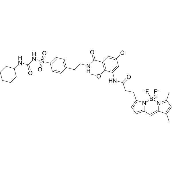
- Formula: C37H42BClF2N6O6S
- Molecular Weight: 783.09
ER-Tracker dye is a derivative of BODIPY series dyes coupled with Glibenclamide (HY-15206), highly selective binding to the endoplasmic reticulum, non-toxic to cells at low concentrations, this type of dye is an environmentally sensitive probe, and formaldehyde treatment can still retain part of the fluorescence, with high fluorescence life, good extinction coefficient and other characteristics. Glibenclamide is an atp-dependent K+ channel blocker (Kir6, KATP) and CFTR Cl-channel blocker that binds in the endoplasmic reticulum. ER-Tracker is not suitable for staining cells after fixation.
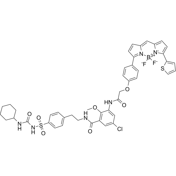
- Formula: C44H42BClF2N6O7S2
- Molecular Weight: 915.23
ER-Tracker dye is a derivative of BODIPY series dyes coupled with Glibenclamide (HY-15206), highly selective binding to the endoplasmic reticulum, non-toxic to cells at low concentrations, this type of dye is an environmentally sensitive probe, and formaldehyde treatment can still retain part of the fluorescence, with high fluorescence life, good extinction coefficient and other characteristics. Glibenclamide is an atp-dependent K+ channel blocker (Kir6, KATP) and CFTR Cl-channel blocker that binds in the endoplasmic reticulum. ER-Tracker is not suitable for staining cells after fixation. Ex/Em=587/615 nm.
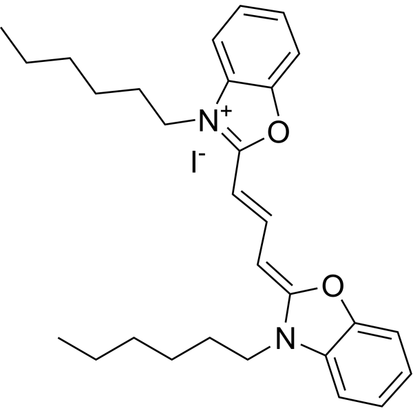
- Formula: C29H37IN2O2
- Molecular Weight: 572.52
3,3'-Dihexyloxacarbocyanine iodide is a carbocyanine dye which can be used to monitor changes in mitochondrial membrane potential.
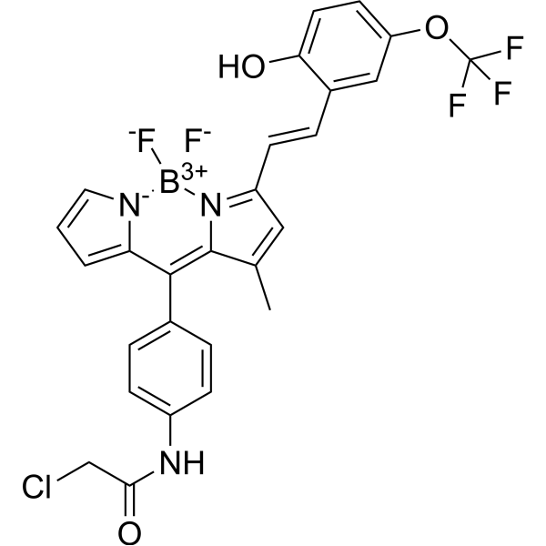
- Formula: C27H20BClF5N3O3
- Molecular Weight: 575.72
ER Tracker Yellow (BDNCA-346) is a Fluorescent probe for targeted visualization of temperature at the endoplasmic reticulum. Its detection mechanism depends on temperature, with fluorescence intensity decreasing at higher temperatures due to preferential non-radiative processes; this intensity reversibly returns to baseline when cooled, provided the probe does not precipitate or degrade. It localizes specifically to the endoplasmic reticulum, where it may covalently attach to biomacromolecules via a thiol-reactive moiety, and its fluorescence remains stable after cell fixation; its fluorescence intensity is unaffected by physiological pH ranges of 5.0–8.0 and free Ca²⁺ concentrations of 0–800 μM. Its excitation/emission wavelength is Ex/Em = 559/581 nm[1].
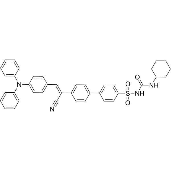
- Formula: C40H36N4O3S
- Molecular Weight: 652.80
AIE-ER is a specific endoplasmic reticulum (ER) fluorescent probe (green channel: λex=405 nm, λem=450~650 nm) that exhibits remarkable photostability, high brightness, and low working concentration. AIE-ER may provide an avenue for studying diseases related to the endoplasmic reticulum.

- Formula: C25H26ClN3O3
- Molecular Weight: 451.95
NRERcl is a Golgi-targeting lipid order probe. NRERcl can be used for imaging the lipid order and polarity of the endoplasmic reticulum. NRERcl carries a propyl chloride targeting group that directs it to the endoplasmic reticulum, and it functions via excited-state charge transfer, emitting colors according to local polarity changes associated with lipid order. NRERcl partitions into lipid membranes, with a higher fluorescence quantum yield in membrane environments than in aqueous buffers; its excitation wavelength is 520 nm, and the emission maximum varies with the environment (Ex/Em = 520/584-653 nm).

- Formula: C53H69F5N4O15
- Molecular Weight: 1097.12
NRERf is an endoplasmic reticulum (ER)-targeted solvatochromic fluorescent probe based on Nile Red (HY-D0718). It possesses ER-targeting capability and can sense changes in local polarity and lipid order of biological membranes. NRERf undergoes excited-state charge transfer, with its emission color changing according to local polarity: the tightly packed, low-polarity liquid-ordered phase (Lo) induces a blue-shifted emission, while the high-polarity liquid-disordered phase (Ld) induces a red-shifted emission. The emission wavelength of NRERf varies with the environment. NRERf can be used to detect changes in lipid order and polarity induced in the ER by cholesterol extraction/enrichment, oxidative stress, and hyperosmotic shock.

- Formula: C33H37N5O4S
- Molecular Weight: 599.74
DCA is a Fluorescent probe for visualization of phase behavior in ER membranes. DCA is an ER-targeting, polarity-responsive NIR ratiometric probe, with its p-toluenesulfonamide group responsible for ER localization; its sensitivity to polarity relies on its donor-π-acceptor (D-π-A) structure, where aniline acts as the donor and dicyanomethylene acts as the acceptor, driving an intramolecular charge transfer (ICT) process upon excitation. In environments with low polarity, such as the closely packed, low water content ERₒ phase of ER membranes, DCA emits at a shorter wavelength, while in high polarity environments like the loosely packed, higher water content ERd phase, ICT leads to a red-shifted emission, allowing discrimination of the two phases via dual NIR emission colors and ratiometric imaging. Ex/Em = 488/570–620 nm and 488/665–735 nm; additional excitation/emission pairs include Ex/Em = 488/631 nm in low polarity 1,4-dioxane and Ex/Em = 488/677 nm in 1,4-dioxane with 30% water, the higher polarity condition. It shows a large Stokes shift of ~170 nm, and pH, viscosity, and biologically relevant species including Cys, GSH, H₂O₂, and metal ions do not exert marked interference on its fluorescence spectra[1].
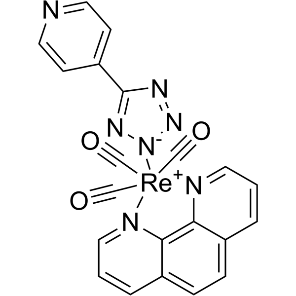
- Formula: C21H12N7O3Re
- Molecular Weight: 596.57
Endoplasmic reticulum dye 1 is a rhenium (I)-based luminescent, membrane-permeable cell imaging reagent with low cytotoxicity, photobleaching resistance, and suitability for multicolor imaging. Endoplasmic reticulum dye 1 localizes to the nuclear membrane, endoplasmic reticulum, and nucleoplasmic reticulum in live cells. Endoplasmic reticulum dye 1 enables detection of exocytosis events, dynamic changes in nuclear membrane-derived phagosomes, and vesicle trafficking processes.

- Formula: C46H59BF2N4O3
- Molecular Weight: 764.79
ER-Fluidity is a fluorescent probe used to detect the membrane fluidity of the endoplasmic reticulum (ER). ER-Fluidity consists of BODIPY, Coumarin (HY-N0709) moieties and a long-chain n-octadecyl group (n-C18). The excitation/emission wavelengths of the Coumarin moiety of ER-Fluidity are Ex/Em = 350/439 nm, while those of the BODIPY moiety are Ex/Em = 500/518 nm. In cell experiments, 488 nm is additionally used as an excitation wavelength for the BODIPY moiety.

- Formula: C39H31N3O4S
- Molecular Weight: 637.75


- Formula: C37H42N8O6
- Molecular Weight: 694.78
ER-Cu (II) is an endoplasmic reticulum-selective fluorescent Cu2+ probe. ER-Cu (II) localizes specifically to the endoplasmic reticulum of hepatocytes.

- Formula: C56H66F5N5O15S6
- Molecular Weight: 1336.53
ER Flipper-TR 28 is a flipper probe with a small molecule fluorophore that can image membrane tension in the endoplasmic reticulum (ER). ER Flipper-TR 28 bears a pentafluorophenyl group and also reacts with protein thiolate on the ER surface facing the cytoplasm.
- 1



