



E722-2648
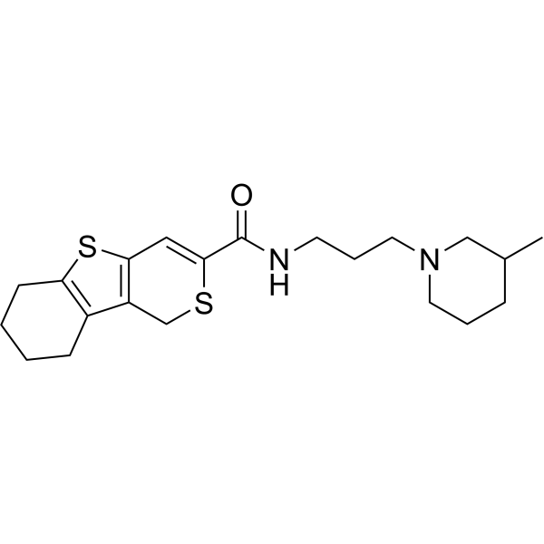
E722-2648 is an inhibitor targeting the BCL9 and β-catenin complex with antitumor activity. E722-2648 blocks complex formation by disrupting the interaction between the two proteins, thereby inhibiting β-catenin-mediated transcriptional activity and downregulating the expression of WNT target genes. E722-2648 effectively inhibits tumor growth in colon cancer xenograft models and colorectal cancer mouse models. E722-2648 can be used for the research of colon cancer and colorectal cancer.
For research use only. We do not sell to patients.
- CAS No.: 931963-55-2
- Formula: C21H30N2OS2
- Molecular Weight:390.61
-
Storage:
Please store the product under the recommended conditions in the Certificate of Analysis.

To place orders, for customer services and technical support, please contact: MedChemExpress USA
Tel: 609-228-6898 E-mail: [email protected] [email protected]


-
 Biological Activity
-
Chemical Information
- Protocols
- Purity & Documentation
- References
-
 Help & FAQs




Biological Activity
Description
In Vitro
E722-2648 (1-20 μM; 16 h) dose-dependently inhibits the formation of endogenous β-catenin/BCL9 complexes in Colo320 and HCT116 colorectal cancer cells, without disrupting the interaction between β-catenin and E-cadherin, and exhibits excellent target specificity[1].
E722-2648 (6.25-25 μM; 16 h) dose-dependently inhibits the transcriptional activity of the Wnt/β-catenin pathway in dual-luciferase reporter assays using Colo320 and HCT116 colorectal cancer cells[1].
E722-2648 (10 μM, 20 μM; 6-48 h) downregulates the mRNA expression levels of Wnt pathway target genes AXIN2 and CD44 in a time- and dose-dependent manner in Colo320 and HCT116 colorectal cancer cells[1].
E722-2648 (25-50 μM; 24 h) dose-dependently downregulates the mRNA expression levels of Wnt pathway target genes AXIN2, CD44, and LGR5 in human neoplastic colon organoids carrying the APC gene c.4778delA mutation[1].
E722-2648 (5-20 μM; 24-48 h) dose-dependently downregulates the protein expression levels of AXIN2, CD44 and active β-catenin, and simultaneously increases the level of cleaved PARP to induce apoptosis in Colo320 and HCT116 colorectal cancer cells, whereas it exerts no such effects on β-catenin-independent RKO cells[1].
E722-2648 (20 μM; 48 h) significantly reduces the viability of HCT116 and Colo320 colorectal cancer cells. The decrease in cell viability becomes more pronounced when combined with the Wnt inhibitor ICG-001, the cholesterol synthesis inhibitor Lovastatin (HY-N0504), and the cholesterol esterification inhibitor Avasimibe (HY-13215). Exogenous cholesterol supplementation partially rescues the reduction in cell viability induced by E722-2648 treatment[1].
MedChemExpress (MCE) has not independently confirmed the accuracy of these methods. They are for reference only. Further protocols information, click here.
-
Cell Line:Human CRC cell lines Colo320, HCT116, DLD-1; murine breast cancer cell line 4T1
-
Concentration:1 μM, 10 μM, 20 μM
-
Incubation Time:16 h
-
Result:Dose-dependently inhibited the formation of endogenous β-catenin/BCL9 complexes in Colo320 and HCT116 cells, with significant inhibitory effect observed at concentrations as low as 1 μM.
In DLD-1 cells with high E-cadherin expression, E722-2648 did not disrupt the interaction between β-catenin and E-cadherin at any tested concentration.
Also dose-dependently inhibited the binding of murine BCL9 to β-catenin in 4T1 cells.
-
Cell Line:Human CRC cell lines Colo320, HCT116, RKO; neoplastic human colon organoids with APC c.4778delA mutation
-
Concentration:5 μM, 10 μM, 20 μM (for CRC cell lines); 25 μM, 50 μM (for colon organoids)
-
Incubation Time:24 h, 48 h, 72 h, 96 h (for CRC cell lines); 24 h, 48 h (for colon organoids)
-
Result:Significantly reduced the proliferation of Colo320 and HCT116 cells in a time- and dose-dependent manner at all tested concentrations.
Had no inhibitory effect on the proliferation of the β-catenin-independent RKO cell line.
In neoplastic human colon organoids, E722-2648 dose-dependently inhibited proliferation and induced obvious apoptosis of organoids after 24 and 48 h of treatment.
In Vivo
E722-2648 (3 mg/kg; intratumoral injection; administered once every other day for 20 days) significantly inhibits subcutaneous tumor growth, reduces tumor weight and GFP fluorescence signals in tumor tissues, downregulates the expression of Wnt pathway target genes AXIN2 and CD44 in tumor tissues, suppresses tumor cell proliferation and tumor angiogenesis, induces tumor cell apoptosis, and decreases the infiltration of pro-tumor M2-like tumor-associated macrophages in the subcutaneous colorectal cancer xenograft model of NCr nude mice bearing HCT116-GFP cells[1].
MedChemExpress (MCE) has not independently confirmed the accuracy of these methods. They are for reference only.
-
Animal Model:NOD scid gamma (NSG) mice (NOD.Cg-Prkdcscid Il2rgtm1Wjl/SzJ, 5-week-old female), established with intraperitoneal colorectal cancer xenograft model via intraperitoneal injection of 2×10⁶ HCT116 cells[1]
-
Dosage:4 mg/kg
-
Administration:Intraperitoneal injection every other day for 20 days
-
Result:Significantly inhibited the growth of intraperitoneal colorectal tumors in NSG mice.
After 20 days of administration, Significantly lowered the weight and volume of the dissected tumors from treated mice than those from the vehicle control group.
-
Animal Model:NCr nude mice (Taconic, 5-week-old female), established with subcutaneous colorectal cancer xenograft model via subcutaneous injection of 2×10⁵ HCT116 cells stably expressing green fluorescent protein (GFP)[1]
-
Dosage:3 mg/kg
-
Administration:Intratumoral injection every other day for 20 days
-
Result:Significantly suppressed the growth of subcutaneous HCT116 tumors in NCr nude mice, with a significant reduction in tumor volume, tumor weight, and GFP fluorescence intensity of tumor tissues compared with the vehicle control group.
Significantly downregulated the protein expression of Wnt target genes AXIN2 and CD44 in tumor tissues, decreased the expression of the proliferation marker Ki-67 and angiogenesis marker CD31, and increased the expression of the apoptosis marker cleaved caspase-3.
Significantly reduced the infiltration of protumorigenic M2-like tumor-associated macrophages marked by CD163 in tumor tissues.


Chemical Information
-
CAS No. 931963-55-2
-
Molecular Weight 390.61
-
Formula C21H30N2OS2
-
SMILES
O=C(C1=CC(SC2=C3CCCC2)=C3CS1)NCCCN4CCCC(C4)C
-
Shipping
Room temperature in continental US; may vary elsewhere.
-
Storage
Please store the product under the recommended conditions in the Certificate of Analysis.

Protocols
-
Dual Luciferin reporter gene assay
Luciferin reporter gene assay is a reporting system to detect the activity of Firefly Luciferase using luciferin as a substrate, which is often used in the research of miRNA target gene verification and promoter transcriptive activity regulation. Dual luciferase usually refers to Firefly luciferase and Renilla luciferase.
-
RNA extraction experimental
By lysing cells, releasing RNA, and removing impurities such as proteins and DNA, high-purity RNA products are finally obtained. The commonly used traditional method is the guanidine isothiocyanate/phenol/chloroform method (Trizol), which is suitable for a variety of animal materials including animal tissues, microorganisms, cultured cells, etc., and most plant materials.
-
Patient-Derived Xenograft (PDX)
Patient-derived xenograft (PDX) models are generated by engrafting primary human tumor tissue directly into immunodeficient mice, allowing in vivo propagation of patient tumor biology without initial in vitro adaptation. These models are used to preserve key histopathological and molecular characteristics of the original tumor and enable assessment of tumor growth dynamics and therapeutic response in a living organism. The biological readout is tumor engraftment and subsequent growth in the murine host, which reflects the ability of human tumor cells to survive, vascularize, and expand in an immunocompromised microenvironment.
-
Patient-Derived Orthotopic Xenograft (PDOX)
Patient-derived orthotopic xenograft (PDOX) modeling implants fresh patient tumor tissue or patient-derived tumor cells into the anatomically corresponding organ or tissue site of immunodeficient mice, usually by surgical orthotopic implantation, to preserve patient tumor histology, local microenvironmental context, invasion, metastatic behavior, and treatment-response features better than subcutaneous implantation. PDOX readouts include tumor engraftment, orthotopic tumor growth, local invasion, metastasis, recurrence after resection, histologic similarity to the donor tumor, biomarker retention, molecular concordance, survival, and response or resistance to therapy. PDOX models are used for preclinical drug testing and individualized therapy evaluation, but engraftment success varies by tumor type and specimen quality.
-
Splenic/Portal-Vein Liver Metastasis Xenograft
Splenic and portal-vein liver metastasis xenograft models deliver tumor cells into the portal circulation so that cells reach the liver first and form hepatic metastatic lesions; splenic injection uses the spleen as an access route to the portal system, while direct portal-vein injection introduces cells into the portal vein without requiring splenectomy. The assay detects liver colonization, intrahepatic tumor growth, tumor distribution, treatment response, survival, and liver-metastasis microenvironment changes; readouts include bioluminescence or fluorescence imaging, gross liver nodule counts, liver weight or tumor burden, histology, and survival.
-
Subcutaneous Cell-Line-Derived Xenograft
Subcutaneous cell-line-derived xenograft (CDX) models are established by implanting cultured human cancer cell lines into immunodeficient mice, where the injected cells form localized tumors that can be monitored in vivo as a measure of tumorigenic potential, growth kinetics, and treatment response. These models are widely used in oncology research because they allow reproducible tumor formation and enable comparative assessment of tumor growth between different cell lines or genetic manipulations in a controlled in vivo microenvironment. Subcutaneous implantation of cancer cells in immunodeficient mice is a standard approach for evaluating tumor growth behavior and therapeutic response across multiple cancer types, including prostate, esophageal, pancreatic, and colon cancer models.
-
Orthotopic Cell-Line Xenograft
Orthotopic cell-line xenograft models involve implantation of human cancer cell lines into the anatomically corresponding organ of immunodeficient mice to reproduce tumor growth within a native microenvironment, enabling more clinically relevant tumor behavior compared with subcutaneous models. These models are widely used because orthotopic placement better recapitulates tumor progression, including invasion and metastatic spread, which are often underrepresented in heterotopic implantation systems. Compared with conventional xenografts, orthotopic implantation is described as more technically complex but provides improved simulation of tumor-microenvironment interactions and metastatic behavior, making it particularly valuable for translational oncology research. Surgical orthotopic implantation approaches have been emphasized as enabling faithful reproduction of clinical cancer features, including metastasis and disease progression patterns that align with the tumor’s organ of origi
-
Intraperitoneal/Peritoneal Dissemination Xenograft
Intraperitoneal (IP) or peritoneal dissemination xenograft models are based on the introduction of human cancer cells into the peritoneal cavity of immunodeficient mice, where they attach to peritoneal surfaces, form multicellular aggregates or spheroids, and progressively generate disseminated tumor nodules that mimic advanced peritoneal metastatic disease. These models are widely used to study ovarian cancer progression, tumor-microenvironment interactions, and intraperitoneal therapeutic responses, often incorporating bioluminescence or fluorescence imaging to longitudinally monitor tumor burden in vivo. The biological principle relies on the capacity of tumor cells such as SKOV3 or related ovarian carcinoma lines to survive in suspension, aggregate within ascites-like fluid, adhere to mesothelial surfaces, and invade peritoneal organs, thereby recapitulating human peritoneal carcinomatosis patterns observed in advanced disease.


Purity & Documentation
References
[1]. Groenewald W, et al. The Role of WNT Pathway Mutations in Cancer Development and an Overview of Therapeutic Options. Cells. 2023;12(7):990. Published 2023 Mar 24. [Content Brief]
[2]. Cao M, et al. Classical Angiogenic Signaling Pathways and Novel Anti-Angiogenic Strategies for Colorectal Cancer. Curr Issues Mol Biol. 2022;44(10):4447-4471. Published 2022 Sep 26. [Content Brief]

Calculators
Concentration (start) × Volume (start) = Concentration (final) × Volume (final)


E722-2648 Related Classifications
HY-148918 Related Classifications
 Powered by Bioz