



HDAC-IN-53
Based on 1 publication(s) in Google Scholar
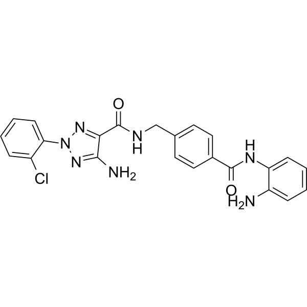
HDAC-IN-53 is an orally active, and selective HDAC1-3 inhibitor with IC50 values of 47 nM, 125 nM, and 450 nM, respectively. HDAC-IN-53 does not inhibit class II HDACs (HDAC4, 5, 6, 7, 9; IC50>10 μM). HDAC-IN-53 induces caspase-dependent apoptosis. HDAC-IN-53 significantly inhibits the growth of human tumor xenografts in nude mice and murine tumor growth in immune-competent mice bearing MC38 colon cancer.
商品は「研究用試薬」です。人や動物の医療用・臨床診断用・食品用の製品ではありません。
研究用途以外に使用した場合、当社は一切の責任を負いかねます。
- 純度 : 95.73%
- CAS 番号: 2921948-27-6
- 分子式: C23H20ClN7O2
- 分子量:461.90
-
保管条件:Powder -20°C, 3 years , 4°C, 2 years ; In solvent -80°C, 6 months , -20°C, 1 month

To place orders, for customer services and technical support, please contact: MedChemExpress USA
Tel: 609-228-6898 E-mail: [email protected] [email protected]


-
 生物活性
生物活性
-
化学情報
-
溶剤 & 溶解度
- プロトコル
- 純度とドキュメンテーション
- 参考文献
-
 Help & FAQs


MedChemExpress(MCE)の使用を引用している文献 HDAC-IN-53
More

生物活性
製品説明
IC50 & Target
[1]|
HDAC1 47 nM (IC50) |
HDAC2 125 nM (IC50) |
HDAC3 450 nM (IC50) |
HDAC4 >10 μM (IC50) |
HDAC5 >10 μM (IC50) |
HDAC6 >10 μM (IC50) |
HDAC7 >10 μM (IC50) |
HDAC8 >10 μM (IC50) |
HDAC9 >10 μM (IC50) |
Cellular Effect
|
Cell Line

|
Type | Value | Description | References |
|---|---|---|---|---|
| A549 | IC50 |
1425 nM
Compound: 19h
|
Antiproliferative activity against human A549 cells assessed as reduction of cell viability incubated for 72 hrs by CCK-8 assay
Antiproliferative activity against human A549 cells assessed as reduction of cell viability incubated for 72 hrs by CCK-8 assay
|
[PMID: 36934335] |
| CAKI-1 | IC50 |
252 nM
Compound: 19h
|
Antiproliferative activity against human CAKI-1 cells assessed as reduction of cell viability incubated for 72 hrs by CCK-8 assay
Antiproliferative activity against human CAKI-1 cells assessed as reduction of cell viability incubated for 72 hrs by CCK-8 assay
|
[PMID: 36934335] |
| DOHH-2 | IC50 |
65 nM
Compound: 19h
|
Antiproliferative activity against human DOHH-2 cells assessed as reduction of cell viability incubated for 72 hrs by CCK-8 assay
Antiproliferative activity against human DOHH-2 cells assessed as reduction of cell viability incubated for 72 hrs by CCK-8 assay
|
[PMID: 36934335] |
| ES-2 | IC50 |
15 nM
Compound: 19h
|
Antiproliferative activity against human ES2 cells assessed as reduction of cell viability incubated for 72 hrs by CCK-8 assay
Antiproliferative activity against human ES2 cells assessed as reduction of cell viability incubated for 72 hrs by CCK-8 assay
|
[PMID: 36934335] |
| HCT-116 | IC50 |
0.56 μM
Compound: 19h
|
Antiproliferative activity against human HCT-116 cells assessed as reduction of cell viability incubated for 72 hrs by CCK-8 assay
Antiproliferative activity against human HCT-116 cells assessed as reduction of cell viability incubated for 72 hrs by CCK-8 assay
|
[PMID: 36934335] |
| HEK293 | IC50 |
1256 nM
Compound: 19h
|
Antiproliferative activity against human HEK293 cells assessed as reduction of cell viability incubated for 72 hrs by CCK-8 assay
Antiproliferative activity against human HEK293 cells assessed as reduction of cell viability incubated for 72 hrs by CCK-8 assay
|
[PMID: 36934335] |
| HeLa | IC50 |
644 nM
Compound: 19h
|
Antiproliferative activity against human HeLa cells assessed as reduction of cell viability incubated for 72 hrs by CCK-8 assay
Antiproliferative activity against human HeLa cells assessed as reduction of cell viability incubated for 72 hrs by CCK-8 assay
|
[PMID: 36934335] |
| Jurkat | IC50 |
126 nM
Compound: 19h
|
Antiproliferative activity against human Jurkat cells assessed as reduction of cell viability incubated for 72 hrs by CCK-8 assay
Antiproliferative activity against human Jurkat cells assessed as reduction of cell viability incubated for 72 hrs by CCK-8 assay
|
[PMID: 36934335] |
| K562 | IC50 |
345 nM
Compound: 19h
|
Antiproliferative activity against human K562 cells assessed as reduction of cell viability incubated for 72 hrs by CCK-8 assay
Antiproliferative activity against human K562 cells assessed as reduction of cell viability incubated for 72 hrs by CCK-8 assay
|
[PMID: 36934335] |
| MC-38 | IC50 |
0.66 μM
Compound: 19h
|
Antiproliferative activity against mouse MC38 cells assessed as reduction of cell viability incubated for 72 hrs by CCK-8 assay
Antiproliferative activity against mouse MC38 cells assessed as reduction of cell viability incubated for 72 hrs by CCK-8 assay
|
[PMID: 36934335] |
| MDA-MB-231 | IC50 |
425 nM
Compound: 19h
|
Antiproliferative activity against human MDA-MB-231 cells assessed as reduction of cell viability incubated for 72 hrs by CCK-8 assay
Antiproliferative activity against human MDA-MB-231 cells assessed as reduction of cell viability incubated for 72 hrs by CCK-8 assay
|
[PMID: 36934335] |
| MIA PaCa-2 | IC50 |
297 nM
Compound: 19h
|
Antiproliferative activity against human MIA PaCa-2 cells assessed as reduction of cell viability incubated for 72 hrs by CCK-8 assay
Antiproliferative activity against human MIA PaCa-2 cells assessed as reduction of cell viability incubated for 72 hrs by CCK-8 assay
|
[PMID: 36934335] |
| NCI-N87 | IC50 |
607 nM
Compound: 19h
|
Antiproliferative activity against human NCI-N87 cells assessed as reduction of cell viability incubated for 72 hrs by CCK-8 assay
Antiproliferative activity against human NCI-N87 cells assessed as reduction of cell viability incubated for 72 hrs by CCK-8 assay
|
[PMID: 36934335] |
| PC-3 | IC50 |
1657 nM
Compound: 19h
|
Antiproliferative activity against human PC-3 cells assessed as reduction of cell viability incubated for 72 hrs by CCK-8 assay
Antiproliferative activity against human PC-3 cells assessed as reduction of cell viability incubated for 72 hrs by CCK-8 assay
|
[PMID: 36934335] |
| PC-9 | IC50 |
249 nM
Compound: 19h
|
Antiproliferative activity against human PC-9 cells assessed as reduction of cell viability incubated for 72 hrs by CCK-8 assay
Antiproliferative activity against human PC-9 cells assessed as reduction of cell viability incubated for 72 hrs by CCK-8 assay
|
[PMID: 36934335] |
| ZR-75-1 | IC50 |
1125 nM
Compound: 19h
|
Antiproliferative activity against human ZR-75-1 cells assessed as reduction of cell viability incubated for 72 hrs by CCK-8 assay
Antiproliferative activity against human ZR-75-1 cells assessed as reduction of cell viability incubated for 72 hrs by CCK-8 assay
|
[PMID: 36934335] |
体外実験
HDAC-IN-53 (compound 19h) has good antiproliferative activity against a panel of cancer cell lines, for example MC38 (IC50=0.66 μM), HCT116 cell (IC50=0.56 μM) [1].
HDAC-IN-53 (0.1-1 μM; 24 h) causes G0/G1 cell cycle arrest in MC38 cells and induces G2/M cell cycle arrest in HCT116 cells[1].
HDAC-IN-53 (0.1-1 μM; 24 h) upregulates the expressions of cleaved caspase-3 and cleaved PARP in a dose-dependent manner[1].
MedChemExpress (MCE) has not independently confirmed the accuracy of these methods. They are for reference only.
-
Cell Line:MC38 and HCT116 cells
-
Concentration:0.1, 0.3, 1 μM
-
Incubation Time:24 h
-
Result:Caused G0/G1 cell cycle arrest in MC38 cells and induced G2/M cell cycle arrest in HCT116 cells.
Significantly decreased the proportion of S phase cells in MC38 cells.
-
Cell Line:MC38 and HCT116 cells
-
Concentration:0.1, 0.3, 1 μM
-
Incubation Time:24 h
-
Result:Upregulated the expressions of cleaved caspase-3 and cleaved PARP in a dose-dependent manner.
体内実験
Pharmacokinetic Parameters of HDAC-IN-53 in Mice[1].
| IV (5 mg/kg) | PO (20 mg/kg) | |
| Tmax (h) | 0.42 | |
| Cmax (ng/mL) | 8129 | 9558 |
| AUC0-t (ng/mL∗h) | 5864 | 15278 |
| t1/2 (h) | 0.85 | 2.49 |
| F (%) | 65.1% |
MedChemExpress (MCE) has not independently confirmed the accuracy of these methods. They are for reference only.
-
Animal Model:C57BL/6 Mice or athymic nude mice (female, 6-8 weeks old) with MC38 cells[1]
-
Dosage:60 or 120 mg/kg
-
Administration:PO; daily for 15 days
-
Result:Yielded TGI values of 60.3 and 87.6%, respectively.
Increased the percentage of CD4+ T cells.


化学情報
-
CAS 番号 2921948-27-6
-
性状 Solid
-
分子量 461.90
-
分子式 C23H20ClN7O2
-
Color White to off-white
-
SMILES
NC1=NN(C2=CC=CC=C2Cl)N=C1C(NCC3=CC=C(C=C3)C(NC4=CC=CC=C4N)=O)=O
-
輸送条件
Room temperature in continental US; may vary elsewhere.
-
保管条件
Powder -20°C 3 years 4°C 2 years In solvent -80°C 6 months -20°C 1 month


Publications (1)
-
Journal Impact Factor
-
Most Recent


溶剤 & 溶解度
体外:
DMSO : 100 mg/mL (216.50 mM; Need ultrasonic; Hygroscopic DMSO has a significant impact on the solubility of product, please use newly opened DMSO)
Please refer to the solubility information to select the appropriate solvent. Once prepared, please aliquot and store the solution to prevent product inactivation from repeated freeze-thaw cycles.
Storage method and period of stock solution: -80°C, 6 months; -20°C, 1 month. When stored at -80°C, please use it within 6 months. When stored at -20°C, please use it within 1 month.
Please refer to the solubility information to select the appropriate solvent. Once prepared, please aliquot and store the solution to prevent product inactivation from repeated freeze-thaw cycles.
Storage method and period of stock solution: -80°C, 6 months; -20°C, 1 month. When stored at -80°C, please use it within 6 months. When stored at -20°C, please use it within 1 month.
濃度 (開始) × 体積 (開始) = 濃度 (終了) × 体積 (終了)


純度とドキュメンテーション
-
データシート (277 KB)
-
SDS (251 KB)
- English - EN (251 KB)
- Français - FR (251 KB)
- Deutsch - DE (251 KB)
- Norwegian - NO (251 KB)
- Español - ES (251 KB)
- Swedish - SV (251 KB)
- Italian - IT (251 KB)
- Korean - KR (251 KB)
- Portuguese - PT (251 KB)
-
取扱説明書 (2659 KB)
参考文献


Complete Stock Solution Preparation Table
Please refer to the solubility information to select the appropriate solvent. Once prepared, please aliquot and store the solution to prevent product inactivation from repeated freeze-thaw cycles.
Storage method and period of stock solution: -80°C, 6 months; -20°C, 1 month. When stored at -80°C, please use it within 6 months. When stored at -20°C, please use it within 1 month.
| Optional Solvent | Concentration Solvent Mass | 1 mg | 5 mg | 10 mg | 25 mg |
|---|---|---|---|---|---|
| DMSO | 1 mM | 2.1650 mL | 10.8249 mL | 21.6497 mL | 54.1243 mL |
| 5 mM | 0.4330 mL | 2.1650 mL | 4.3299 mL | 10.8249 mL | |
| 10 mM | 0.2165 mL | 1.0825 mL | 2.1650 mL | 5.4124 mL | |
| 15 mM | 0.1443 mL | 0.7217 mL | 1.4433 mL | 3.6083 mL | |
| 20 mM | 0.1082 mL | 0.5412 mL | 1.0825 mL | 2.7062 mL | |
| 25 mM | 0.0866 mL | 0.4330 mL | 0.8660 mL | 2.1650 mL | |
| 30 mM | 0.0722 mL | 0.3608 mL | 0.7217 mL | 1.8041 mL | |
| 40 mM | 0.0541 mL | 0.2706 mL | 0.5412 mL | 1.3531 mL | |
| 50 mM | 0.0433 mL | 0.2165 mL | 0.4330 mL | 1.0825 mL | |
| 60 mM | 0.0361 mL | 0.1804 mL | 0.3608 mL | 0.9021 mL | |
| 80 mM | 0.0271 mL | 0.1353 mL | 0.2706 mL | 0.6766 mL | |
| 100 mM | 0.0216 mL | 0.1082 mL | 0.2165 mL | 0.5412 mL |


HDAC-IN-53 関連分類
HY-149208 関連分類
 Powered by Bioz