



HDAC-IN-9
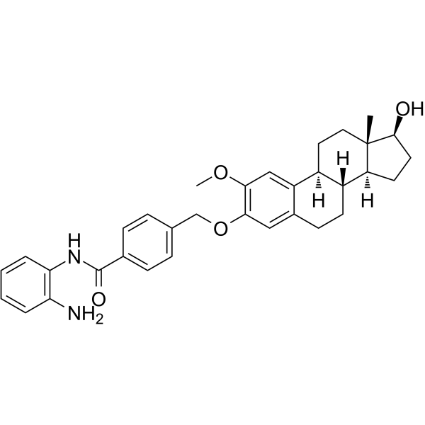
HDAC-IN-9 is a potent and selective tubulin and HDAC dual inhibitor. HDAC-IN-9 inhibits the invasion and migration of A549 cells. HDAC-IN-9 shows potent antitumor and antiangiogenic effect in vitro and in vivo.
Para uso exclusivo en investigación. No vendemos a pacientes.
- Fòrmula: C33H38N2O4
- Peso molecular:526.67
-
Almacenamiento:
Please store the product under the recommended conditions in the Certificate of Analysis.

To place orders, for customer services and technical support, please contact: MedChemExpress USA
Tel: 609-228-6898 E-mail: [email protected] [email protected]


-
 Actividad biológica
-
Chemical Information
- Protocolo
- Pureza y Documentación
- Referencias
-
 Help & FAQs




Actividad biológica
Descripciòn
IC50 & Target
[1]|
HDAC1 6.8 μM (IC50) |
HDAC2 0.06 μM (IC50) |
HDAC6 0.12 μM (IC50) |
HDAC8 8.5 μM (IC50) |
Cellular Effect
|
Cell Line

|
Type | Value | Description | References |
|---|---|---|---|---|
| A549 | IC50 |
1.78 μM
Compound: 17
|
Cytotoxicity against human A549 cells
Cytotoxicity against human A549 cells
|
[PMID: 38823265] |
| U-937 | IC50 |
0.371 μM
Compound: 17
|
Cytotoxicity against human U-937 cells
Cytotoxicity against human U-937 cells
|
[PMID: 38823265] |
In Vitro
HDAC-IN-9 (compound 4s) shows anti-proliferative activity (IC50s of 1.821, 2.538, 4.84, 1.782, 2.525, 0.371 µM for MCF-7, MGC-803, HeLa, A549, HepG2, U937 cells, respectively)[1].
HDAC-IN-9 displays 6.1, 3.2 and 4.6-fold selectivity ratios for 293 T, macrophages, and L02 cells respectively (IC50s of 10.89, 5.63, 8.14 µM for 293 T, macrophages, and L02 cells, respectively)[1].
HDAC-IN-9 shows inhibition activities against HDAC isoforms (IC50s of 6.8, 0.06, 0.12, 8.5 µM for HDAC 1, HDAC 2, HDAC 6, HDAC 10, respectively)[1].
HDAC-IN-9 (1, 2, 4 µM; 72 h; A459 cells) induces cell cycle arrest at the G2/M phase in a concentration-dependent manner[1].
HDAC-IN-9 (1, 2, 4 µM; 72 h) induces apoptosis via a Caspase-dependent intrinsic mitochondrial-mediated pathway in A549 cells[1].
HDAC-IN-9 (0, 0.5, 1, 2 µM; 48 h) shows anti-proliferative activity by the inhibition of angiogenesis and the reduction of endothelial cell metastasis in A549 cells[1].
HDAC-IN-9 (0, 1, 2, 4 µM, 48 h) inhibits the invasion and migration of A549 cells[1].
MedChemExpress (MCE) has not independently confirmed the accuracy of these methods. They are for reference only.
-
Cell Line:MCF-7, MGC-803, HeLa, A549, HepG2, U937 cells
-
Concentration:
-
Incubation Time:72 h
-
Result:Showed anti-proliferative activity (IC50s of 1.821, 2.538, 4.84, 1.782, 2.525, 0.371 µM for MCF-7, MGC-803, HeLa, A549, HepG2, U937 cells, respectively).
-
Cell Line:A459 cells
-
Concentration:0, 1, 2, 4 µM
-
Incubation Time:72 h
-
Result:Cells were arrested at the G2/M phase with a dose-dependent manner.
-
Cell Line:A459 cells
-
Concentration:0, 1, 2, 4 µM
-
Incubation Time:72 h
-
Result:Induced apoptosis via a Caspase-dependent intrinsic mitochondrial-mediated pathway in A549 cells.
-
Cell Line:A459 cells
-
Concentration:0, 1, 2, 4 µM
-
Incubation Time:72 h
-
Result:Showed down regulation of Cyclin B1 and Pcdc2 levels.
In Vivo
HDAC-IN-9 (0, 2.5, 5, 10 µM; 3 days) inhibits the proliferation and metastasis of A549 cells in zebrafish xenograft models[1].
MedChemExpress (MCE) has not independently confirmed the accuracy of these methods. They are for reference only.
-
Animal Model:embryos of Tg(flk:EGFP) transgenic zebrafish (transgenic zebrafish model Tg(flk1:EGFP))[1]
-
Dosage:0, 10, 20, 40 µM
-
Administration:
-
Result:Suppressed the formation of ISVs in zebrafish embryos.
-
Animal Model:zebrafish xenograft models[1]
-
Dosage:0, 2.5, 5, 10 µM
-
Administration:3 days
-
Result:Inhibited the proliferation and metastasis of A549 cells in zebrafish xenograft models.


Chemical Information
-
Peso molecular 526.67
-
Fòrmula C33H38N2O4
-
SMILES
[H][C@]12CC[C@@]3([C@H](CC[C@]3([C@@]1(CCC4=CC(OCC5=CC=C(C=C5)C(NC6=C(C=CC=C6)N)=O)=C(C=C42)OC)[H])[H])O)C
-
Envío
Room temperature in continental US; may vary elsewhere.
-
Almacenamiento
Please store the product under the recommended conditions in the Certificate of Analysis.


Pureza y Documentación
Referencias

Calculators
Concentration (start) × Volume (start) = Concentration (final) × Volume (final)


HDAC-IN-9 Related Classifications
HY-115941 Related Classifications
 Powered by Bioz