



HDAC6-IN-4
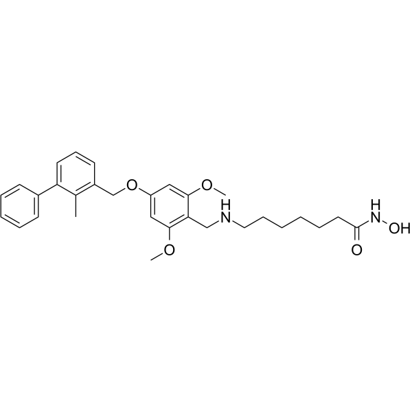
HDAC6-IN-4 (C10) is a potent, orally active and highly selective HDAC6 inhibitor with an IC50 value of 23 nM. HDAC6-IN-4 induces cancer cells apoptosis and shows significant antitumor efficacy, without obvious toxicity.
For research use only. We do not sell to patients.
- CAS No.: 2709103-20-6
- Formula: C30H38N2O5
- Molecular Weight:506.63
-
Storage:
Please store the product under the recommended conditions in the Certificate of Analysis.

To place orders, for customer services and technical support, please contact: MedChemExpress USA
Tel: 609-228-6898 E-mail: [email protected] [email protected]


-
 Biological Activity
-
Chemical Information
- Protocols
- Purity & Documentation
- References
-
 Help & FAQs




Biological Activity
Description
IC50 & Target
|
HDAC6 23 nM (IC50) |
HDAC3 46 nM (IC50) |
HDAC2 172 nM (IC50) |
HDAC8 2175 nM (IC50) |
HDAC1 3604 nM (IC50) |
Cellular Effect
|
Cell Line

|
Type | Value | Description | References |
|---|---|---|---|---|
| A549 | IC50 |
5.77 μM
Compound: C10
|
Antiproliferative activity against human A549 cells after 72 hrs by MTT assay
Antiproliferative activity against human A549 cells after 72 hrs by MTT assay
|
[PMID: 35245830] |
| B16 | IC50 |
1.52 μM
Compound: C10
|
Antiproliferative activity against mouse B16 cells after 72 hrs by MTT assay
Antiproliferative activity against mouse B16 cells after 72 hrs by MTT assay
|
[PMID: 35245830] |
| CT26 | IC50 |
2.09 μM
Compound: C10
|
Antiproliferative activity against mouse CT26 cells after 72 hrs by MTT assay
Antiproliferative activity against mouse CT26 cells after 72 hrs by MTT assay
|
[PMID: 35245830] |
| HepG2 | IC50 |
2.36 μM
Compound: C10
|
Antiproliferative activity against human HepG2 cells after 72 hrs by MTT assay
Antiproliferative activity against human HepG2 cells after 72 hrs by MTT assay
|
[PMID: 35245830] |
In Vitro
HDAC6-IN-4 (C10) (0-50 µM, 72 h) shows strong antiproliferative activity against different cancer cells with low cytotoxicity[1].
HDAC6-IN-4 (0-6 µM, 24 h) exhibits significant selectivity for HDAC6 over HDAC1[1].
HDAC6-IN-4 inhibits migration activity in a time-dependent and dose-dependent way in B16 and CT26 cells[1].
HDAC6-IN-4 (0-8 µM, 24 h) induces B16 cell apoptosis in a dose-dependent manner[1].
HDAC6-IN-4 exhibits significant plasma stability in humans (97% retention after 6 h), and exhibits significant metabolic stability in human (half-life of 101.91 min) and mouse liver (half-life of 67.94 min) microsomes[1].
MedChemExpress (MCE) has not independently confirmed the accuracy of these methods. They are for reference only.
-
Cell Line:B16, HepG2, A549, and CT26 cells
-
Concentration:0-50 µM
-
Incubation Time:72 h
-
Result:Showed antiproliferative activity with IC50 values of 1.52, 2.36, 5.77, and 2.09 µM against B16, HepG2, A549, and CT26 cells, respectively.
-
Cell Line:B16 and CT26 cancer cells
-
Concentration:2, 4, and 6 µM
-
Incubation Time:2, 4, 8, 12, and 24 h
-
Result:Dramatically increased the level of Ac-Tub (acetyl-α-tubulin) in a dose-dependent and time-dependent manner. Had almost no effect on the content of Ac-H3 (acetyl-H3).
-
Cell Line:B16 cells
-
Concentration:4, 6, and 8 µM
-
Incubation Time:24 h
-
Result:Caused moderate to potent induction of apoptosis in the B16 cell line in a dose-dependent manner. Upregulated the expression of apoptotic protein cleaved PARP.
In Vivo
MedChemExpress (MCE) has not independently confirmed the accuracy of these methods. They are for reference only.
-
Animal Model:Five-week-old C57BL/6 mice (immune-related CT26 xenograft model)[1].
-
Dosage:50 and 100 mg/kg
-
Administration:Oral gavage, once daily for 21 days
-
Result:Resulted in a substantial tumor growth and tumor tissue size inhibition in a dose-dependent way. Showed significantly high antitumor activity (TGI = 75%) at 100 mg/kg. Raised the plasma IFN-g level and the numbers of CD+ and CD3+CD+ (activated cytotoxic T) cells. Decreased CD4+CD25+CD127low/- T regulatory cells. Showed no obvious toxicity.


Chemical Information
-
CAS No. 2709103-20-6
-
Molecular Weight 506.63
-
Formula C30H38N2O5
-
SMILES
O=C(NO)CCCCCCNCC1=C(OC)C=C(OCC2=C(C)C(C3=CC=CC=C3)=CC=C2)C=C1OC
-
Shipping
Room temperature in continental US; may vary elsewhere.
-
Storage
Please store the product under the recommended conditions in the Certificate of Analysis.


Purity & Documentation
References

Calculators
Concentration (start) × Volume (start) = Concentration (final) × Volume (final)


HDAC6-IN-4 Related Classifications
HY-144395 Related Classifications
 Powered by Bioz