



ME-344

Based on 1 Customer Validation
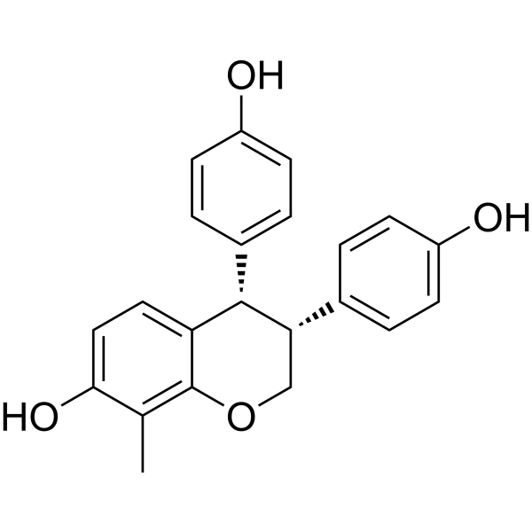
ME-344 is an Isoflavone. ME-344 increases mitochondrial ROS generation. ME-344 inhibits tubulin polymerization. ME-344 inhibits HO-1 and impacts its mitochondrial translocation. ME-344 induces Apoptosis through Caspase 3 activation. ME-344 synergizes with Vinblastine in leukemia cells. ME-344 displays anti-tumor activity against leukemia and lung tumor. ME-344 can be used in the research of lung cancer, acute myeloid leukemia, and HER2-negative breast cancer.
For research use only. We do not sell to patients.
- Purity : 99.80%
- CAS No.: 1374524-68-1
- Formula: C22H20O4
- Molecular Weight:348.39
-
Storage:Powder -20°C, 3 years , 4°C, 2 years ; In solvent -80°C, 6 months , -20°C, 1 month

To place orders, for customer services and technical support, please contact: MedChemExpress USA
Tel: 609-228-6898 E-mail: [email protected] [email protected]


-
 Biological Activity
-
Chemical Information
- Protocols
- Purity & Documentation
- References
-
 Help & FAQs


All Caspase Isoforms
More

Biological Activity
Description
IC50 & Target
[1]|
Caspase 3 |
Caspase 8 |
Caspase 9 |
HO-1 |
In Vitro
ME-344 (24 h) induces apoptosis in sensitive H460 and SHP-77 human lung cancer cells via activation of Caspase 3 and subsequent cleavage of PARP[1].
ME-344 (10-10000 nM; 72 h) is cytotoxic to NB4, U937, K562, OCI-AML2, KG1a, HL-60, and TEX leukemia cell lines with IC50 values ranging from 70 to 260 nM after 72 h of treatment[2].
ME-344 (10 μM) potently inhibits tubulin polymerization in a cell-free biochemical assay[2].
ME-344 (8 h) suppresses the de novo purine biosynthesis pathway in MV4-11 AML cells after 8 h of treatment, significantly reducing levels of AICAR and IMP[4].
MedChemExpress (MCE) has not independently confirmed the accuracy of these methods. They are for reference only.
-
Cell Line:H460, SHP-77
-
Concentration:IC50 concentrations
-
Incubation Time:24 h
-
Result:Induced marked cleavage of caspase 8, caspase 9, caspase 3, and PARP in H460 and SHP-77 cells. Showed higher levels of cleaved caspase 8 in H460 cells compared to SHP-77 cells. Showed higher levels of cleaved caspase 9 in SHP-77 cells compared to H460 cells.
In Vivo
MedChemExpress (MCE) has not independently confirmed the accuracy of these methods. They are for reference only.
-
Animal Model:SCID mice (male)[2]
-
Dosage:50 mg/kg; 75 mg/kg; 100 mg/kg
-
Administration:i.p.; every other day; 11 days
-
Result:Significantly reduced tumor growth relative to vehicle control at all tested doses. Produced a significant decrease in tumor weight at 100 mg/kg relative to 50 mg/kg. Showed no evidence of toxicity, as measured by stable body weight, unaltered behavior, and normal gross and histologic appearance of organs at necropsy.
Clinical Trial
| NCT Number | Sponsor | Condition | Start Date |
Phase

|
|---|---|---|---|---|
| NCT01329991 | Plexxikon| | 2011-05 | PHASE1 |


Chemical Information
-
CAS No. 1374524-68-1
-
Appearance Solid
-
Molecular Weight 348.39
-
Formula C22H20O4
-
Color White to off-white
-
SMILES
OC1=CC=C2[C@H](C3=CC=C(O)C=C3)[C@H](C4=CC=C(O)C=C4)COC2=C1C
-
Shipping
Room temperature in continental US; may vary elsewhere.
-
Storage
Powder -20°C 3 years 4°C 2 years In solvent -80°C 6 months -20°C 1 month


Purity & Documentation
-
Data Sheet (275 KB)
-
SDS (251 KB)
- English - EN (251 KB)
- Français - FR (251 KB)
- Deutsch - DE (251 KB)
- Norwegian - NO (251 KB)
- Español - ES (251 KB)
- Swedish - SV (251 KB)
- Italian - IT (251 KB)
- Korean - KR (251 KB)
- Portuguese - PT (251 KB)
-
Handling Instructions (2659 KB)
References
[1]. Zhang L, et al. Isoflavone ME-344 Disrupts Redox Homeostasis and Mitochondrial Function by Targeting Heme Oxygenase 1. Cancer Res. 2019;79(16):4072-4085. [Content Brief]
[2]. Jeyaraju DV, et al. A novel isoflavone, ME-344, targets the cytoskeleton in acute myeloid leukemia. Oncotarget. 2016;7(31):49777-49785. [Content Brief]
[3]. Quintela-Fandino M, et al. Randomized Phase 0/I Trial of the Mitochondrial Inhibitor ME-344 or Placebo Added to Bevacizumab in Early HER2-Negative Breast Cancer. Clin Cancer Res. 2020;26(1):35-45. [Content Brief]

Calculators
Concentration (start) × Volume (start) = Concentration (final) × Volume (final)


ME-344 Related Classifications
HY-112749 Related Classifications
 Powered by Bioz
Keywords
- ME-344
- 1374524-68-1
- ME344
- ME 344
- Heme Oxygenase (HO)
- Reactive Oxygen Species (ROS)
- Mitochondrial Metabolism
- Microtubule/Tubulin
- Caspase
- Apoptosis
- lung cancer
- acute myeloid leukemia
- tubulin
- mitochondrial complex I
- rough endoplasmic reticulum
- reactive oxygen species
- colchicine-binding site
- Nrf2
- apoptosis
- HO-1
- Inhibitor
- inhibitor
- inhibit