



PDGF-DD Protein, Mouse (His-SUMO)

Based on 1 Customer Validation
PDGF-DD protein is an important growth factor that controls embryonic development, cell proliferation, migration, survival and chemotaxis, and has a potent mitogenic effect on mesenchymal cells. It plays a key role in wound healing and has carcinogenic potential, contributing to tumor formation. PDGF-DD Protein, Mouse (His-SUMO) is the recombinant mouse-derived PDGF-DD protein, expressed by E. coli , with N-His, N-SUMO labeled tag.
- Species: Mouse
- Source: E. coli
-
Storage:Stored at -20°C for 2 years from date of receipt. After reconstitution, it is stable at 4°C for 1 week or -20°C for longer (with carrier protein). It is recommended to freeze aliquots at -20°C or -80°C for extended storage.


To place orders, for customer services and technical support, please contact: MedChemExpress USA
Tel: 609-228-6898 E-mail: [email protected] [email protected]


-
 Biological Activity
-
Technical Parameters
-
Product Properties
- Documentation
- Help & FAQs




Biological Activity
Description
PDGF-DD protein is an important growth factor that controls embryonic development, cell proliferation, migration, survival and chemotaxis, and has a potent mitogenic effect on mesenchymal cells. It plays a key role in wound healing and has carcinogenic potential, contributing to tumor formation. PDGF-DD Protein, Mouse (His-SUMO) is the recombinant mouse-derived PDGF-DD protein, expressed by E. coli , with N-His, N-SUMO labeled tag.
Background
PDGF-DD Protein emerges as a crucial growth factor governing embryonic development, cell proliferation, migration, survival, and chemotaxis. Renowned for its potent mitogenic effects on mesenchymal cells, PDGF-DD assumes a pivotal role in wound healing and, intriguingly, harbors oncogenic potential capable of inducing tumor formation. Its influence extends to the intricate orchestration of events during angiogenesis, promoting macrophage recruitment, heightened interstitial pressure, and blood vessel maturation. PDGF-DD's involvement is notable in the initiation of processes leading to mesangial proliferative glomerulonephritis, marked by monocyte and macrophage influx, as well as extracellular matrix production. Structurally, PDGF-DD exists as a homodimer linked by disulfide bonds, engaging in interactions with PDGFRB homodimers and heterodimers formed by PDGFRA and PDGFRB, contributing to its versatile regulatory mechanisms.
Verified Bioactivity
Measured in a cell proliferation assay using NIH-3T3 mouse fibroblast cells. The ED50 for this effect is 62.82 ng/mL, corresponding to a specific activity is 1.59×10⁴ units/mg.
MCE Validation Data
-
Bioactivity - Cell-Based Assay
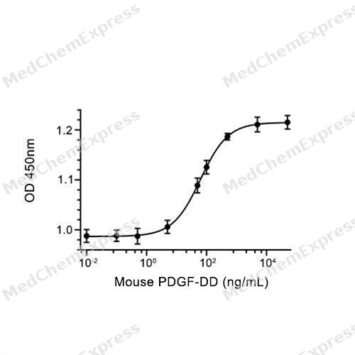
Bioactivity - Cell-Based Assay


Technical Parameters
-
Species Mouse
-
Source E. coli
-
Tag N-SUMO;N-6*His
-
Accession
Q925I7-1 (T24-R370)
-
Molecular Construction
-
N-term
-
6*His-SUMO
-
PDGF-DD (T24-R370)
Accession # Q925I7-1 -
C-term
-
-
Protein Length
Full Length of Isoform-1 Mature Protein
-
Synonyms
PDGFD; Platelet-Derived Growth Factor D; Platelet Derived Growth Factor D; Iris-Expressed Growth Factor; SCDGF-B; SCDGFB; IEGF; PDGF-D; MSTP036; Spinal Cord Derived Growth Factor B; Spinal Cord-Derived Growth Factor B
-
AA Sequence
TPQRASIKALRNANLRRDESNHLTDLYQREENIQVTSNGHVQSPRFPNSYPRNLLLTWWLRSQEKTRIQLSFDHQFGLEEAENDICRYDFVEVEEVSESSTVVRGRWCGHKEIPPRITSRTNQIKITFKSDDYFVAKPGFKIYYSFVEDFQPEAASETNWESVTSSFSGVSYHSPSITDPTLTADALDKTVAEFDTVEDLLKHFNPVSWQDDLENLYLDTPHYRGRSYHDRKSKVDLDRLNDDVKRYSCTPRNHSVNLREELKLTNAVFFPRCLLVQRCGGNCGCGTVNWKSCTCSSGKTVKKYHEVLKFEPGHFKRRGKAKNMALVDIQLDHHERCDCICSSRPPR
-
Predicted Molecular Mass
56.2 kDa
-
Molecular Weight
Approximately 66 kDa, based on SDS-PAGE under reducing conditions.
-
Purity
≥ 90%, as determined by reducing SDS-PAGE.


Product Properties
Lyophilized powder.
Lyophilized from a 0.22 μm filtered solution of 10 mM Tris-HCl, 1 mM EDTA, 6% trehalose, pH 8.0.
Note: For SPR assay, please replace the buffer. Primary amine components (e.g., Tris, imidazole) can affect protein-coupled chips.
<1 EU/μg, determined by LAL method.
It is not recommended to reconstitute to a concentration less than 100 μg/mL in ddH2O.
Stored at -20°C for 2 years from date of receipt. After reconstitution, it is stable at 4°C for 1 week or -20°C for longer (with carrier protein). It is recommended to freeze aliquots at -20°C or -80°C for extended storage.
Room temperature in continental US; may vary elsewhere.


Documentation
-
Data Sheet (237 KB)
-
SDS (251 KB)
- English - EN (251 KB)
- Français - FR (251 KB)
- Deutsch - DE (251 KB)
- Norwegian - NO (251 KB)
- Español - ES (251 KB)
- Swedish - SV (251 KB)
- Italian - IT (251 KB)
- Korean - KR (251 KB)
- Portuguese - PT (251 KB)
-
Handling Instructions (2659 KB)

Calculators
Concentration (start) × Volume (start) = Concentration (final) × Volume (final)


 Powered by Bioz

