From 11:00 pm to 12:00 pm EST ( 8:00 pm to 9:00 pm PST ) on January 6th, the website will be under maintenance. We are sorry for the inconvenience. Please arrange your schedule properly.
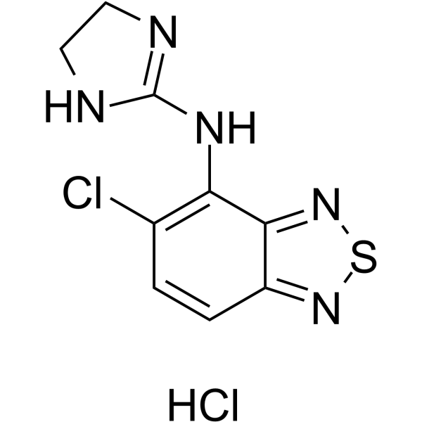
Tizanidine hydrochloride, a skeletal muscle relaxant, is an orally effective central α2-adrenoceptor agonist (IC50 = 6.9 nmol). Tizanidine hydrochloride primarily exerts muscle relaxation effects by inhibiting the release of excitatory amino acids (glutamate and aspartate) from the presynaptic terminals of spinal cord interneurons. Tizanidine hydrochloride has anti-injury activity and can inhibit gastrointestinal (GI) transport. Tizanidine hydrochloride can inhibit the proliferation, migration, and invasion of lung cancer cells and induce cell apoptosis by upregulating Nischarin and inhibiting the AKT and Wnt3a/β-catenin signaling pathways. Tizanidine hydrochloride can be used to treat spasticity caused by diseases such as multiple sclerosis (MS), stroke, and spinal cord injury (SCI) [3] .
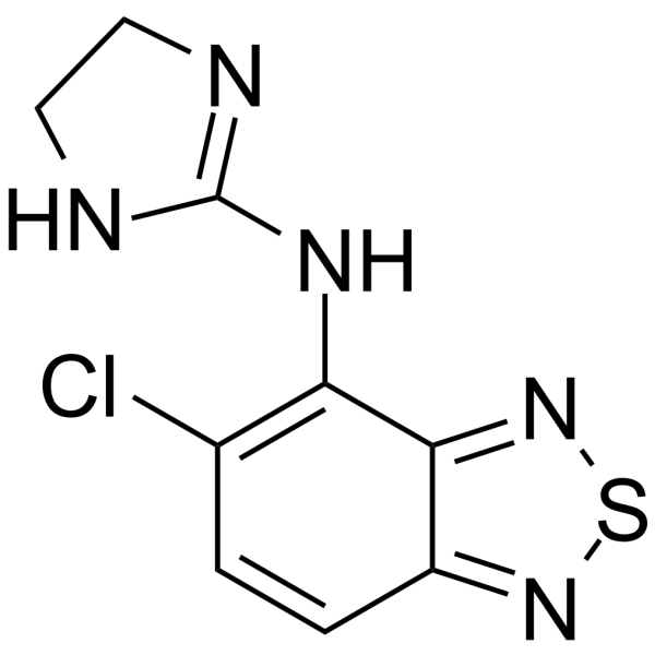
Tizanidine, a skeletal muscle relaxant, is an orally effective central α2-adrenoceptor agonist (IC50 = 6.9 nmol). Tizanidine primarily exerts muscle relaxation effects by inhibiting the release of excitatory amino acids (glutamate and aspartate) from the presynaptic terminals of spinal cord interneurons. Tizanidine has anti-injury activity and can inhibit gastrointestinal (GI) transport. Tizanidine can inhibit the proliferation, migration, and invasion of lung cancer cells and induce cell apoptosis by upregulating Nischarin and inhibiting the AKT and Wnt3a/β-catenin signaling pathways. Tizanidine can be used to treat spasticity caused by diseases such as multiple sclerosis (MS), stroke, and spinal cord injury (SCI) [3] .
Zamaporvint (RXC004) is an orally active and selective inhibitor of Wnt. Zamaporvint targete membrane-bound o-acyltransferase Porcupine and inhibited Wnt ligand palmitoylation, secretion, and pathway activation. Zamaporvint displays a favorable pharmacokinetic profile and shows potent antiproliferative effects in Wnt ligand-dependent colorectal and pancreatic cell lines. Zamaporvint possesses multiple antitumor mechanisms and can be used in cancer research .
ABC99 is an N-hydroxyhydantoin (NHH) carbamate that selectively inhibits the Wnt-deacylating enzyme NOTUM (IC50=13 nM). ABC99 preserves Wnt3A signaling in the presence of NOTUM .
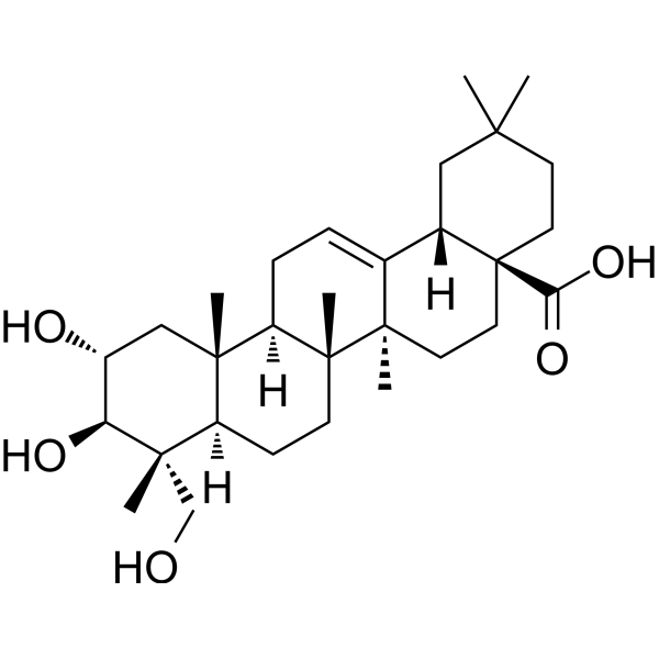
Arjunolic acid is an orally active, multifunctional bioactive compound. Arjunolic acid exhibits free radical scavenging activity, as well as fungal and bacterial activities. Arjunolic acid induces apoptosis (Apoptosis) in various cancer cells. Arjunolic acid protects hepatocytes against induced oxidative stress and apoptosis by reducing reactive oxygen species and inhibiting NF-κB activation. Arjunolic acid regulates pancreatic dysfunction in type 2 diabetic rats by blocking the activation of the TLR-4/MyD88 and canonical Wnt pathways. Arjunolic acid inhibits neuroinflammation and ameliorates depressive behaviors via the SIRT1/AMPK/Notch1 signaling pathway in microglia. Arjunolic acid improves Crohn's disease-like colitis by restoring gut microbiota composition and inhibiting TLR4 signaling. Arjunolic acid suppresses osteosarcoma progression by inhibiting Wnt3a-mediated M2 polarization of macrophages. Arjunolic acid ameliorates diabetic retinopathy via the autophagy pathway regulated by AMPK/mTOR/HO-1. Arjunolic acid is applicable to research related to type 2 diabetes, organ toxicity, depression, Crohn's disease, osteosarcoma, diabetic retinopathy, and testicular dysfunction [3] .
Anti-pan-Glypican Antibody (HS20) is a kind of human IgG1 κ human antibody, targeting to human pan-Glypican. Anti-pan-Glypican Antibody (HS20) can neutralize the heparan sulfate (HS) chains on GPC3 thereby disrupting the Wnt3a-GPC3 interactions leading to blockade of the Wnt3a/β-catenin signaling. Anti-pan-Glypican Antibody (HS20) can be used for the research of cancer, such as hepatocellular carcinoma (HCC) .
TFMB-(S)-2-HG is a potent TET2 inhibitor. TFMB-(S)-2-HG also inhibits the EglN prolyl hydroxylases. TFMB-(S)-2-HG downregulates Wnt3a, β-catenin (intranuclear) protein expression. TFMB-(S)-2-HG inhibits osteogenic differentiation of cells. TFMB-(S)-2-HG has the potential for the research of acute myeloid leukemia (AML) .
Wnt/β-catenin-IN-2 (Compound 3235-0367) is a Wnt/β-catenin signaling pathway inhibitor, with IC50 and KD values of 7.1 and 2.5 μM, respectively. Wnt/β-catenin-IN-2 can be used for the research of cancer .
Tizanidine-d4 is the deuterium labeled Tizanidine (HY-B0194). Tizanidine, a skeletal muscle relaxant, is an orally effective central α2-adrenoceptor agonist (IC50 = 6.9 nmol). Tizanidine primarily exerts muscle relaxation effects by inhibiting the release of excitatory amino acids (glutamate and aspartate) from the presynaptic terminals of spinal cord interneurons. Tizanidine has anti-injury activity and can inhibit gastrointestinal (GI) transport. Tizanidine can inhibit the proliferation, migration, and invasion of lung cancer cells and induce cell apoptosis by upregulating Nischarin and inhibiting the AKT and Wnt3a/β-catenin signaling pathways. Tizanidine can be used to treat spasticity caused by diseases such as multiple sclerosis (MS), stroke, and spinal cord injury (SCI).
WNT3A Human Pre-designed siRNA Set A contains three designed siRNAs for WNT3A gene (Human), as well as a negative control, a positive control, and a FAM-labeled negative control.
FZD7 antagonist 1 (peptide 34) is a dFz7-21 analogue. FZD7 antagonist 1 is an FZD7 antagonist that inhibits the wnt3a with IC50 value of 9.2 nM. FZD7 antagonist 1 blocks TcdB−FZD interaction via targeting FZD receptors .
Wnt3a Mouse Pre-designed siRNA Set A contains three designed siRNAs for Wnt3a gene (Mouse), as well as a negative control, a positive control, and a FAM-labeled negative control.
Wnt3a Rat Pre-designed siRNA Set A contains three designed siRNAs for Wnt3a gene (Rat), as well as a negative control, a positive control, and a FAM-labeled negative control.
Tizanidine (Standard) is the analytical standard of Tizanidine (HY-B0194). This product is intended for research and analytical applications. Tizanidine, a skeletal muscle relaxant, is an orally effective central α2-adrenoceptor agonist (IC50 = 6.9 nmol). Tizanidine primarily exerts muscle relaxation effects by inhibiting the release of excitatory amino acids (glutamate and aspartate) from the presynaptic terminals of spinal cord interneurons. Tizanidine has anti-injury activity and can inhibit gastrointestinal (GI) transport. Tizanidine can inhibit the proliferation, migration, and invasion of lung cancer cells and induce cell apoptosis by upregulating Nischarin and inhibiting the AKT and Wnt3a/β-catenin signaling pathways. Tizanidine can be used to treat spasticity caused by diseases such as multiple sclerosis (MS), stroke, and spinal cord injury (SCI).
Tizanidine hydrochloride (Standard) is the analytical standard of Tizanidine hydrochloride (HY-B0194A). This product is intended for research and analytical applications. Tizanidine hydrochloride, a skeletal muscle relaxant, is an orally effective central α2-adrenoceptor agonist (IC50 = 6.9 nmol). Tizanidine hydrochloride primarily exerts muscle relaxation effects by inhibiting the release of excitatory amino acids (glutamate and aspartate) from the presynaptic terminals of spinal cord interneurons. Tizanidine hydrochloride has anti-injury activity and can inhibit gastrointestinal (GI) transport. Tizanidine hydrochloride can inhibit the proliferation, migration, and invasion of lung cancer cells and induce cell apoptosis by upregulating Nischarin and inhibiting the AKT and Wnt3a/β-catenin signaling pathways. Tizanidine hydrochloride can be used to treat spasticity caused by diseases such as multiple sclerosis (MS), stroke, and spinal cord injury (SCI).
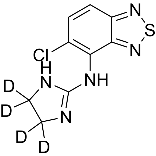
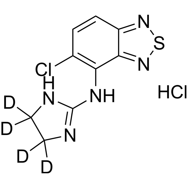
Tizanidine-d4 hydrochloride is deuterium labeled Tizanidine hydrochloride (HY-B0194A). Tizanidine hydrochloride, a skeletal muscle relaxant, is an orally effective central α2-adrenoceptor agonist (IC50 = 6.9 nmol). Tizanidine hydrochloride primarily exerts muscle relaxation effects by inhibiting the release of excitatory amino acids (glutamate and aspartate) from the presynaptic terminals of spinal cord interneurons. Tizanidine hydrochloride has anti-injury activity and can inhibit gastrointestinal (GI) transport. Tizanidine hydrochloride can inhibit the proliferation, migration, and invasion of lung cancer cells and induce cell apoptosis by upregulating Nischarin and inhibiting the AKT and Wnt3a/β-catenin signaling pathways. Tizanidine hydrochloride can be used to treat spasticity caused by diseases such as multiple sclerosis (MS), stroke, and spinal cord injury (SCI).
UM206_L is a linear Wnt fragment peptide derived from conserved Wnt3a/Wnt5a sequences, inactive in soluble form but capable of activating canonical Wnt/β-catenin signaling when conjugated to magnetic nanoparticles and exposed to a high-gradient, time-varying magnetic field or immobilized on glass surfaces .
WNT3Ap is a polypeptide corresponding to the hairpin 2 domain of the WNT3A protein. WNT3Ap undergoes palmitoylation modification catalyzed by PORCN. WNT3Ap is a key compound for elucidating the molecular mechanism of the Wnt signaling pathway and for developing PORCN inhibitors .
UM206_C is a circular Wnt fragment peptide derived from conserved Wnt3a/Wnt5a sequences, inactive in soluble form but capable of activating canonical Wnt/β-catenin signaling when conjugated to magnetic nanoparticles and exposed to a high-gradient, time-varying magnetic field or immobilized on glass surfaces .
Picoberin is a low picomolar inhibitor of Hedgehog-induced osteoblast differentiation. Picoberin is a potent aryl hydrocarbon receptor (AhR) agonist. Picoberin inhibits Wnt3A-induced osteoblast differentiation of C3H10T1/2 cells. Picoberin can upregulate genes encoding phase I and II metabolic enzymes .
BChE-IN-49 is an orally active BChE inhibitor with IC50 values of 0.73 μM and 6.43 μM against BChE and AChE, respectively. BChE-IN-49 reduces AChE levels to inhibit the degradation of acetylcholine. By inhibiting GSK-3β, BChE-IN-49 potently activates the Wnt/β-catenin signaling pathway, exerting potent anti-Aβ, antioxidant, anti-neuroinflammatory and anti-apoptotic effects. BChE-IN-49 can be used in research related to Alzheimer's disease .
FZD7 antagonist 1 (peptide 34) is a dFz7-21 analogue. FZD7 antagonist 1 is an FZD7 antagonist that inhibits the wnt3a with IC50 value of 9.2 nM. FZD7 antagonist 1 blocks TcdB−FZD interaction via targeting FZD receptors .
UM206_L is a linear Wnt fragment peptide derived from conserved Wnt3a/Wnt5a sequences, inactive in soluble form but capable of activating canonical Wnt/β-catenin signaling when conjugated to magnetic nanoparticles and exposed to a high-gradient, time-varying magnetic field or immobilized on glass surfaces .
WNT3Ap is a polypeptide corresponding to the hairpin 2 domain of the WNT3A protein. WNT3Ap undergoes palmitoylation modification catalyzed by PORCN. WNT3Ap is a key compound for elucidating the molecular mechanism of the Wnt signaling pathway and for developing PORCN inhibitors .
UM206_C is a circular Wnt fragment peptide derived from conserved Wnt3a/Wnt5a sequences, inactive in soluble form but capable of activating canonical Wnt/β-catenin signaling when conjugated to magnetic nanoparticles and exposed to a high-gradient, time-varying magnetic field or immobilized on glass surfaces .
Anti-pan-Glypican Antibody (HS20) is a kind of human IgG1 κ human antibody, targeting to human pan-Glypican. Anti-pan-Glypican Antibody (HS20) can neutralize the heparan sulfate (HS) chains on GPC3 thereby disrupting the Wnt3a-GPC3 interactions leading to blockade of the Wnt3a/β-catenin signaling. Anti-pan-Glypican Antibody (HS20) can be used for the research of cancer, such as hepatocellular carcinoma (HCC) .
Arjunolic acid is an orally active, multifunctional bioactive compound. Arjunolic acid exhibits free radical scavenging activity, as well as fungal and bacterial activities. Arjunolic acid induces apoptosis (Apoptosis) in various cancer cells. Arjunolic acid protects hepatocytes against induced oxidative stress and apoptosis by reducing reactive oxygen species and inhibiting NF-κB activation. Arjunolic acid regulates pancreatic dysfunction in type 2 diabetic rats by blocking the activation of the TLR-4/MyD88 and canonical Wnt pathways. Arjunolic acid inhibits neuroinflammation and ameliorates depressive behaviors via the SIRT1/AMPK/Notch1 signaling pathway in microglia. Arjunolic acid improves Crohn's disease-like colitis by restoring gut microbiota composition and inhibiting TLR4 signaling. Arjunolic acid suppresses osteosarcoma progression by inhibiting Wnt3a-mediated M2 polarization of macrophages. Arjunolic acid ameliorates diabetic retinopathy via the autophagy pathway regulated by AMPK/mTOR/HO-1. Arjunolic acid is applicable to research related to type 2 diabetes, organ toxicity, depression, Crohn's disease, osteosarcoma, diabetic retinopathy, and testicular dysfunction [3] .
Wnt-3a protein is a Frizzled receptor ligand that activates TCF/LEF transcription factors in the canonical Wnt pathway, which is critical for embryonic mesoderm development, somite formation, neural tube morphogenesis, and stem cell self-renewal in vitro. Formation of a 1:1 complex with AFM ensures prolonged biological activity and may represent its physiological form. Wnt3a Protein, Mouse (His) is the recombinant mouse-derived Wnt3a, expressed by E. coli, with N-10*His labeled tag.
Wnt3a Surrogate protein is a Wnt recombination protein that plays an important role in embryonic development, adult tissue homeostasis and injury repair. Wnt3a Surrogate protein, consisting of the domain and Fc portion of Wnt3a, helps organoids expand and maintain a source of Wnt. Organoids are a rapidly evolving model system for tissue development, and Wnt3a is considered as the essential protein for organoid growth. Wnt3a or the small molecule glycogen synthase kinase 3 (GSK3) inhibitor CHIR are commonly used as Wnt sources in experiments. However, the application of Wnt3a is hampered by Wnt lipidation, and cross-reactivity with Wnt frizzled (Fzd). Therefore, Wnt3a Surrogate protein was used instead of Wnt3a, as a water-soluble and Fzd-specific alternative Wnt agonist. And the Wnt3a Surrogate protein exhibits tissue selectivity consistent with Fzd isoform specificity. Wnt3a Surrogate Protein, Human (HEK293, C-hFc) is the recombinant human-derived Wnt3a Surrogate, expressed by HEK293, with C-hFc labeled tag.
Wnt3a Surrogate protein is a Wnt recombination protein that plays an important role in embryonic development, adult tissue homeostasis and injury repair. Wnt3a Surrogate protein, consisting of the domain and Fc portion of Wnt3a, helps organoids expand and maintain a source of Wnt. Organoids are a rapidly evolving model system for tissue development, and Wnt3a is considered as the essential protein for organoid growth. Wnt3a or the small molecule glycogen synthase kinase 3 (GSK3) inhibitor CHIR are commonly used as Wnt sources in experiments. However, the application of Wnt3a is hampered by Wnt lipidation, and cross-reactivity with Wnt frizzled (Fzd). Therefore, Wnt3a Surrogate protein was used instead of Wnt3a, as a water-soluble and Fzd-specific alternative Wnt agonist. And the Wnt3a Surrogate protein exhibits tissue selectivity consistent with Fzd isoform specificity. Wnt3a Surrogate Protein, Human (HEK293, Fc) is a recombinant Wnt3a protein of human origin, expressed by HEK293 cells with an Fc tag.
Wnt3a Surrogate protein is a Wnt recombination protein that plays an important role in embryonic development, adult tissue homeostasis and injury repair. Wnt3a Surrogate protein, consisting of the domain and Fc portion of Wnt3a, helps organoids expand and maintain a source of Wnt. Organoids are a rapidly evolving model system for tissue development, and Wnt3a is considered as the essential protein for organoid growth. Wnt3a or the small molecule glycogen synthase kinase 3 (GSK3) inhibitor CHIR are commonly used as Wnt sources in experiments. However, the application of Wnt3a is hampered by Wnt lipidation, and cross-reactivity with Wnt frizzled (Fzd). Therefore, Wnt3a Surrogate protein was used instead of Wnt3a, as a water-soluble and Fzd-specific alternative Wnt agonist. And the Wnt3a Surrogate protein exhibits tissue selectivity consistent with Fzd isoform specificity. Wnt3a Surrogate Protein, Human (HEK293, N-hFc) is the recombinant human-derived Wnt3a Surrogate, expressed by HEK293, with N-hFc labeled tag.
Tizanidine-d4 is the deuterium labeled Tizanidine (HY-B0194). Tizanidine, a skeletal muscle relaxant, is an orally effective central α2-adrenoceptor agonist (IC50 = 6.9 nmol). Tizanidine primarily exerts muscle relaxation effects by inhibiting the release of excitatory amino acids (glutamate and aspartate) from the presynaptic terminals of spinal cord interneurons. Tizanidine has anti-injury activity and can inhibit gastrointestinal (GI) transport. Tizanidine can inhibit the proliferation, migration, and invasion of lung cancer cells and induce cell apoptosis by upregulating Nischarin and inhibiting the AKT and Wnt3a/β-catenin signaling pathways. Tizanidine can be used to treat spasticity caused by diseases such as multiple sclerosis (MS), stroke, and spinal cord injury (SCI).
Tizanidine-d4 hydrochloride is deuterium labeled Tizanidine hydrochloride (HY-B0194A). Tizanidine hydrochloride, a skeletal muscle relaxant, is an orally effective central α2-adrenoceptor agonist (IC50 = 6.9 nmol). Tizanidine hydrochloride primarily exerts muscle relaxation effects by inhibiting the release of excitatory amino acids (glutamate and aspartate) from the presynaptic terminals of spinal cord interneurons. Tizanidine hydrochloride has anti-injury activity and can inhibit gastrointestinal (GI) transport. Tizanidine hydrochloride can inhibit the proliferation, migration, and invasion of lung cancer cells and induce cell apoptosis by upregulating Nischarin and inhibiting the AKT and Wnt3a/β-catenin signaling pathways. Tizanidine hydrochloride can be used to treat spasticity caused by diseases such as multiple sclerosis (MS), stroke, and spinal cord injury (SCI).
MGC119418; MGC119419; MGC119420; Protein Wnt3a Precursor; Protein Wnt-3a; Wingless type MMTV integration site family member 3a; Wnt3a; Wnt3a; Wnt3a protein; Wnt3a_HUMAN.
WB
Human
Wnt3a Antibody (YA5319) is a Mouse-derived and non-conjugated monoclonal antibody, targeting to Wnt3a.
WNT3A Human Pre-designed siRNA Set A contains three designed siRNAs for WNT3A gene (Human), as well as a negative control, a positive control, and a FAM-labeled negative control.
Wnt3a Mouse Pre-designed siRNA Set A contains three designed siRNAs for Wnt3a gene (Mouse), as well as a negative control, a positive control, and a FAM-labeled negative control.
Wnt3a Rat Pre-designed siRNA Set A contains three designed siRNAs for Wnt3a gene (Rat), as well as a negative control, a positive control, and a FAM-labeled negative control.
Inquiry Online
Your information is safe with us. * Required Fields.
Western blot analysis of extracts from THP-1(lane 2(20μg), Jurkat (lane 3(20μg) and NIH3T3(lane 4(20μg) using FOXO1A (HY-P80132) Rabbit mAb. Proteins were transferred
to a PVDF membrane and blocked with 5% non-fat milk in TBST for 2 hour at room temperature. The primary antibody (1/1000) and Loading control antibody (Beta Actin, HY-P80438, 1/10000) was
used in 5% non-fat milk in TBST at 4°C overnight. Goat Anti-Mouse/Rabbit IgG-HRP Secondary Antibody (1/10000) was used for 1 hour at room temperature.
Western blot analysis of extracts from THP-1(lane 2(20μg), Jurkat (lane 3(20μg) and NIH3T3(lane 4(20μg) using FOXO1A (HY-P80132) Rabbit mAb. Proteins were transferred
to a PVDF membrane and blocked with 5% non-fat milk in TBST for 2 hour at room temperature. The primary antibody (1/1000) and Loading control antibody (Beta Actin, HY-P80438, 1/10000) was
MedchemExpress Validation 03
Western blot analysis of extracts from THP-1(lane 2(20μg), Jurkat (lane 3(20μg) and NIH3T3(lane 4(20μg) using FOXO1A (HY-P80132) Rabbit mAb. Proteins were transferred
MedchemExpress Validation 04
Western blot analysis of extracts from THP-1(lane 2(20μg), Jurkat (lane 3(20μg) and NIH3T3(lane 4(20μg) using FOXO1A (HY-P80132) Rabbit mAb. Proteins were transferred
to a PVDF membrane and blocked with 5% non-fat milk in TBST for 2 hour at room temperature. The primary antibody (1/1000) and Loading control antibody (Beta Actin, HY-P80438, 1/10000) was
used in 5% non-fat milk in TBST at 4°C overnight. Goat Anti-Mouse/Rabbit IgG-HRP Secondary Antibody (1/10000) was used for 1 hour at room temperature.
MedchemExpress Validation
Western blot analysis of extracts from THP-1(lane 2(20μg), Jurkat (lane 3(20μg) and NIH3T3(lane 4(20μg) using FOXO1A (HY-P80132) Rabbit mAb. Proteins were transferred
to a PVDF membrane and blocked with 5% non-fat milk in TBST for 2 hour at room temperature. The primary antibody (1/1000) and Loading control antibody (Beta Actin, HY-P80438, 1/10000) was
used in 5% non-fat milk in TBST at 4°C overnight. Goat Anti-Mouse/Rabbit IgG-HRP Secondary Antibody (1/10000) was used for 1 hour at room temperature.
MedchemExpress Validation
Western blot analysis of extracts from THP-1(lane 2(20μg), Jurkat (lane 3(20μg) and NIH3T3(lane 4(20μg) using FOXO1A (HY-P80132) Rabbit mAb. Proteins were transferred
to a PVDF membrane and blocked with 5% non-fat milk in TBST for 2 hour at room temperature. The primary antibody (1/1000) and Loading control antibody (Beta Actin, HY-P80438, 1/10000) was
used in 5% non-fat milk in TBST at 4°C overnight. Goat Anti-Mouse/Rabbit IgG-HRP Secondary Antibody (1/10000) was used for 1 hour at room temperature.
MedChemExpress values your privacy and your trust is important to us. We use cookies to enhance your website experience. Some cookies are necessary to run the website.
Privacy and Cookie Policy