



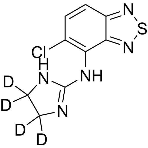
Tizanidine-d4

Based on 1 Customer Validation
Tizanidine-d4 is the deuterium labeled Tizanidine (HY-B0194). Tizanidine, a skeletal muscle relaxant, is an orally effective central α2-adrenoceptor agonist (IC50 = 6.9 nmol). Tizanidine primarily exerts muscle relaxation effects by inhibiting the release of excitatory amino acids (glutamate and aspartate) from the presynaptic terminals of spinal cord interneurons. Tizanidine has anti-injury activity and can inhibit gastrointestinal (GI) transport. Tizanidine can inhibit the proliferation, migration, and invasion of lung cancer cells and induce cell apoptosis by upregulating Nischarin and inhibiting the AKT and Wnt3a/β-catenin signaling pathways. Tizanidine can be used to treat spasticity caused by diseases such as multiple sclerosis (MS), stroke, and spinal cord injury (SCI).
For research use only. We do not sell to patients.
- Purity : 99.93%
- CAS No.: 1188331-19-2
- Formula: C9H4D4ClN5S
- Molecular Weight:257.74
-
Storage:Powder -20°C, 3 years , 4°C, 2 years ; In solvent -80°C, 6 months , -20°C, 1 month

To place orders, for customer services and technical support, please contact: MedChemExpress USA
Tel: 609-228-6898 E-mail: [email protected] [email protected]


-
 Biological Activity
-
Chemical Information
-
Solvent & Solubility
- Protocols
- Purity & Documentation
- References
-
 Help & FAQs


All Adrenergic Receptor Isoforms
More

Biological Activity
Description
Application
1. This compound can be used as a tracer
2. This compound can be used as an internal standard for quantitative analysis by NMR, GC-MS, or LC-MS.


Chemical Information
-
CAS No. 1188331-19-2
-
Unlabeled CAS 64461-82-1
-
Appearance Solid
-
Molecular Weight 257.74
-
Formula C9H4D4ClN5S
-
Color Yellow to orange
-
SMILES
N(C=1C=2C(C=CC1Cl)=NSN2)C=3NC(C(N3)([2H])[2H])([2H])[2H]
-
Shipping
Room temperature in continental US; may vary elsewhere.
-
Storage
Powder -20°C 3 years 4°C 2 years In solvent -80°C 6 months -20°C 1 month


Solvent & Solubility
In Vitro:
DMSO : 22.22 mg/mL (86.21 mM; Need ultrasonic and warming; Hygroscopic DMSO has a significant impact on the solubility of product, please use newly opened DMSO)
Please refer to the solubility information to select the appropriate solvent. Once prepared, please aliquot and store the solution to prevent product inactivation from repeated freeze-thaw cycles.
Storage method and period of stock solution: -80°C, 6 months; -20°C, 1 month. When stored at -80°C, please use it within 6 months. When stored at -20°C, please use it within 1 month.
Please refer to the solubility information to select the appropriate solvent. Once prepared, please aliquot and store the solution to prevent product inactivation from repeated freeze-thaw cycles.
Storage method and period of stock solution: -80°C, 6 months; -20°C, 1 month. When stored at -80°C, please use it within 6 months. When stored at -20°C, please use it within 1 month.
Concentration (start) × Volume (start) = Concentration (final) × Volume (final)


Purity & Documentation
-
Data Sheet (279 KB)
-
SDS (251 KB)
- English - EN (251 KB)
- Français - FR (251 KB)
- Deutsch - DE (251 KB)
- Norwegian - NO (251 KB)
- Español - ES (251 KB)
- Swedish - SV (251 KB)
- Italian - IT (251 KB)
- Korean - KR (251 KB)
- Portuguese - PT (251 KB)
-
Handling Instructions (2659 KB)
References
[1]. Russak EM, et al. Impact of Deuterium Substitution on the Pharmacokinetics of Pharmaceuticals. Ann Pharmacother. 2019;53(2):211-216. [Content Brief]
[2]. Kamen L, et al. A practical overview of tizanidine use for spasticity secondary to multiple sclerosis, stroke, and spinal cord injury. Curr Med Res Opin. 2008 Feb;24(2):425-39. [Content Brief]


Complete Stock Solution Preparation Table
Please refer to the solubility information to select the appropriate solvent. Once prepared, please aliquot and store the solution to prevent product inactivation from repeated freeze-thaw cycles.
Storage method and period of stock solution: -80°C, 6 months; -20°C, 1 month. When stored at -80°C, please use it within 6 months. When stored at -20°C, please use it within 1 month.
| Optional Solvent | Concentration Solvent Mass | 1 mg | 5 mg | 10 mg | 25 mg |
|---|---|---|---|---|---|
| DMSO | 1 mM | 3.8799 mL | 19.3994 mL | 38.7988 mL | 96.9970 mL |
| 5 mM | 0.7760 mL | 3.8799 mL | 7.7598 mL | 19.3994 mL | |
| 10 mM | 0.3880 mL | 1.9399 mL | 3.8799 mL | 9.6997 mL | |
| 15 mM | 0.2587 mL | 1.2933 mL | 2.5866 mL | 6.4665 mL | |
| 20 mM | 0.1940 mL | 0.9700 mL | 1.9399 mL | 4.8498 mL | |
| 25 mM | 0.1552 mL | 0.7760 mL | 1.5520 mL | 3.8799 mL | |
| 30 mM | 0.1293 mL | 0.6466 mL | 1.2933 mL | 3.2332 mL | |
| 40 mM | 0.0970 mL | 0.4850 mL | 0.9700 mL | 2.4249 mL | |
| 50 mM | 0.0776 mL | 0.3880 mL | 0.7760 mL | 1.9399 mL | |
| 60 mM | 0.0647 mL | 0.3233 mL | 0.6466 mL | 1.6166 mL | |
| 80 mM | 0.0485 mL | 0.2425 mL | 0.4850 mL | 1.2125 mL |


Tizanidine-d4 Related Classifications
HY-B0194S Related Classifications
 Powered by Bioz