Delivery of temperature sensitive items including proteins and kits will be paused on 6/19 for the Juneteenth holiday. For urgent orders please contact customer service.
UDP-xylose disodium is an endogenous sugar nucleotide and a catalytic substrate of UDP-xylose disodium synthase (UXS). UDP-xylose disodium is a sugar donor for the synthesis of glycoproteins, polysaccharides, various metabolites and oligosaccharides in plants, vertebrates and fungi, and participates in the synthesis of proteoglycans as a glycosyl donor. UDP-xylose disodium participates in the regulation of the synthesis of extracellular matrix components and can be used to study the mechanism of proteoglycan biosynthesis in glycobiology and related diseases (such as connectivetissue diseases)[1][2].
FG-3019 (Pamrevlumab) is a recombinant human antibody that binds to connectivetissue growth factor (CTGF). FG-3019 can be used for the research of idiopathic pulmonary fibrosis (IPF) .
Bevonescein (ALM-488) is a fluorescein-conjugated peptide that facilitates the targeted delivery of a fluorescent moiety (5-FAM (HY-66022)) to nerves after intravenous (IV) administration. Bevonescein binds nerve-associated connectivetissue, labels peripheral nerves under real-time fluorescence imaging (FL) in living mice and human ex vivo nerve tissue. Bevonescein is a peptide-linked tracer which fluorescently labeled both intact and degenerated nerves (Ex/Em = 480/530 nm) .
JNJ-7777120 is a potent and selective histamine H4 receptor antagonist (Ki=4.5 nM). JNJ-7777120 effectively blocks histamine-induced migration of mouse tracheal mast cells from connectivetissue to epithelial cells. JNJ-7777120 also significantly blocks neutrophil infiltration in a mouse Zymosan-induced peritonitis model. JNJ-7777120 has a good potential to study antipruritic and anti-inflammatory .
UDP-xylose is an endogenous sugar nucleotide and a catalytic substrate of UDP-xylose synthase (UXS). UDP-xylose is a sugar donor for the synthesis of glycoproteins, polysaccharides, various metabolites and oligosaccharides in plants, vertebrates and fungi, and participates in the synthesis of proteoglycans as a glycosyl donor. UDP-xylose participates in the regulation of the synthesis of extracellular matrix components and can be used to study the mechanism of proteoglycan biosynthesis in glycobiology and related diseases (such as connectivetissue diseases)[1][2].
(rel)-AR234960 is a selective and competitive agonist of the G protein-coupled receptor MAS. (rel)-AR234960 binds to the MAS receptor to activate the downstream ERK1/2 signaling pathway, inducing the expression of connectivetissue growth factor (CTGF) and its downstream collagen subtype genes (such as COL1A1, COL3A1). (rel)-AR234960 promotes collagen synthesis in cardiac fibroblasts through the MAS-ERK1/2-CTGF pathway and aggravates extracellular matrix remodeling. (rel)-AR234960's in vitro effect can be blocked by the MAS inverse agonist AR244555 and MEK1 inhibitor. (rel)-AR234960 regulates the expression of cardiac fibrosis-related genes and can be used in the study of heart failure .
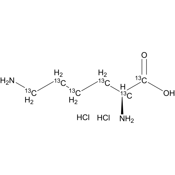
L-Lysine- 13C6 (dihydrochloride) is the 13C-labeled L-Lysine dihydrochloride. L-lysine dihydrochloride is an essential amino acid with important roles in connectivetissues and carnitine synthesis, energy production, growth in children, and maintenance of immune functions .
Agar, meets USP testing specifications is a high-quality selective growth support and substrate for non-adherent cells. Agar, meets USP testing specifications effectively supports the growth, colony formation and metachromatic matrix production of chondrocytes, and also facilitates the isolation and differentiation of pure chondrocyte strains by restricting the proliferation of fibroblast-like cells. Chondrocytes grown in Agar, meets USP testing specifications can be successfully transferred to a liquid suspension culture system, where they continue to proliferate while retaining the characteristics exhibited during growth in agar .
Firefly Luciferase mRNA is a reporter mRNA that can be transfected into cells to express firefly luciferase protein. Firefly Luciferase mRNA induces cytotoxicity in cancer cells at low concentrations. In cancer cells, the expression level of luciferase shows a non-linear relationship with the dose of Firefly Luciferase mRNA. When combined with the H2S-responsive bioluminescent probe (H-Luc), Firefly Luciferase mRNA enables bioluminescence-based detection of endogenous hydrogen sulfide in non-transgenic NAFLD cell models and NAFLD mouse models. Firefly Luciferase mRNA can be used in studies related to non-alcoholic fatty liver disease .
Hyaluronic acid tetrasaccharide, 98% is a naturally occurring polysaccharide found in the extracellular matrix of connectivetissues. Hyaluronic acid tetrasaccharide can be used as a substrate for enzyme assays to characterize hyaluronidases and other hyaluronic acid-degrading enzymes.
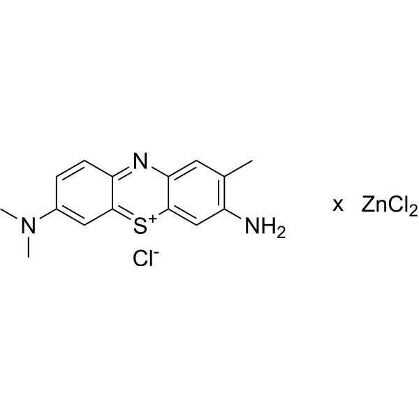
Toluidine blue (ZnCl2) is a basic thiazine dye commonly used as a biological stain for microscopy. It has a deep bluish-purple color and is commonly used to stain nucleic acids such as DNA and RNA, as well as to stain mast cells, cartilage, and other connectivetissues. Toluidine blue (ZnCl2) stains the acidic components of these tissues, such as sulfated or carboxylated mucopolysaccharides. It is frequently used in histology, cytology, and pathology applications to aid in the diagnosis of various diseases and conditions. The dye is usually applied to tissue sections prior to microscopic examination and can be differentiated using an acidic alcohol solution. Toluidine blue (ZnCl2) is a relatively simple and inexpensive stain with good reproducibility, making it a popular choice for many laboratories.
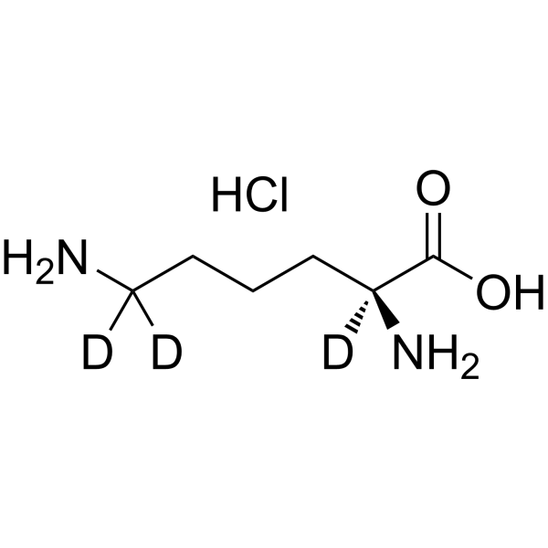
L-Lysine-d3 (hydrochloride) is the deuterium labeled L-Lysine. L-lysine is an essential amino acid with important roles in connectivetissues and carnitine synthesis, energy production, growth in children, and maintenance of immune functions .
Antifibrotic agent 2 (Compound 636) is a polycyclic pyridinone derivative with antifibrotic activity. Antifibrotic agent 2 reduces the pathological accumulation of fibrosis-related proteins such as fibronectin and collagen, prevents excessive fibrous connectivetissue from depositing in organs or tissues, and reverses or delays the remodeling of tissue fibrosisby regulating the abnormal proliferation and activation of fibroblasts. Antifibrotic agent 2 can be used for research on pulmonary fibrosis .
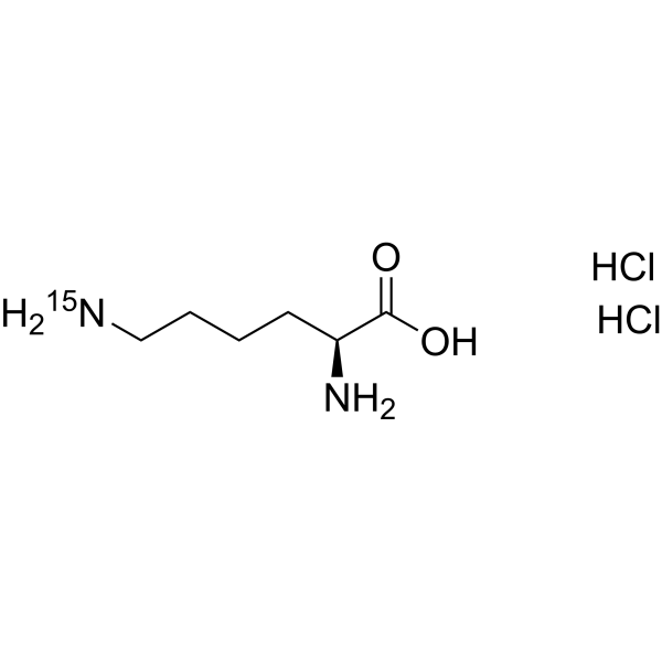
L-Lysine- 15N-1 (dihydrochloride) is the 15N-labeled L-Lysine. L-lysine is an essential amino acid with important roles in connectivetissues and carnitine synthesis, energy production, growth in children, and maintenance of immune functions .
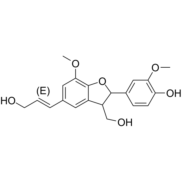
(E)-Dehydrodiconiferyl alcohol behaves as good hCA IX and hCA XII dual inhibitors . And (E)-Dehydrodiconiferyl alcohol suppresses the NF-κB nuclear translocation in connectivetissue of healing area .
JNJ-7777120 (Standard) is the analytical standard of JNJ-7777120. This product is intended for research and analytical applications. JNJ-7777120 is a potent and selective histamine H4 receptor antagonist (Ki=4.5 nM). JNJ-7777120 effectively blocks histamine-induced migration of mouse tracheal mast cells from connectivetissue to epithelial cells. JNJ-7777120 also significantly blocks neutrophil infiltration in a mouse Zymosan-induced peritonitis model. JNJ-7777120 has a good potential to study antipruritic and anti-inflammatory .
Seletinoid G is a retinoic acid receptor (RAR) agonist. Seletinoid G can repair altered connectivetissue in old skin and inhibit UV-induced collagen deficiency in young skin. Seletinoid G can be used for skin aging and photoaging research .
Aniline blue diammonium is a component of commonly used polychrome stains. Aniline blue diammonium is used to stain collagen fibers in tissue sections using Masson′s trichrome protocol for staining multiple components. Collagen is stained blue by this method. The dye is suitable for selective staining of callose in plant specimens and staining histones for assessing nuclear maturity. Aniline blue diammonium is used in Gomori′s one-step trichrome stain and Mallory′s connectivetissue stain for tissue including kidney and intestine.
SHR-1906 is a selective fully humanized monoclonal IgG1 inhibitory antibody targeting CTGF. SHR-1906 specifically binds to CTGF, thereby blocking the interaction between CTGF and TGF-B1 with an inhibition rate of 55%. SHR-1906increases the survival rate in a pulmonary fibrosis model by reducing TGF-β1 levels and inhibiting fibrotic lesions in lung tissue in Bleomycin (HY-108345)-induced pulmonary fibrosis.SHR-1906 can be used for pulmonary fibrosis (IPF) research. Recommend Isotype Controls: Human IgG1 kappa, Isotype Control (HY-P99001) .
Elastase, Rat (EC 3.4.21.35) is a form of elastase that is produced in the acinar cells of the pancreas, initially produced as an inactive zymogen and later activated in the duodenum by trypsin. Elastases form a subfamily of serine proteases, characterized by a distinctive structure consisting of two beta-barrel domains converging at the active site that hydrolyze amides and esters amongst many proteins in addition to elastin, a type of connectivetissue that holds organs together.
Neuropeptides are small proteins produced and released by neurons through the regulation of secretory pathways, expressed in neurons and have transmitter or co-transmitter functions, and are used as nerve substrates. Neuropeptides are by far the largest and most diverse signaling molecules in the brain and have been implicated in the development of diseases and drugs. Neuropeptides are involved in inflammatory and immune diseases and have an impact on epithelial, vascular, and connectivetissue cells proliferation and tissue repair. Studies have shown that neuropeptides are particularly important when the nervous system is challenged, such as stress, injury, or substance abuse. Substance P is a neuropeptide that acts as a neurotransmitter and neuromodulator in the central nervous system and is currently in clinical research and has been shown to be involved in inflammatory processes and pain.
MCE can provide 126 neuropeptides that can be used for scientific research.
Toluidine blue (ZnCl2) is a basic thiazine dye commonly used as a biological stain for microscopy. It has a deep bluish-purple color and is commonly used to stain nucleic acids such as DNA and RNA, as well as to stain mast cells, cartilage, and other connectivetissues. Toluidine blue (ZnCl2) stains the acidic components of these tissues, such as sulfated or carboxylated mucopolysaccharides. It is frequently used in histology, cytology, and pathology applications to aid in the diagnosis of various diseases and conditions. The dye is usually applied to tissue sections prior to microscopic examination and can be differentiated using an acidic alcohol solution. Toluidine blue (ZnCl2) is a relatively simple and inexpensive stain with good reproducibility, making it a popular choice for many laboratories.
Aniline blue diammonium is a component of commonly used polychrome stains. Aniline blue diammonium is used to stain collagen fibers in tissue sections using Masson′s trichrome protocol for staining multiple components. Collagen is stained blue by this method. The dye is suitable for selective staining of callose in plant specimens and staining histones for assessing nuclear maturity. Aniline blue diammonium is used in Gomori′s one-step trichrome stain and Mallory′s connectivetissue stain for tissue including kidney and intestine.
Agar, meets USP testing specifications is a high-quality selective growth support and substrate for non-adherent cells. Agar, meets USP testing specifications effectively supports the growth, colony formation and metachromatic matrix production of chondrocytes, and also facilitates the isolation and differentiation of pure chondrocyte strains by restricting the proliferation of fibroblast-like cells. Chondrocytes grown in Agar, meets USP testing specifications can be successfully transferred to a liquid suspension culture system, where they continue to proliferate while retaining the characteristics exhibited during growth in agar .
Hyaluronic acid tetrasaccharide, 98% is a naturally occurring polysaccharide found in the extracellular matrix of connectivetissues. Hyaluronic acid tetrasaccharide can be used as a substrate for enzyme assays to characterize hyaluronidases and other hyaluronic acid-degrading enzymes.
Bevonescein (ALM-488) is a fluorescein-conjugated peptide that facilitates the targeted delivery of a fluorescent moiety (5-FAM (HY-66022)) to nerves after intravenous (IV) administration. Bevonescein binds nerve-associated connectivetissue, labels peripheral nerves under real-time fluorescence imaging (FL) in living mice and human ex vivo nerve tissue. Bevonescein is a peptide-linked tracer which fluorescently labeled both intact and degenerated nerves (Ex/Em = 480/530 nm) .
GRGESP is a collagen gel contraction inhibitor. GRGESP inhibits the spreading of human fibroblasts inside collagen gels and markedly decreased gel contraction. GRGDSP can be used for the research of connectivetissue morphogenesis .
MCE Masson Staining Kit can simultaneously stain various tissue components, such as cell nuclei, collagen fibers, and muscle fibers. It features low toxicity, environmental friendliness, simple operation, and stable performance. The staining results show clear coloration and high contrast. The stained sections can be stored for long periods with minimal fading, facilitating long-term preservation and image analysis. This kit is widely used in studies of connectivetissue, muscle tissue, and collagen fibers, and is suitable for histological observation and related pathological analyses.
MCE Modified Masson Staining Kit employs Celestine Blue hematoxylin for light nuclear staining, offering shorter differentiation time compared to conventional methods. It features low toxicity, environmental friendliness, simple operation, and stable performance. The staining results show clear coloration and high contrast. The stained sections can be stored for long periods with minimal fading, facilitating long-term preservation and image analysis. This kit is widely used in studies of connectivetissue, muscle tissue, and collagen fibers, and is suitable for histological observation and related pathological analyses.
MCE Modified Van Gieson Staining Kit employs Celestine Blue and Mayer’s hematoxylin for nuclear staining, providing clearer and more stable nuclear visualization while facilitating longer preservation of stained sections. Ponceau S is used for collagen fiber staining, offering stable coloration and strong resistance to fading. This method enables effective differentiation between collagen fibers and muscle fibers, and can assist in distinguishing collagen fiber–derived tumors from myogenic tumors to a certain extent. It is also suitable for observing tissue or organ injury, repair processes, and the degree of fibrosis.
FG-3019 (Pamrevlumab) is a recombinant human antibody that binds to connectivetissue growth factor (CTGF). FG-3019 can be used for the research of idiopathic pulmonary fibrosis (IPF) .
SHR-1906 is a selective fully humanized monoclonal IgG1 inhibitory antibody targeting CTGF. SHR-1906 specifically binds to CTGF, thereby blocking the interaction between CTGF and TGF-B1 with an inhibition rate of 55%. SHR-1906increases the survival rate in a pulmonary fibrosis model by reducing TGF-β1 levels and inhibiting fibrotic lesions in lung tissue in Bleomycin (HY-108345)-induced pulmonary fibrosis.SHR-1906 can be used for pulmonary fibrosis (IPF) research. Recommend Isotype Controls: Human IgG1 kappa, Isotype Control (HY-P99001) .
UDP-xylose disodium is an endogenous sugar nucleotide and a catalytic substrate of UDP-xylose disodium synthase (UXS). UDP-xylose disodium is a sugar donor for the synthesis of glycoproteins, polysaccharides, various metabolites and oligosaccharides in plants, vertebrates and fungi, and participates in the synthesis of proteoglycans as a glycosyl donor. UDP-xylose disodium participates in the regulation of the synthesis of extracellular matrix components and can be used to study the mechanism of proteoglycan biosynthesis in glycobiology and related diseases (such as connectivetissue diseases)[1][2].
UDP-xylose is an endogenous sugar nucleotide and a catalytic substrate of UDP-xylose synthase (UXS). UDP-xylose is a sugar donor for the synthesis of glycoproteins, polysaccharides, various metabolites and oligosaccharides in plants, vertebrates and fungi, and participates in the synthesis of proteoglycans as a glycosyl donor. UDP-xylose participates in the regulation of the synthesis of extracellular matrix components and can be used to study the mechanism of proteoglycan biosynthesis in glycobiology and related diseases (such as connectivetissue diseases)[1][2].
(E)-Dehydrodiconiferyl alcohol behaves as good hCA IX and hCA XII dual inhibitors . And (E)-Dehydrodiconiferyl alcohol suppresses the NF-κB nuclear translocation in connectivetissue of healing area .
The CCN2/CTGF protein is a major connective tissue mitochondrial attractant secreted by vascular endothelial cells and critical for chondrocyte proliferation and differentiation. Additionally, it mediates heparin- and divalent cation-dependent cell adhesion in various cell types, including fibroblasts, myofibroblasts, endothelial cells, and epithelial cells. CCN2/CTGF Protein, Rat (HEK293) is the recombinant rat-derived CCN2/CTGF protein, expressed by HEK293 , with tag free.
CCN2/CTGF Protein, secreted by vascular endothelial cells, crucially promotes chondrocyte proliferation and differentiation, and mediates heparin- and divalent cation-dependent cell adhesion in diverse cell types. Additionally, it enhances fibroblast growth factor-induced DNA synthesis while existing as a monomer and interacting with TSKU. CCN2/CTGF Protein, Human (His) is the recombinant human-derived CCN2/CTGF protein, expressed by E. coli , with N-6*His labeled tag.
rHuconnectivetissue growth factor/CTGF; connectivetissue growth factor; CCN family member 2; Hypertrophic chondrocyte-specific protein 24; Insulin-like growth factor-binding protein 8; IBP-8; IGF-binding protein 8; IGFBP-8
CCN2/CTGF (Connective Tissue Growth Factor) is a protein produced primarily by endothelial cells in blood vessels. It plays an important role in various cellular processes. CCN2/CTGF Protein, Human (HEK293) is the recombinant human-derived CCN2/CTGF protein, expressed by HEK293 , with tag free.
connectivetissue growth factor; CCN family member 2; Hypertrophic chondrocyte-specific protein 24; Insulin-like growth factor-binding protein 8; CTGF; IGFBP8
CCN2/CTGF (Connective Tissue Growth Factor) is a protein produced primarily by endothelial cells in blood vessels. It plays an important role in various cellular processes. CCN2/CTGF Protein, Human (HEK293, Fc) is the recombinant human-derived CCN2/CTGF protein, expressed by HEK293 , with C-hFc labeled tag.
CCN2/CTGF protein is a major connective tissue mitochondrial attractant secreted by vascular endothelial cells and promotes chondrocyte proliferation and differentiation. It mediates heparin- and divalent cation-dependent cell adhesion in various cell types, including fibroblasts, myofibroblasts, endothelial cells, and epithelial cells. CCN2/CTGF Protein, Mouse (GST) is the recombinant mouse-derived CCN2/CTGF protein, expressed by E. coli , with N-GST labeled tag.
CCN2/CTGF protein is a major connective tissue mitochondrial attractant secreted by vascular endothelial cells and promotes chondrocyte proliferation and differentiation. It mediates heparin- and divalent cation-dependent cell adhesion in various cell types, including fibroblasts, myofibroblasts, endothelial cells, and epithelial cells. CCN2/CTGF Protein, Mouse (HEK293, His) is the recombinant mouse-derived CCN2/CTGF protein, expressed by HEK293, with C-His labeled tag.
L-Lysine- 13C6 (dihydrochloride) is the 13C-labeled L-Lysine dihydrochloride. L-lysine dihydrochloride is an essential amino acid with important roles in connectivetissues and carnitine synthesis, energy production, growth in children, and maintenance of immune functions .
L-Lysine-d3 (hydrochloride) is the deuterium labeled L-Lysine. L-lysine is an essential amino acid with important roles in connectivetissues and carnitine synthesis, energy production, growth in children, and maintenance of immune functions .
L-Lysine- 15N-1 (dihydrochloride) is the 15N-labeled L-Lysine. L-lysine is an essential amino acid with important roles in connectivetissues and carnitine synthesis, energy production, growth in children, and maintenance of immune functions .
connectivetissue growth factor; CCN family member 2; Hypertrophic chondrocyte-specific protein 24; Insulin-like growth factor-binding protein 8; IBP-8; IGF-binding protein 8; IGFBP-8;
WB, IP, ELISA
Human, Mouse, Rat
CTGF Antibody (YA6274) is a Rabbit-derived and non-conjugated IgG monoclonal antibody, targeting to CTGF.
Firefly Luciferase mRNA is a reporter mRNA that can be transfected into cells to express firefly luciferase protein. Firefly Luciferase mRNA induces cytotoxicity in cancer cells at low concentrations. In cancer cells, the expression level of luciferase shows a non-linear relationship with the dose of Firefly Luciferase mRNA. When combined with the H2S-responsive bioluminescent probe (H-Luc), Firefly Luciferase mRNA enables bioluminescence-based detection of endogenous hydrogen sulfide in non-transgenic NAFLD cell models and NAFLD mouse models. Firefly Luciferase mRNA can be used in studies related to non-alcoholic fatty liver disease .
Inquiry Online
Your information is safe with us. * Required Fields.
Western blot analysis of extracts from THP-1(lane 2(20μg), Jurkat (lane 3(20μg) and NIH3T3(lane 4(20μg) using FOXO1A (HY-P80132) Rabbit mAb. Proteins were transferred
to a PVDF membrane and blocked with 5% non-fat milk in TBST for 2 hour at room temperature. The primary antibody (1/1000) and Loading control antibody (Beta Actin, HY-P80438, 1/10000) was
used in 5% non-fat milk in TBST at 4°C overnight. Goat Anti-Mouse/Rabbit IgG-HRP Secondary Antibody (1/10000) was used for 1 hour at room temperature.
Western blot analysis of extracts from THP-1(lane 2(20μg), Jurkat (lane 3(20μg) and NIH3T3(lane 4(20μg) using FOXO1A (HY-P80132) Rabbit mAb. Proteins were transferred
to a PVDF membrane and blocked with 5% non-fat milk in TBST for 2 hour at room temperature. The primary antibody (1/1000) and Loading control antibody (Beta Actin, HY-P80438, 1/10000) was
MedchemExpress Validation 03
Western blot analysis of extracts from THP-1(lane 2(20μg), Jurkat (lane 3(20μg) and NIH3T3(lane 4(20μg) using FOXO1A (HY-P80132) Rabbit mAb. Proteins were transferred
MedchemExpress Validation 04
Western blot analysis of extracts from THP-1(lane 2(20μg), Jurkat (lane 3(20μg) and NIH3T3(lane 4(20μg) using FOXO1A (HY-P80132) Rabbit mAb. Proteins were transferred
to a PVDF membrane and blocked with 5% non-fat milk in TBST for 2 hour at room temperature. The primary antibody (1/1000) and Loading control antibody (Beta Actin, HY-P80438, 1/10000) was
used in 5% non-fat milk in TBST at 4°C overnight. Goat Anti-Mouse/Rabbit IgG-HRP Secondary Antibody (1/10000) was used for 1 hour at room temperature.
MedchemExpress Validation
Western blot analysis of extracts from THP-1(lane 2(20μg), Jurkat (lane 3(20μg) and NIH3T3(lane 4(20μg) using FOXO1A (HY-P80132) Rabbit mAb. Proteins were transferred
to a PVDF membrane and blocked with 5% non-fat milk in TBST for 2 hour at room temperature. The primary antibody (1/1000) and Loading control antibody (Beta Actin, HY-P80438, 1/10000) was
used in 5% non-fat milk in TBST at 4°C overnight. Goat Anti-Mouse/Rabbit IgG-HRP Secondary Antibody (1/10000) was used for 1 hour at room temperature.
MedchemExpress Validation
Western blot analysis of extracts from THP-1(lane 2(20μg), Jurkat (lane 3(20μg) and NIH3T3(lane 4(20μg) using FOXO1A (HY-P80132) Rabbit mAb. Proteins were transferred
to a PVDF membrane and blocked with 5% non-fat milk in TBST for 2 hour at room temperature. The primary antibody (1/1000) and Loading control antibody (Beta Actin, HY-P80438, 1/10000) was
used in 5% non-fat milk in TBST at 4°C overnight. Goat Anti-Mouse/Rabbit IgG-HRP Secondary Antibody (1/10000) was used for 1 hour at room temperature.
MedChemExpress values your privacy and your trust is important to us. We use cookies to enhance your website experience. Some cookies are necessary to run the website.
Privacy and Cookie Policy