



UDP-xylose disodium
Based on 2 publication(s) in Google Scholar
UDP-xylose disodium is an endogenous sugar nucleotide and a catalytic substrate of UDP-xylose disodium synthase (UXS). UDP-xylose disodium is a sugar donor for the synthesis of glycoproteins, polysaccharides, various metabolites and oligosaccharides in plants, vertebrates and fungi, and participates in the synthesis of proteoglycans as a glycosyl donor. UDP-xylose disodium participates in the regulation of the synthesis of extracellular matrix components and can be used to study the mechanism of proteoglycan biosynthesis in glycobiology and related diseases (such as connective tissue diseases)[1][2].
For research use only. We do not sell to patients.
- Purity : 99.90%
- CAS No.: 108320-89-4
- Formula: C14H20N2Na2O16P2
- Molecular Weight:580.24
-
Storage:
-20°C, sealed storage, away from moisture
* In solvent : -80°C, 6 months; -20°C, 1 month (sealed storage, away from moisture)

To place orders, for customer services and technical support, please contact: MedChemExpress USA
Tel: 609-228-6898 E-mail: [email protected] [email protected]


-
 Biological Activity
-
Chemical Information
-
Solvent & Solubility
- Protocols
- Purity & Documentation
- References
-
 Help & FAQs


-
HY-N7033
UDP-glucuronic acid trisodiumGlycoconjugates Precursor -
HY-114365
UDP-GalNAc disodiumEpsC115 Substrate -
HY-114364
UDP-Galactose disodiumP2Y14 Receptor Agonist -
HY-101916
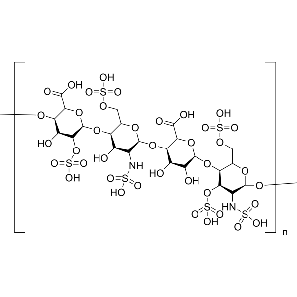
Heparan SulfateLinear Polysaccharide -
HY-N8278
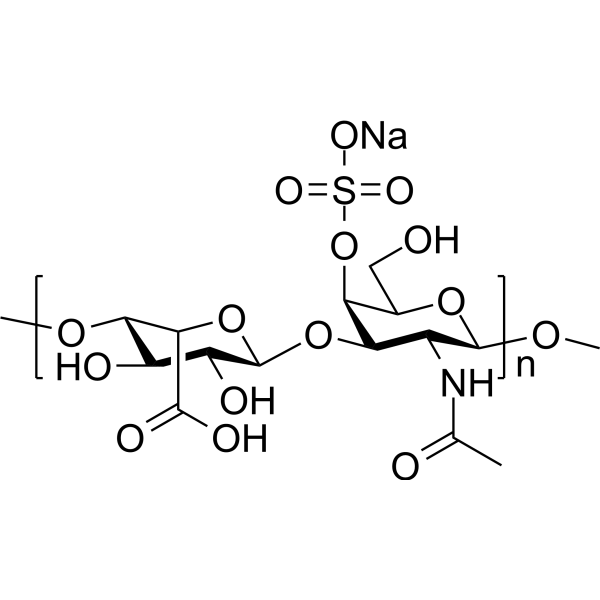
Dermatan sulphate sodiumThrombin Inactivator -
HY-N7389A
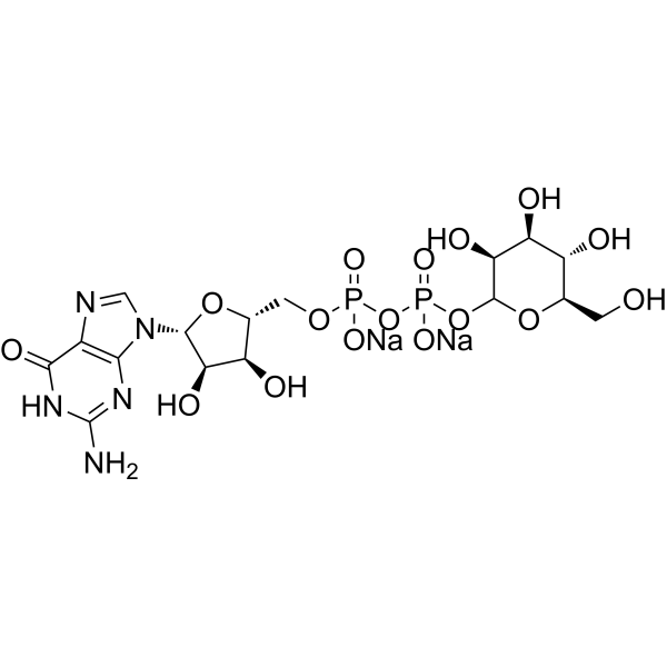
GDP-D-mannose disodiumGDP-L-fucose Precursor
Publications Citing Use of MedChemExpress (MCE) UDP-xylose disodium
MoreAll Endogenous Metabolite Isoforms
More

Biological Activity
Description
In Vitro
UDP-xylose disodium can be generated from UDP-Glc in recombinant yeast cytoplasmic extracts catalyzed by AtUGD1 and AtUXS3[1].
MedChemExpress (MCE) has not independently confirmed the accuracy of these methods. They are for reference only. Further protocols information, click here.


Chemical Information
-
CAS No. 108320-89-4
-
Appearance Solid
-
Molecular Weight 580.24
-
Formula C14H20N2Na2O16P2
-
Color White to off-white
-
SMILES
O[C@H]([C@H]([C@@H](CO1)O)O)[C@H]1OP(OP(OC[C@@H]2[C@@H](O)[C@@H](O)[C@H](N3C(NC(C=C3)=O)=O)O2)(O[Na])=O)(O[Na])=O
-
Structure Classification
-
Initial Source
-
Shipping
Room temperature in continental US; may vary elsewhere.
-
Storage
-20°C, sealed storage, away from moisture
* In solvent : -80°C, 6 months; -20°C, 1 month (sealed storage, away from moisture)


Publications (2)
-
Journal Impact Factor
-
Most Recent
-
Mol Plant
2025 Dec 13:S1674-2052(25)00435-6. PMID: 41392480 -
bioRxiv
Biochemical and structural characterization of glycosyltransferase family 61 proteins reveal a key determinant of sugar donor specificity. [Abstract]2025 Jul 19:2025.07.18.665579. PMID: 40791416


Solvent & Solubility
In Vitro:
H2O : 100 mg/mL (172.34 mM; Need ultrasonic)
Please refer to the solubility information to select the appropriate solvent. Once prepared, please aliquot and store the solution to prevent product inactivation from repeated freeze-thaw cycles.
Storage method and period of stock solution: -80°C, 6 months; -20°C, 1 month (sealed storage, away from moisture). When stored at -80°C, please use it within 6 months. When stored at -20°C, please use it within 1 month.
* Note: If you choose water as the stock solution, please dilute it to the working solution, then filter and sterilize it with a 0.22 μm filter before use.
Please refer to the solubility information to select the appropriate solvent. Once prepared, please aliquot and store the solution to prevent product inactivation from repeated freeze-thaw cycles.
Storage method and period of stock solution: -80°C, 6 months; -20°C, 1 month (sealed storage, away from moisture). When stored at -80°C, please use it within 6 months. When stored at -20°C, please use it within 1 month.
* Note: If you choose water as the stock solution, please dilute it to the working solution, then filter and sterilize it with a 0.22 μm filter before use.
Concentration (start) × Volume (start) = Concentration (final) × Volume (final)

Protocols
-
Fibrosis/Collagen Morphometry
Fibrosis and collagen morphometry is based on the quantitative visualization of fibrillar collagen deposition in tissue sections using histochemical stains such as Sirius Red (Picrosirius Red) or Masson's trichrome, followed by image-based or polarization-enhanced analysis to estimate collagen proportional area as a surrogate of extracellular matrix accumulation during fibrotic remodeling. Sirius Red combined with polarized light microscopy enhances detection of collagen fibers due to birefringence properties, enabling more specific visualization of collagen type I and III fibrils compared to conventional bright-field histology, while whole-section or region-restricted digital morphometry reduces field-selection bias in fibrosis assessment. Alternative quantitative approaches include second harmonic generation (SHG) and two-photon excited fluorescence microscopy, which enable label-free detection of fibrillar collagen and have been validated against histological staining and biochemica
-
Collagen: Sirius Red Staining
Sirius Red or picrosirius red staining is a histochemical method for visualizing collagen-rich extracellular matrix in tissue sections, and collagen fibers are detected as red-stained structures under bright-field microscopy with enhanced birefringence under polarized light. Picrosirius red is useful for assessing total collagen organization, distribution, and fibrosis burden, but polarized color should not be interpreted as a definitive collagen type I versus type III readout because color is affected by fiber orientation, thickness, and packing.
-
ECM-Embedded Organoid (Matrigel/Dome) Culture
ECM-embedded organoid dome culture embeds epithelial stem cells, crypts, organoid fragments, or tumor-derived epithelial cells in a basement-membrane-like hydrogel such as Matrigel, allowing 3D growth, self-organization, lumen formation, budding or cystic morphogenesis, and lineage maintenance under defined niche-factor-containing medium. The primary readouts are organoid establishment efficiency, growth, morphology, passaging capacity, lineage-marker expression, and, when fluorescently labeled lines are used, microscopy- or flow-cytometry-based quantification of population behavior in 3D culture.
-
Connective Tissue: Masson's Trichrome/Collagen Trichrome Staining
Masson’s Trichrome (collagen/trichrome staining) is a histological technique that differentially stains tissue compartments using sequential acidic dyes to distinguish collagen from muscle and cytoplasmic components based on dye affinity and tissue permeability differences, enabling visualization of fibrosis and connective tissue architecture in histological sections. The classical formulation typically uses Weigert's iron hematoxylin for nuclear staining, Biebrich scarlet-acid fuchsin for cytoplasm and muscle, and aniline blue (or light green variants) for collagen, producing a characteristic blue/green collagen signal contrasted against red cytoplasm and dark nuclei. The staining principle relies on selective displacement of smaller dye molecules by larger anionic dyes in collagen-rich regions under controlled acidified conditions, which enhances collagen-specific dye retention. This property makes the method widely used for fibrosis assessment in organs such as heart, liver, lung, a
-
Matrigel/ECM Transwell Invasion Assay
The Matrigel/ECM Transwell invasion assay measures the ability of cells to move toward a chemoattractant while crossing an extracellular-matrix barrier placed on a porous membrane; therefore, the readout reflects both chemotactic motility and matrix invasion rather than migration alone. Matrigel is a basement-membrane-rich matrix derived from Engelbreth-Holm-Swarm mouse sarcoma and has been used as a reconstituted basement membrane barrier in chemoinvasion assays. The assay readout is generated by quantifying cells that reach the underside of the insert membrane or lower compartment after incubation, commonly by staining and counting invaded cells or by fluorescence-based quantification.
-
Matrigel Transwell/Boyden Chamber Invasion Assay
Matrigel Transwell/Boyden chamber invasion assay measures the ability of cells to degrade or traverse an extracellular matrix-coated porous membrane and move from an upper chamber toward a chemoattractant in a lower chamber. Invasion is distinguished from migration by coating the membrane with Matrigel or basement membrane matrix; uncoated inserts measure migration, while coated inserts require cells to cross an ECM barrier before reaching the underside of the membrane.


Purity & Documentation
-
Data Sheet (276 KB)
-
SDS (393 KB)
- English - EN (393 KB)
- Français - FR (393 KB)
- Deutsch - DE (393 KB)
- Norwegian - NO (393 KB)
- Español - ES (393 KB)
- Swedish - SV (393 KB)
- Italian - IT (393 KB)
- Korean - KR (393 KB)
- Portuguese - PT (393 KB)
-
Handling Instructions (2659 KB)
References
[1]. Oka T, et al. Reconstruction of de novo pathway for synthesis of UDP-glucuronic acid and UDP-xylose from intrinsic UDP-glucose in Saccharomyces cerevisiae. FEBS J. 2006 Jun;273(12):2645-57. [Content Brief]
[2]. Eixelsberger T, et al. Structure and mechanism of human UDP-xylose synthase: evidence for a promoting role of sugar ring distortion in a three-step catalytic conversion of UDP-glucuronic acid. J Biol Chem. 2012 Sep 7;287(37):31349-58. [Content Brief]


Complete Stock Solution Preparation Table
Please refer to the solubility information to select the appropriate solvent. Once prepared, please aliquot and store the solution to prevent product inactivation from repeated freeze-thaw cycles.
Storage method and period of stock solution: -80°C, 6 months; -20°C, 1 month (sealed storage, away from moisture). When stored at -80°C, please use it within 6 months. When stored at -20°C, please use it within 1 month.
| Optional Solvent | Concentration Solvent Mass | 1 mg | 5 mg | 10 mg | 25 mg |
|---|---|---|---|---|---|
| H2O | 1 mM | 1.7234 mL | 8.6171 mL | 17.2342 mL | 43.0856 mL |
| 5 mM | 0.3447 mL | 1.7234 mL | 3.4468 mL | 8.6171 mL | |
| 10 mM | 0.1723 mL | 0.8617 mL | 1.7234 mL | 4.3086 mL | |
| 15 mM | 0.1149 mL | 0.5745 mL | 1.1489 mL | 2.8724 mL | |
| 20 mM | 0.0862 mL | 0.4309 mL | 0.8617 mL | 2.1543 mL | |
| 25 mM | 0.0689 mL | 0.3447 mL | 0.6894 mL | 1.7234 mL | |
| 30 mM | 0.0574 mL | 0.2872 mL | 0.5745 mL | 1.4362 mL | |
| 40 mM | 0.0431 mL | 0.2154 mL | 0.4309 mL | 1.0771 mL | |
| 50 mM | 0.0345 mL | 0.1723 mL | 0.3447 mL | 0.8617 mL | |
| 60 mM | 0.0287 mL | 0.1436 mL | 0.2872 mL | 0.7181 mL | |
| 80 mM | 0.0215 mL | 0.1077 mL | 0.2154 mL | 0.5386 mL | |
| 100 mM | 0.0172 mL | 0.0862 mL | 0.1723 mL | 0.4309 mL |
* Note: If you choose water as the stock solution, please dilute it to the working solution, then filter and sterilize it with a 0.22 μm filter before use.


UDP-xylose disodium Related Classifications
HY-N11287A Related Classifications
 Powered by Bioz