



Swainsonine
Based on 1 publication(s) in Google Scholar
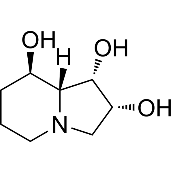
Swainsonine (Tridolgosir) is an natural indolizidine alkaloid, a potent and reversible α-mannosidase inhibitor. Swainsonine induces apoptosis and cell cycle arrest at G2/M phase. Swainsonine shows anti-tumor activity.
For research use only. We do not sell to patients.
- Purity : 99.95%
- CAS No.: 72741-87-8
- Formula: C8H15NO3
- Molecular Weight:173.21
-
Storage:Powder -20°C, 3 years ; In solvent -80°C, 6 months , -20°C, 1 month

To place orders, for customer services and technical support, please contact: MedChemExpress USA
Tel: 609-228-6898 E-mail: [email protected] [email protected]


-
 Biological Activity
-
Chemical Information
-
Solvent & Solubility
- Protocols
- Purity & Documentation
- References
-
 Help & FAQs


-
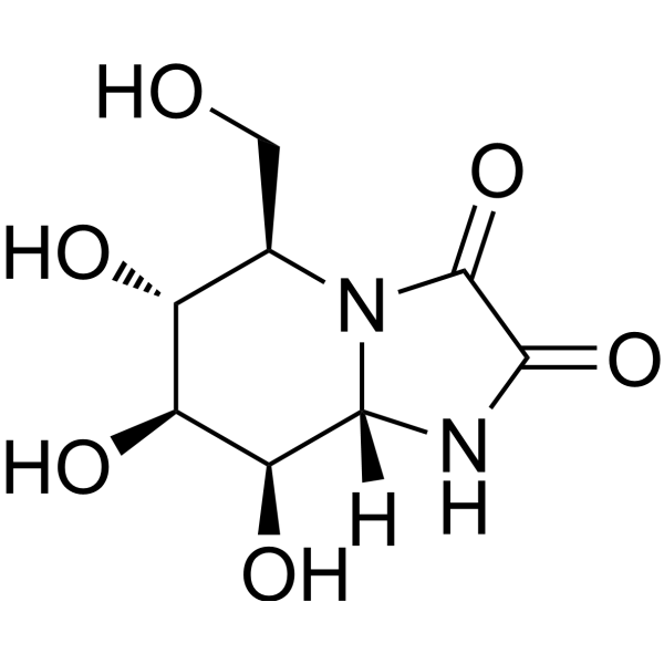
HY-19332
Kifunensineα-Mannosidases Inhibitor -
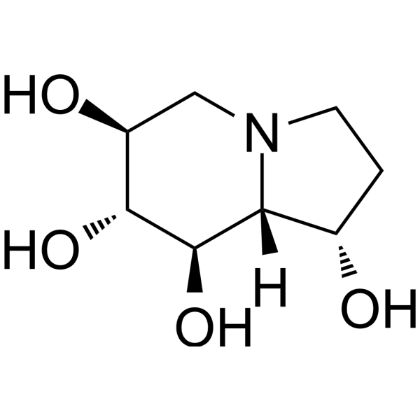
HY-N2022
Castanospermineα/β-Glucosidases Inhibitor -
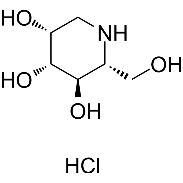
HY-W009783
1-Deoxymannojirimycin hydrochlorideα1,2-mannosidase I Inhibitor -
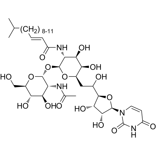
HY-A0098
TunicamycinER Stress Inducer -

HY-10219
RapamycinmTOR Inhibitor -
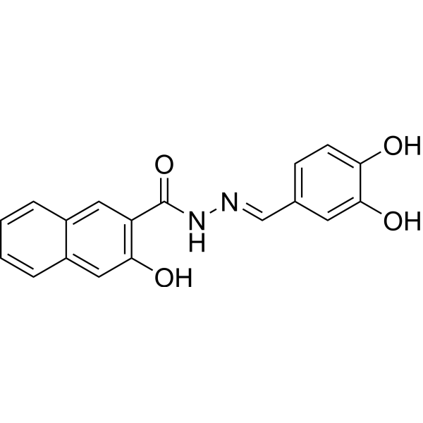
HY-15304
DynasoreAutophagy Inducer -
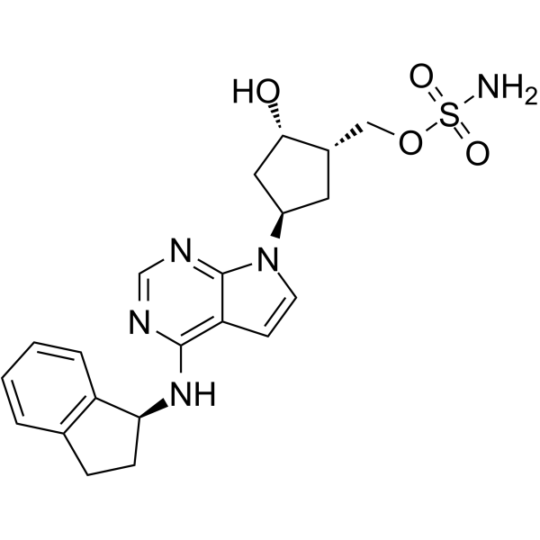
HY-70062
PevonedistatNAE Inhibitor -
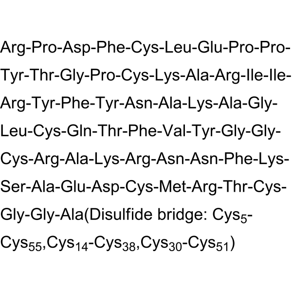
HY-P0017
AprotininBPTI Inhibitor -

HY-10583
Y-27632 dihydrochlorideROCK Inhibitor -
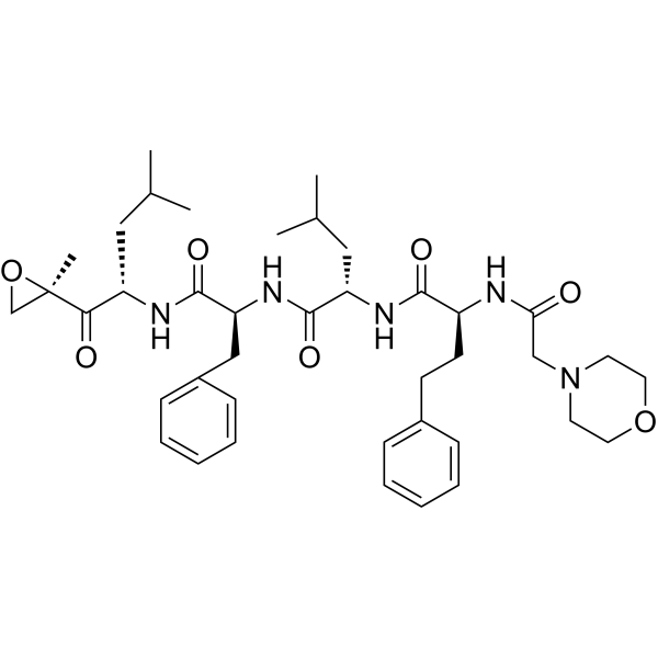
HY-10455
CarfilzomibProteasome Inhibitor -

HY-19332R
Kifunensine (Standard)α-Mannosidases Inhibitor -

HY-P2929
PNGase FGlycosidase
Publications Citing Use of MedChemExpress (MCE) Swainsonine
MoreAll Antibiotic Isoforms
More

Biological Activity
Description
In Vitro
Swainsonine (0-40 μM; 12 h) shows antiproliferative activity in U251 and LN444 cells[2].
Swainsonine (30 μM; 12 h) induces apoptosis and cell cycle arrest at G2/M phase, and decreases the expression of CyclinD1 and CDK4 protein levels, increases the expression of p16 protein level[2].
Swainsonine (30 μM; 12 h) reduces glioma cell migration and invasion[2].
MedChemExpress (MCE) has not independently confirmed the accuracy of these methods. They are for reference only.
-
Cell Line:U251, LN444 cells
-
Concentration:0, 10, 20, 30, 40 µM
-
Incubation Time:12 h
-
Result:Inhibited cell viability in a dose-dependent manner, and shows no cytotoxicity in normal brain cells.
-
Cell Line:U251 cells
-
Concentration:30 µM
-
Incubation Time:12 h
-
Result:Induced cell cycle arrest at G2/M phase.
-
Cell Line:U251 cells
-
Concentration:30 µM
-
Incubation Time:12 h
-
Result:Induced apoptosis with the cleaved-Caspase-3 and cleaved-Caspase-9 expression were obviously increased.
In Vivo
MedChemExpress (MCE) has not independently confirmed the accuracy of these methods. They are for reference only.
-
Animal Model:Eighty-seven sixty-day-old male C57BL/6 mice[3]
-
Dosage:1 mg/kg (cisplatin; 0.25 mg/kg, i.p., every other day for 10 days)
-
Administration:I.p.; twice daily for ten days
-
Result:Showed a significant reduction in ascites volume of 63.5% was observed in mice and a significant reduction in the percentage of cells in the G0/G1, S and G2 phases.


Chemical Information
-
CAS No. 72741-87-8
-
Appearance Solid
-
Molecular Weight 173.21
-
Formula C8H15NO3
-
Color White to yellow
-
SMILES
O[C@@H]1[C@H](O)CN2CCC[C@@H](O)[C@]12[H]
-
Synonyms
Tridolgosir
-
Structure Classification
-
Initial Source
-
Shipping
Room temperature in continental US; may vary elsewhere.
-
Storage
Powder -20°C 3 years In solvent -80°C 6 months -20°C 1 month


Publications (1)
-
Journal Impact Factor
-
Most Recent
-
Microbiome
An iron-binding protein of entomopathogenic fungus suppresses the proliferation of host symbiotic bacteria. [Abstract]2024 Oct 15;12(1):202. PMID: 39407320


Solvent & Solubility
In Vitro:
DMSO : 10 mg/mL (57.73 mM; Need ultrasonic and warming; Hygroscopic DMSO has a significant impact on the solubility of product, please use newly opened DMSO)
H2O : 4.17 mg/mL (24.07 mM; Need ultrasonic)
Please refer to the solubility information to select the appropriate solvent. Once prepared, please aliquot and store the solution to prevent product inactivation from repeated freeze-thaw cycles.
Storage method and period of stock solution: -80°C, 6 months; -20°C, 1 month. When stored at -80°C, please use it within 6 months. When stored at -20°C, please use it within 1 month.
* Note: If you choose water as the stock solution, please dilute it to the working solution, then filter and sterilize it with a 0.22 μm filter before use.
Please refer to the solubility information to select the appropriate solvent. Once prepared, please aliquot and store the solution to prevent product inactivation from repeated freeze-thaw cycles.
Storage method and period of stock solution: -80°C, 6 months; -20°C, 1 month. When stored at -80°C, please use it within 6 months. When stored at -20°C, please use it within 1 month.
* Note: If you choose water as the stock solution, please dilute it to the working solution, then filter and sterilize it with a 0.22 μm filter before use.
Concentration (start) × Volume (start) = Concentration (final) × Volume (final)


Purity & Documentation
-
Data Sheet (280 KB)
-
SDS (393 KB)
- English - EN (393 KB)
- Français - FR (393 KB)
- Deutsch - DE (393 KB)
- Norwegian - NO (393 KB)
- Español - ES (393 KB)
- Swedish - SV (393 KB)
- Italian - IT (393 KB)
- Korean - KR (393 KB)
- Portuguese - PT (393 KB)
-
Handling Instructions (2659 KB)
References
[1]. Dorling PR, et al. Inhibition of lysosomal alpha-mannosidase by swainsonine, an indolizidine alkaloid isolated from Swainsona canescens. Biochem J. 1980 Nov 1;191(2):649-51. [Content Brief]
[2]. Sun L, et al. Swainsonine represses glioma cell proliferation, migration and invasion by reduction of miR-92a expression. BMC Cancer. 2019 Mar 19;19(1):247. doi: 10.1186/s12885-019-5425-7. [Content Brief]
[3]. Santos FM, et al. Increased antitumor efficacy by the combined administration of swainsonine and cisplatin in vivo. Phytomedicine. 2011 Sep 15;18(12):1096-101. [Content Brief]


Complete Stock Solution Preparation Table
Please refer to the solubility information to select the appropriate solvent. Once prepared, please aliquot and store the solution to prevent product inactivation from repeated freeze-thaw cycles.
Storage method and period of stock solution: -80°C, 6 months; -20°C, 1 month. When stored at -80°C, please use it within 6 months. When stored at -20°C, please use it within 1 month.
| Optional Solvent | Concentration Solvent Mass | 1 mg | 5 mg | 10 mg | 25 mg |
|---|---|---|---|---|---|
| H2O / DMSO | 1 mM | 5.7733 mL | 28.8667 mL | 57.7334 mL | 144.3335 mL |
| 5 mM | 1.1547 mL | 5.7733 mL | 11.5467 mL | 28.8667 mL | |
| 10 mM | 0.5773 mL | 2.8867 mL | 5.7733 mL | 14.4333 mL | |
| 15 mM | 0.3849 mL | 1.9244 mL | 3.8489 mL | 9.6222 mL | |
| 20 mM | 0.2887 mL | 1.4433 mL | 2.8867 mL | 7.2167 mL | |
| DMSO | 25 mM | 0.2309 mL | 1.1547 mL | 2.3093 mL | 5.7733 mL |
| 30 mM | 0.1924 mL | 0.9622 mL | 1.9244 mL | 4.8111 mL | |
| 40 mM | 0.1443 mL | 0.7217 mL | 1.4433 mL | 3.6083 mL | |
| 50 mM | 0.1155 mL | 0.5773 mL | 1.1547 mL | 2.8867 mL |
* Note: If you choose water as the stock solution, please dilute it to the working solution, then filter and sterilize it with a 0.22 μm filter before use.


Swainsonine Related Classifications
HY-N6722 Related Classifications
 Powered by Bioz