



Cilengitide
Based on 78 publication(s) in Google Scholar
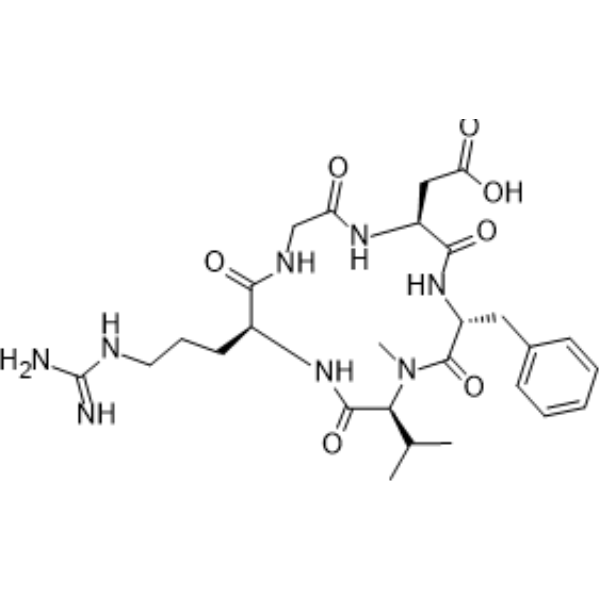
Cilengitide (EMD 121974) is an integrin (integrin) inhibitor with blood-brain barrier permeability, with IC50 values against human targets as follows: 0.61 nM for αvβ3, 8.4 nM for αvβ5, 14.9 nM for α5β1, 5400 nM for αIIbβ3, 2050 nM for αvβ6, 2350 nM for αvβ8. Cilengitide inhibits the binding of integrins to vitronectin, fibronectin, fibrinogen and LAP (TGF-β), and serves as an internal standard for solid-phase integrin binding assays. Cilengitide inhibits tumor cell viability, induces apoptosis, reduces the phosphorylation levels of STAT3, AKT and mTOR, downregulates the expression of PD-L1, inhibits cell viability and angiogenesis, regulates anti-tumor immune responses and slows tumor growth. Cilengitide can be used in research related to glioblastoma, melanoma, advanced solid tumors and refractory brain tumors.
Nur für Forschungszwecke. Wir verkaufen nicht an Patienten.
- Reinheit : 99.91%
- CAS. Nr.: 188968-51-6
- Formel: C27H40N8O7
- Molecular Weight:588.66
-
Speicherung:Powder -20°C, 3 years ; In solvent -80°C, 1 year , -20°C, 6 months

To place orders, for customer services and technical support, please contact: MedChemExpress USA
Tel: 609-228-6898 E-mail: [email protected] [email protected]


-
 Biologische Aktivität
-
Chemical Information
-
Lösungsmittel & Löslichkeit
- Protokoll
- Reinheit & Dokumentation
- Verweise
-
 Help & FAQs


-
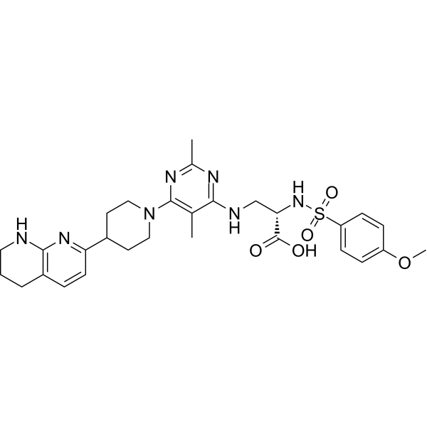
HY-100506
GLPG0187Integrin Receptor Antagonist -
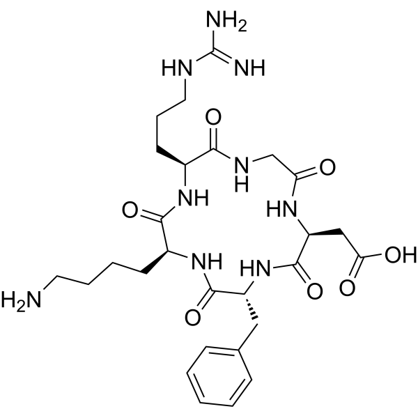
HY-P0023
Cyclo(-RGDfK)αvβ3 Integrin Inhibitor -
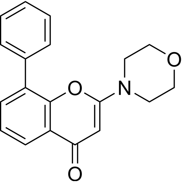
HY-10108
LY294002PI3K Inhibitor -

HY-10583
Y-27632 dihydrochlorideROCK Inhibitor -
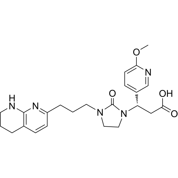
HY-15102
MK-0429αvβ3 Integrin Inhibitor -
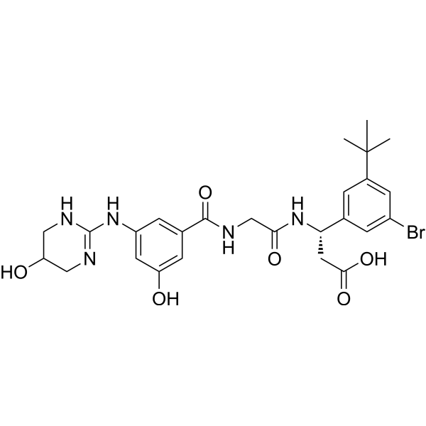
HY-18644
CWHM-12αV Integrins Inhibitor -
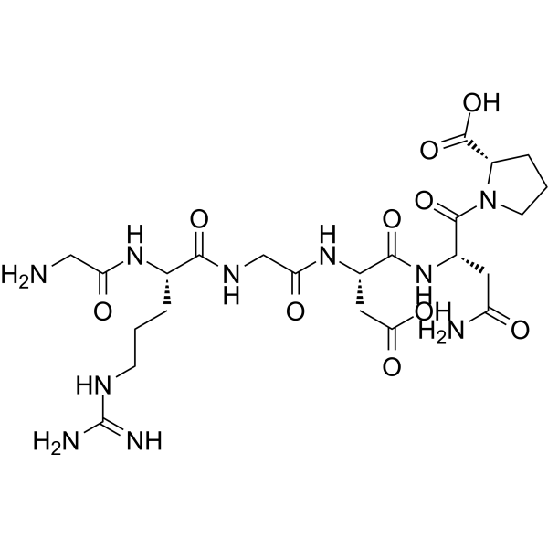
HY-P1740
RGD peptide (GRGDNP)Integrin-Iigand Interaction Inhibitor -

HY-100558
Bafilomycin A1V-ATPase Inhibitor -
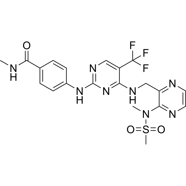
HY-12289
DefactinibFAK Inhibitor -
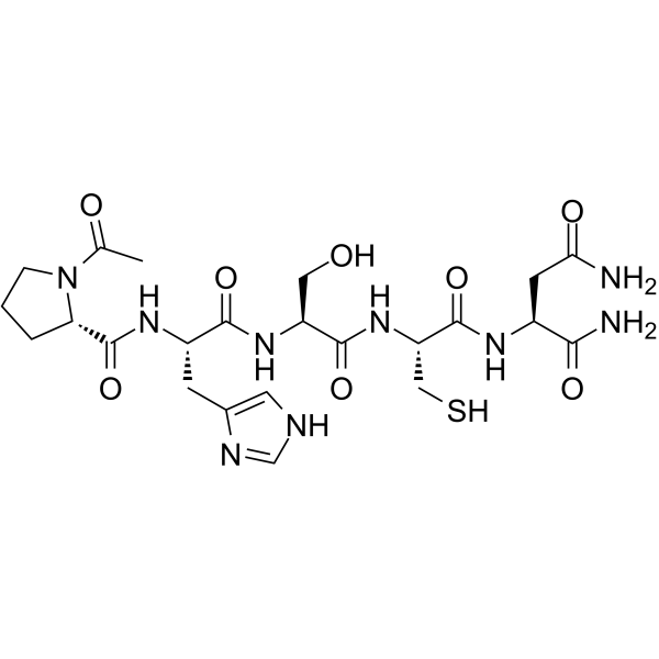
HY-13535
ATN-161α5β1/αvβ3 Integrin Binder -
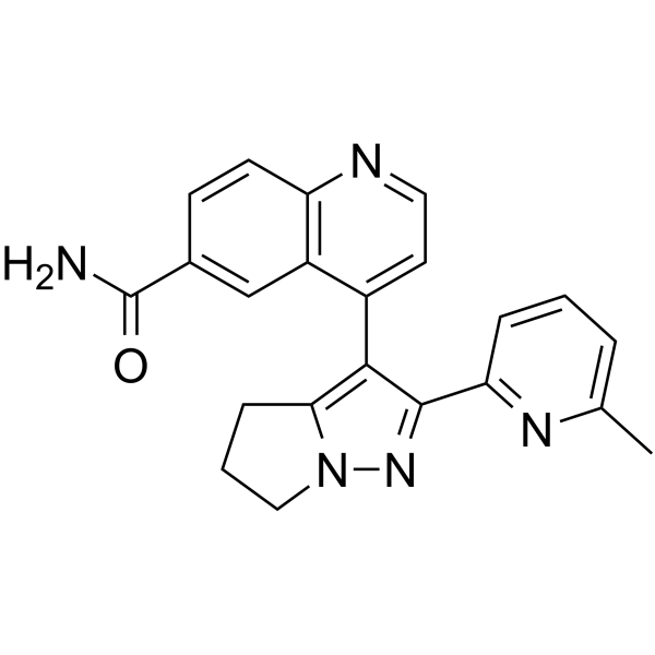
HY-13226
GalunisertibTGFβRI Inhibitor -
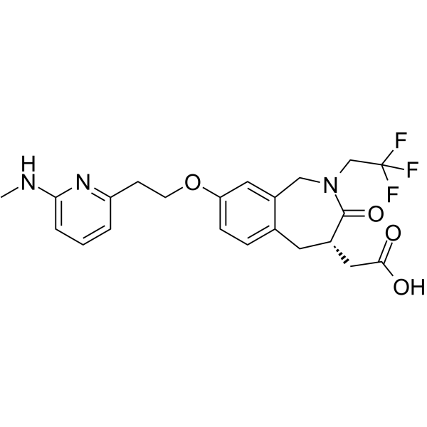
HY-19307
SB-273005αvβ3/αvβ5 Receptor Antagonist
Publications Citing Use of MedChemExpress (MCE) Cilengitide
More- Cancer Cell. 2021 Nov 8;39(11):1531-1547.e10. [Abstract]
- Cell. 2020 Aug 6;182(3):545-562.e23. [Abstract]
- Nat Mater. 2025 Sep 1. [Abstract]
- Nat Cell Biol. 2020 Mar;22(3):289-296. [Abstract]
- J Clin Invest. 2025 Oct 16:e188822. [Abstract]
- Adv Sci (Weinh). 2025 Oct 30:e01602. [Abstract]
- Adv Sci (Weinh). 2025 May;12(20):e2504647. [Abstract]
- Cell Rep Med. 2025 Feb 18;6(2):101922. [Abstract]
- Sci Adv. 2026 Jan 2;12(1):eady4112. [Abstract]
- Sci Adv. 2026 Jan 2;12(1):eadx2768. [Abstract]
- Chem Eng J. 2025 Dec 20.
- Cell Death Dis. 2025 Dec 12;16(1):887. [Abstract]
- Cell Death Dis. 2022 Dec 7;13(12):1028. [Abstract]
- Engineering. 8 October 2020.
- Int J Biol Sci. 2025 May 27;21(8):3573-3596. [Abstract]
- J Immunother Cancer. 2020 Mar;8(1):e000111. [Abstract]
- J Transl Med. 2018 Dec 12;16(1):352. [Abstract]
- Oncogene. 2025 Jul;44(28):2396-2412. [Abstract]
- Cell Mol Gastroenterol Hepatol. 2025;19(10):101548. [Abstract]
- Free Radic Biol Med. 2024 Dec 9:227:296-311. [Abstract]
- Clin Transl Med. 2021 Oct;11(10):e548. [Abstract]
- Br J Cancer. 2023 Mar;128(7):1344-1359. [Abstract]
- Cell Rep. 2026 Jul 28;45(7):117650.
- Cell Rep. 2022 Sep 27;40(13):111422. [Abstract]
- Environ Pollut. 2023 Aug 1:330:121817. [Abstract]
- Blood Rev. 2023 May:59:101038. [Abstract]
- Front Immunol. 2018 Jun 1:9:1207. [Abstract]
- Mol Cancer Ther. 2022 Sep 6;21(9):1485-1496. [Abstract]
- JCI Insight. 2026 Aug 11;11(18):e198555.
- Biochem Pharmacol. 2025 Jun:236:116870. [Abstract]
- J Chem Inf Model. 2023 Oct 23;63(20):6302-6315. [Abstract]
- Oncoimmunology. 2025 Dec;14(1):2508057. [Abstract]
- J Cell Biol. 2023 Aug 7;222(8):e202303107. [Abstract]
- Colloids Surf B Biointerfaces. 2026 Sep:265:115699. [Abstract]
- Commun Biol. 2024 Dec 30;7(1):1713. [Abstract]
- Respir Res. 2025 Jul 2;26(1):229. [Abstract]
- Int Immunopharmacol. 2024 Sep 10:138:112545. [Abstract]
- Stem Cell Reports. 2017 Dec 12;9(6):1948-1960. [Abstract]
- Front Pharmacol. 2021 Feb 24:12:585778. [Abstract]
- Am J Physiol Cell Physiol. 2018 Apr 1;314(4):C415-C427. [Abstract]
- Biochim Biophys Acta Mol Basis Dis. 2025 Jan;1871(1):167484. [Abstract]
- Obesity. 2015 Apr;23(4):779-85. [Abstract]
- Cancers (Basel). 2023 Mar 12;15(6):1729. [Abstract]
- Mol Cell Biochem. 2025 Oct 20. [Abstract]
- FASEB J. 2023 Feb;37(2):e22726. [Abstract]
- Lung. 2020 Dec;198(6):947-955. [Abstract]
- Cell Adh Migr. 2019 Dec;13(1):152-163. [Abstract]
- Differentiation. 2026 May-Jun:149:100962. [Abstract]
- J Cell Physiol. 2024 Jun;239(6):e31267. [Abstract]
- Cell Biol Int. 2020 Apr;44(4):966-974. [Abstract]
- Cancer Med. 2018 Feb;7(2):408-419. [Abstract]
- Atheroscler Plus. 2022 Oct 19:50:57-64. [Abstract]
- J Integr Neurosci. 2024 Jul 25;23(7):140. [Abstract]
- Biomed Res Int. 2021 Nov 30:2021:5954757. [Abstract]
- PLoS One. 2016 Feb 3;11(2):e0148333. [Abstract]
- PLoS One. 2014 Oct 13;9(10):e110453. [Abstract]
- J Mol Histol. 2020 Apr;51(2):147-159. [Abstract]
- J Oral Pathol Med. 2025 Aug;54(7):597-606. [Abstract]
- Bratisl Lek Listy. 2026 Aug 10.
- Scand Cardiovasc J. 2021 Oct;55(5):287-296. [Abstract]
- J Vis Exp. 2025 May 23:(219). [Abstract]
- University of Technology Sydney. 2026.
- bioRxiv. 2026 Jun 1.
- Res Sq. 2026 May 3.
- Universidad de Granada. 2026.
- Res Sq. 2026 Feb 15.
- bioRxiv. 2026 Jan 05.
- bioRxiv. 2025 Sep 21.
- SSRN. 2025 Jul 7.
- medRxiv. 2025 Mar 23.
- SSRN. 2025 Feb 19.
- bioRxiv. 2024 Aug 28:2024.08.27.609975. [Abstract]
- Patent. US20220378739A1.
- Bioengineered. 2022 Feb;13(2):4557-4572. [Abstract]
- Research Square Preprint. 2022 Jan.
- bioRxiv. 2020 Apr.
- Patent. US20180263995A1.
- Santa Clara University. 2014 Jun 12.
 Customer Validation & Images

-
In Vivo Efficacy Study
-
In Vivo Efficacy Study
-
In Vivo Efficacy Study
-
WB

-
WB


Biologische Aktivität
Beschreibung
|
αvβ3 0.61 nM (IC50) |
αvβ5 8.4 nM (IC50) |
α5β1 14.9 nM (IC50) |
αIIbβ3 5400 nM (IC50) |
αvβ6 2050 nM (IC50) |
αvβ8 2350 nM (IC50) |
Cellular Effect
|
Cell Line

|
Type | Value | Description | References |
|---|---|---|---|---|
| CHO | IC50 |
0.54 nM
Compound: Cilengitide
|
Inhibition of vitronectin binding to recombinant human integrin alphaV (Phe31 to Val992 residues) beta3 (Gly27 to Asp718 residues) expressed in CHO cells by ELISA based solid phase binding assay
Inhibition of vitronectin binding to recombinant human integrin alphaV (Phe31 to Val992 residues) beta3 (Gly27 to Asp718 residues) expressed in CHO cells by ELISA based solid phase binding assay
|
[PMID: 32094009] |
| CHO | IC50 |
15.4 nM
Compound: Cilengitide
|
Inhibition of fibronectin binding to recombinant human integrin alpha5 (Phe42 to Tyr995 residues) beta1 (Gln21 to Asp728 residues) expressed in CHO cells by ELISA based solid phase binding assay
Inhibition of fibronectin binding to recombinant human integrin alpha5 (Phe42 to Tyr995 residues) beta1 (Gln21 to Asp728 residues) expressed in CHO cells by ELISA based solid phase binding assay
|
[PMID: 32094009] |
| CHO | IC50 |
8 nM
Compound: Cilengitide
|
Inhibition of vitronectin binding to recombinant human integrin alphaV (Phe31 to Val992 residues) beta5 (Gly24 to Asn719 residues) expressed in CHO cells by ELISA based solid phase binding assay
Inhibition of vitronectin binding to recombinant human integrin alphaV (Phe31 to Val992 residues) beta5 (Gly24 to Asn719 residues) expressed in CHO cells by ELISA based solid phase binding assay
|
[PMID: 32094009] |
| HEK-293T | IC50 |
0.51 nM
Compound: c(RGDfV)
|
Binding affinity to soluble truncated human recombinant Fc-tagged alphaVbeta3 and integrins were expressed in HEK293T cells after 2 hrs by competition ELISA-like assay
Binding affinity to soluble truncated human recombinant Fc-tagged alphaVbeta3 and integrins were expressed in HEK293T cells after 2 hrs by competition ELISA-like assay
|
[PMID: 24095096] |
| M21 | IC50 |
0.4 nM
Compound: c[RGDf(Me)V]; Cilengitide
|
Binding affinity to integrin alphav/beta3 heterodimer in human M21 cells assessed as inhibition of integrin-mediated human M21 cell adhesion to vitronectin after 1 hr in presence of MnCl2
Binding affinity to integrin alphav/beta3 heterodimer in human M21 cells assessed as inhibition of integrin-mediated human M21 cell adhesion to vitronectin after 1 hr in presence of MnCl2
|
[PMID: 26753814] |
| M21 | IC50 |
0.4 μM
Compound: 2
|
Inhibition of integrin alpha-v/beta-3 mediated M21 cell adhesion to vitronectin with monoclonal antibody P1F6
Inhibition of integrin alpha-v/beta-3 mediated M21 cell adhesion to vitronectin with monoclonal antibody P1F6
|
[PMID: 11855984] |
In Vitro
Cilengitide (5 µg/mL; 12 h) alters the transcriptome of B16 melanoma cells, regulates genes and pathways involved in cell growth, apoptosis and integrin signaling pathways, including reducing the phosphorylation levels of AKT and mTOR[2].
Cilengitide (5 µg/mL; 12 h) downregulates PD-L1 expression in B16 and A375 melanoma cells by reducing the phosphorylation level of STAT3, while IL-6-induced STAT3 activation reverses this PD-L1 downregulation[2].
Cilengitide (2 μM) inhibits integrin-mediated adhesion of human umbilical vein endothelial cells to vitronectin, with an IC50 of 2 μM[3].
MedChemExpress (MCE) has not independently confirmed the accuracy of these methods. They are for reference only.
-
Cell Line:B16, A375 melanoma cell lines
-
Concentration:0, 1, 10, 100, 1000 µg/mL
-
Incubation Time:24 h; 48 h; 72 h
-
Result:Inhibited B16 and A375 cell growth in a time- and dose-dependent manner.
Reduced B16 cell viability with IC50 values of 12 µg/mL (24 h), 10 µg/mL (48 h), 8 µg/mL (72 h).
Reduced A375 cell viability with IC50 values of 4 µg/mL (24 h), 1.5 µg/mL (48 h), 0.5 µg/mL (72 h).
-
Cell Line:B16, A375 melanoma cell lines
-
Concentration:0, 5, 10 µg/mL
-
Incubation Time:12 h
-
Result:Increased B16 cell apoptosis rate from ~7% (untreated) to ~15.27% (5 µg/mL) and ~21.71% (10 µg/mL).
Increased A375 cell apoptosis rate from ~3% (untreated) to ~14.89% (5 µg/mL) and ~36.6% (10 µg/mL).
-
Cell Line:B16, A375 melanoma cell lines
-
Concentration:5 µg/mL
-
Incubation Time:12 h
-
Result:Downregulated PD-L1 expression in B16 and A375 cells, as shown by reduced fluorescent signal compared to untreated controls.
-
Cell Line:B16, A375 melanoma cell lines
-
Concentration:0, 5, 10, 20 µg/mL
-
Incubation Time:12 h
-
Result:Reduced PD-L1 protein levels by ~50% (5 µg/mL), ~60% (10 µg/mL), and ~70% (20 µg/mL) in both cell lines.
Reduced STAT3 (Tyr705) phosphorylation by ~30% (5 µg/mL), ~40% (10 µg/mL), and ~60% (20 µg/mL) in both cell lines.
Caused no significant change in total STAT3 protein levels across groups.
-
Cell Line:B16, A375 melanoma cell lines
-
Concentration:5 µg/mL (alone or combined with 20 ng/mL IL-6)
-
Incubation Time:12 h
-
Result:Reduced PD-L1 expression and STAT3 phosphorylation when used alone.
Reversed IL-6-induced increases in STAT3 phosphorylation and PD-L1 expression, restoring PD-L1 levels and STAT3 phosphorylation to near-control levels in co-treatment groups.
In Vivo
Cilengitide (100 μg; i.p.) results in 100% survival rate and nearly complete tumor clearance in nude mice bearing medulloblastoma and glioblastoma xenografts at day 28[3].
MedChemExpress (MCE) has not independently confirmed the accuracy of these methods. They are for reference only.
-
Animal Model:C57BL/6[2]
-
Dosage:50 mg/kg
-
Administration:i.p.; daily; 7 days
-
Result:Reduced PD-L1 protein expression in subcutaneous B16 melanoma tumors.
Showed moderate antitumor activity, with smaller tumor volumes compared to control at day 20 post-implantation.
Increased intratumoral CD8+ T cell infiltration and tumor granzyme B and IFN-γ levels relative to control.
Reduced bioluminescent tumor signal strength relative to control at day 20 post-implantation.
When combined with anti-PD1 therapy, caused significantly weaker tumor signal than with either monotherapy.
-
Animal Model:Nude mice[3]
-
Dosage:10 μg; 50 μg; 250 μg
-
Administration:i.p.; three times per week
-
Result:Reduced tumor volume by 55% and tumor weight by 23%.
Reduced tumor volume by 75% and tumor weight by 38%.
Reduced tumor volume by 89% and tumor weight by 61%.
-
Animal Model:Nude mice[3]
-
Dosage:100 μg
-
Administration:i.p.
-
Result:Resulted in 100% survival of mice at 28 days.
Induced almost complete histological disappearance of tumors.
- Western Blot
- RNA extraction experimental
- Human pluripotent stem cell endothelial-cell differentiation
- Mammalian live/dead viability and cytotoxicity staining
- Vascular/Branching Fractal Analysis
- Annexin V plus membrane-impermeant dye apoptosis staining
- Patient-Derived Orthotopic Xenograft (PDOX)
- CCK-8/WST-8 Cell Proliferation Assay
- Cell Viability Determination by MTT Colorimetric Assay
- Kinase activity and phosphorylation assays
- Detection of Apoptosis
- TUNEL staining for apoptotic DNA fragmentation
- Cell Cytotoxicity Assay
- Endothelial Tube Formation Assay
- Patient-Derived Xenograft (PDX)
- Transepithelial/transendothelial electrical resistance assay
- Apoptosis Solutions
- Protocol for Kinase activity and phosphorylation assays
- MTT Cell Proliferation Assay
- Subcutaneous Cell-Line-Derived Xenograft
- Endothelial Cell Migration/Angiogenic Sprouting Assay
- Orthotopic Cell-Line Xenograft
Clinical Trial
| NCT Number | Sponsor | Condition | Start Date |
Phase
|
|---|---|---|---|---|
| NCT01329991 | Plexxikon| | 2011-05 | PHASE1 |


Chemical Information
-
CAS. Nr. 188968-51-6
-
Appearance Solid
-
Molecular Weight 588.66
-
Formel C27H40N8O7
-
Color White to light yellow
-
SMILES
O=C(NCC(N[C@H](C(N[C@H](CC1=CC=CC=C1)C(N([C@H]2C(C)C)C)=O)=O)CC(O)=O)=O)[C@H](CCCNC(N)=N)NC2=O
-
Synonyms
EMD 121974
-
Versand
Room temperature in continental US; may vary elsewhere.
-
Speicherung
Powder -20°C 3 years In solvent -80°C 1 year -20°C 6 months


Publications (78)
-
Journal Impact Factor
-
Most Recent
-
Cancer Cell
2021 Nov 8;39(11):1531-1547.e10. PMID: 34624218 -
Cell
2020 Aug 6;182(3):545-562.e23. PMID: 32621799 -
-
Nat Cell Biol
Crosstalk with lung epithelial cells regulates Sfrp2-mediated latency in breast cancer dissemination. [Abstract]2020 Mar;22(3):289-296. PMID: 32094692 -
J Clin Invest
Excessive collagen type VII mediates pleural fibrosis via increasing extracellular matrix stiffness. [Abstract]2025 Oct 16:e188822. PMID: 41100460 -
Adv Sci (Weinh)
Cancer Immunotherapy via Disruption of Integrin αvβ3 and CD47 Costabilization on Cancer Cell Surface. [Abstract]2025 Oct 30:e01602. PMID: 41168993 -
Adv Sci (Weinh)
Drug Screening of Primary Human Endometriotic Cells Based on Micro-Encapsulating Microfluidic Chip. [Abstract]2025 May;12(20):e2504647. PMID: 40289897 -
Cell Rep Med
MFGE8 induces anti-PD-1 therapy resistance by promoting extracellular vesicle sorting of PD-L1. [Abstract]2025 Feb 18;6(2):101922. PMID: 39842432 -
Sci Adv
Integrin inhibition facilitates fibrocartilaginous transformation in connective tissue in osteoarthritis. [Abstract]2026 Jan 2;12(1):eady4112. PMID: 41481715 -
Sci Adv
Dynamic adaptive coassembled sericin protein orchestrating stem cell development for nucleus pulposus regeneration. [Abstract]2026 Jan 2;12(1):eadx2768. PMID: 41477862 -
-
Cell Death Dis
2025 Dec 12;16(1):887. PMID: 41387479 -
Cell Death Dis
Laminins in tumor-derived exosomes upregulated by ETS1 reprogram omental macrophages to promote omental metastasis of ovarian cancer. [Abstract]2022 Dec 7;13(12):1028. PMID: 36477408 -
-
Int J Biol Sci
Integrative Single-Cell and Spatial Transcriptomics Analysis Reveals ECM-remodeling Cancer-associated Fibroblast-Derived POSTN as a Key Mediator in Pancreatic Ductal Adenocarcinoma Progression. [Abstract]2025 May 27;21(8):3573-3596. PMID: 40520021 -
J Immunother Cancer
Downregulation of RIG-I mediated by ITGB3/c-SRC/STAT3 signaling confers resistance to interferon-α-induced apoptosis in tumor-repopulating cells of melanoma. [Abstract]2020 Mar;8(1):e000111. PMID: 32152220 -
J Transl Med
Integrin ανβ5 in vitro inhibition limits pro-fibrotic response in cardiac fibroblasts of spontaneously hypertensive rats. [Abstract]2018 Dec 12;16(1):352. PMID: 30541573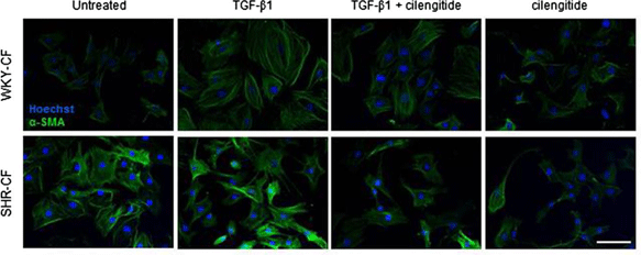
Cilengitide purchased from MedChemExpress. Usage Cited in: J Transl Med. 2018 Dec 12;16(1):352. [Abstract]
Representative images of immunofluorescence performed for α-SMA on WKY- and SHR-CF in the treatment of TGF-β1, cilengitide or both.
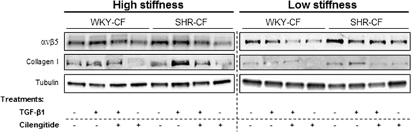
Cilengitide purchased from MedChemExpress. Usage Cited in: J Transl Med. 2018 Dec 12;16(1):352. [Abstract]
Representative Western blot images of ανβ5 and collagen I protein expression in WKY- and SHR-CF cultured on substrates with two different stiffness (high and low) and contemporary treated with 5 ng/ml TGF-β1, TGF-β1 + 0.5 μM cilengitide, or cilengitide.
-
Oncogene
Fibroblast Activation Protein (FAP)+ cancer-associated fibroblasts induce macrophage M2-like polarization via the Fibronectin 1-Integrin α5β1 axis in breast cancer. [Abstract]2025 Jul;44(28):2396-2412. PMID: 40263422 -
Cell Mol Gastroenterol Hepatol
MFAP4 Deficiency Attenuates Liver Fibrosis by Regulating Hepatic Stellate Cell Fate through Inhibition of the FAK/PI3K/NFκB Signaling Pathway. [Abstract]2025;19(10):101548. PMID: 40449846
Cilengitide purchased from MedChemExpress. Usage Cited in: Cell Mol Gastroenterol Hepatol. 2025;19(10):101548. [Abstract]
The fibrotic and apoptotic phenotypes induced by rMFAP4 incubation in LX-2 cells and by MFAP4 overexpression in LX-2 cells can be partially reversed by the integrin αvβ3 inhibitors Cilengitide TFA (1 μM).
Cilengitide purchased from MedChemExpress. Usage Cited in: Cell Mol Gastroenterol Hepatol. 2025;19(10):101548. [Abstract]
The integrin αvβ3 inhibitor Cilengitide TFA (1 μM) partially reverses the activation of the FAK/PI3K/NFκB signaling pathway induced by rMFAP4 incubation or OE-MFAP4 in LX-2 cells.
-
Free Radic Biol Med
2024 Dec 9:227:296-311. PMID: 39653130 -
Clin Transl Med
Targeting integrin αvβ3 with indomethacin inhibits patient-derived xenograft tumour growth and recurrence in oesophageal squamous cell carcinoma. [Abstract]2021 Oct;11(10):e548. PMID: 34709754 -
Br J Cancer
Transcriptome analysis of newly established carboplatin-resistant ovarian cancer cell model reveals genes shared by drug resistance and drug-induced EMT. [Abstract]2023 Mar;128(7):1344-1359. PMID: 36717670 -
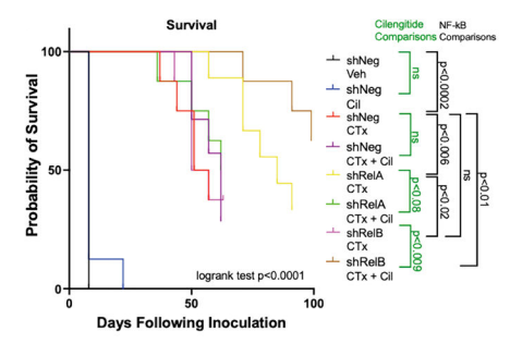
Cilengitide purchased from MedChemExpress. Usage Cited in: Cell Rep. 2026 Jul 28;45(7):117650.
Mice inoculated with control cells (shNeg) were taken down at day 15, and those receiving Cilengitide TFA alone were taken a week later. Probability of survival using Kaplan-Meier survival analysis.
Cilengitide purchased from MedChemExpress. Usage Cited in: Cell Rep. 2026 Jul 28;45(7):117650.
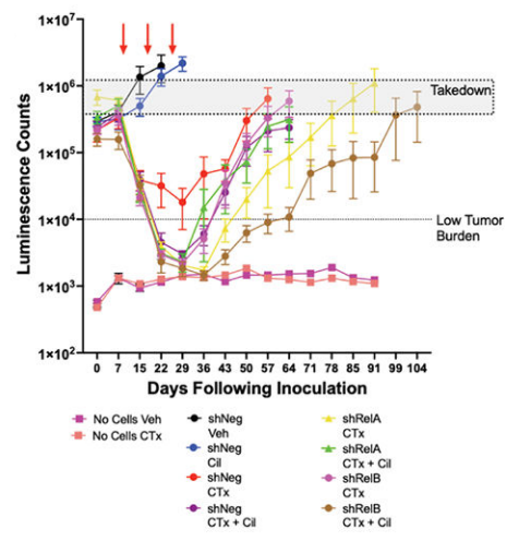
Mice inoculated with control cells (shNeg) were taken down at day 15, and those receiving Cilengitide TFA alone were taken a week later. IVIS quantification of luminescent counts over time highlighting takedown (5e5 luminescence counts) and low tumor burden (1e4 luminescence counts) windows.
Cilengitide purchased from MedChemExpress. Usage Cited in: Cell Rep. 2026 Jul 28;45(7):117650.
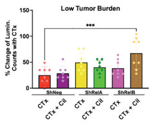
Inhibition of RelB in mice that received chemotherapy and cilengitide led to the greatest number of days with low luminescent counts.
-
Cell Rep
2022 Sep 27;40(13):111422. PMID: 36170814 -
Environ Pollut
Low-dose graphene oxide promotes tumor cells proliferation by activating PI3K-AKT-mTOR signaling via cellular membrane protein integrin αV. [Abstract]2023 Aug 1:330:121817. PMID: 37182579 -
Blood Rev
Fetal and neonatal alloimmune thrombocytopenia: Current pathophysiological insights and perspectives for future diagnostics and treatment. [Abstract]2023 May:59:101038. PMID: 36581513 -
Front Immunol
Adaptive Regulation of Osteopontin Production by Dendritic Cells Through the Bidirectional Interaction With Mesenchymal Stromal Cells. [Abstract]2018 Jun 1:9:1207. PMID: 29910810 -
Mol Cancer Ther
Inhibition of Integrin αVβ3 Signaling Improves the Antineoplastic Effect of Bexarotene in Cutaneous T-Cell Lymphoma. [Abstract]2022 Sep 6;21(9):1485-1496. PMID: 35793463 -
-
Biochem Pharmacol
Irisin alleviates steroid-induced vascular dysfunction by regulating the αVβ5-c-Abl-Caveolin-1 signaling pathway. [Abstract]2025 Jun:236:116870. PMID: 40086515 -
J Chem Inf Model
Molecular View on the i RGD Peptide Binding Mechanism: Implications for Integrin Activity and Selectivity Profiles. [Abstract]2023 Oct 23;63(20):6302-6315. PMID: 37788340 -
Oncoimmunology
Loss of the extracellular protease ADAMTS1 reveals an antitumorigenic program involving the action of NIDOGEN-1 on macrophage polarization. [Abstract]2025 Dec;14(1):2508057. PMID: 40401531 -
J Cell Biol
Reticular adhesions are assembled at flat clathrin lattices and opposed by active integrin α5β1. [Abstract]2023 Aug 7;222(8):e202303107. PMID: 37233325 -
Colloids Surf B Biointerfaces
Guiding macrophage temporal evolution via integrin targeting on peptide-functionalized titanium surfaces to optimize the host response. [Abstract]2026 Sep:265:115699. PMID: 41967441 -
Commun Biol
Abnormal cytoskeletal remodeling but normal neuronal excitability in a mouse model of the recurrent developmental and epileptic encephalopathy-susceptibility KCNB1-p.R312H variant. [Abstract]2024 Dec 30;7(1):1713. PMID: 39738805 -
Respir Res
The MFGE8/integrin β3 axis mitigates experimental neutrophilic asthma by suppressing NLRP3-Caspase-1 pathway-mediated NETosis. [Abstract]2025 Jul 2;26(1):229. PMID: 40605028 -
Int Immunopharmacol
Irisin attenuates acute glaucoma-induced neuroinflammation by activating microglia-integrin αVβ5/AMPK and promoting autophagy. [Abstract]2024 Sep 10:138:112545. PMID: 38955026 -
Stem Cell Reports
Inhibition of Farnesyltransferase Potentiates NOTCH-Targeted Therapy against Glioblastoma Stem Cells. [Abstract]2017 Dec 12;9(6):1948-1960. PMID: 29198824 -
Front Pharmacol
Antagonizing αvβ3 Integrin Improves Ischemia-Mediated Vascular Normalization and Blood Perfusion by Altering Macrophages. [Abstract]2021 Feb 24:12:585778. PMID: 33716733 -
Am J Physiol Cell Physiol
Expression of CTGF/CCN2 in response to LPA is stimulated by fibrotic extracellular matrix via the integrin/FAK axis. [Abstract]2018 Apr 1;314(4):C415-C427. PMID: 29351412
Cilengitide purchased from MedChemExpress. Usage Cited in: Am J Physiol Cell Physiol. 2018 Apr 1;314(4):C415-C427. [Abstract]
C2C12 cells are pre-incubated with different concentrations of Cilengitide prior to LPA 20 μg/mL addition and incubation for 3 hours. CTGF levels are analyzed.
-
Biochim Biophys Acta Mol Basis Dis
CCL2/CCR2 axis promotes perineural invasion of salivary adenoid cystic carcinoma via ITGβ5-mediated nerve-tumor interaction. [Abstract]2025 Jan;1871(1):167484. PMID: 39222826 -
Obesity
Immunological blockade of adipocyte inflammation caused by increased matrix metalloproteinase-cleaved osteopontin in obesity. [Abstract]2015 Apr;23(4):779-85. PMID: 25776538 -
Cancers (Basel)
Sandwich Culture Platforms to Investigate the Roles of Stiffness Gradients and Cell-Matrix Adhesions in Cancer Cell Migration. [Abstract]2023 Mar 12;15(6):1729. PMID: 36980615 -
Mol Cell Biochem
Irisin protects against atherosclerosis in ApoE-/- mice by suppressing the migration of vascular smooth muscle cell via PI3K-Akt-cofilin. [Abstract]2025 Oct 20. PMID: 41114763 -
FASEB J
Periostin increased by mechanical stress upregulates interleukin-6 expression in the ligamentum flavum. [Abstract]2023 Feb;37(2):e22726. PMID: 36583686 -
Lung
The Integrin Inhibitor Cilengitide and Bleomycin-Induced Pulmonary Fibrosis : Cilengitide and Bleomycin-Induced Pulmonary Fibrosis. [Abstract]2020 Dec;198(6):947-955. PMID: 33146772 -
Cell Adh Migr
Fluid shear stress induces cell migration and invasion via activating autophagy in HepG2 cells. [Abstract]2019 Dec;13(1):152-163. PMID: 30663937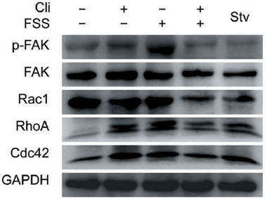
Cilengitide purchased from MedChemExpress. Usage Cited in: Cell Adh Migr. 2019 Dec;13(1):152-163. [Abstract]
HepG2 cells are loaded with FSS at 1 dyn/cm2 for 0.5 h, with or without treatment of 0.5 μM Cli for 6 h prior to FSS application. Lysates are probed with antibodies as indicated.
-
Differentiation
Osteoclast-derived DEL1 promotes pathological bone formation in ankylosing spondylitis by regulating RUNX2 expression in osteoblasts. [Abstract]2026 May-Jun:149:100962. PMID: 42128469 -
J Cell Physiol
FNDC5 inhibits malignant growth of human cervical cancer cells via restraining PI3K/AKT pathway. [Abstract]2024 Jun;239(6):e31267. PMID: 38558303 -
Cell Biol Int
Protective effects of cilengitide on inflammation in chondrocytes under excessive mechanical stress. [Abstract]2020 Apr;44(4):966-974. PMID: 31876323 -
Cancer Med
The synergistic role of ATP-dependent drug efflux pump and focal adhesion signaling pathways in vinorelbine resistance in lung cancer. [Abstract]2018 Feb;7(2):408-419. PMID: 29318780 -
Atheroscler Plus
Periostin contributes to the adventitial remodeling of atherosclerosis by activating adventitial fibroblasts. [Abstract]2022 Oct 19:50:57-64. PMID: 36643802 -
J Integr Neurosci
Milk Fat Globule-EGF Factor 8 (MFGE8) Mitigates Cognitive Impairment in Rats with Sepsis-Associated Encephalopathy: An fMRI Study. [Abstract]2024 Jul 25;23(7):140. PMID: 39082291 -
Biomed Res Int
Cilengitide Inhibits Neovascularization in a Rabbit Abdominal Aortic Plaque Model by Impairing the VEGF Signaling. [Abstract]2021 Nov 30:2021:5954757. PMID: 34888383 -
PLoS One
Inhibition of Cellular Adhesion by Immunological Targeting of Osteopontin Neoepitopes Generated through Matrix Metalloproteinase and Thrombin Cleavage. [Abstract]2016 Feb 3;11(2):e0148333. PMID: 26840958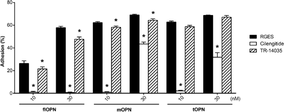
Cilengitide purchased from MedChemExpress. Usage Cited in: PLoS One. 2016 Feb 3;11(2):e0148333. [Abstract]
Blockade of cellular adhesion of HEK 293 cells at 10 or 30 nM coated recombinant OPN forms with 1μM antagonistic integrin inhibitors. RGES (black bars) is used as a control peptide. Cilengitide (white bars) inhibits the integrins αVβ3, αVβ5, and α5β1. TR-14035 (hatched bars) inhibits the integrins α4β7 and α4β1. Depicted are the means ± SEM of 3 independent experiments. * indicate signif
-
PLoS One
A novel 2.5D culture platform to investigate the role of stiffness gradients on adhesion-independent cell migration. [Abstract]2014 Oct 13;9(10):e110453. PMID: 25310593 -
J Mol Histol
Gingipains promote RANKL-induced osteoclastogenesis through the enhancement of integrin β3 in RAW264.7 cells. [Abstract]2020 Apr;51(2):147-159. PMID: 32193744 -
J Oral Pathol Med
2025 Aug;54(7):597-606. PMID: 40589419 -
-
Scand Cardiovasc J
Dual integrin αvβ3 and αvβ5 blockade attenuates cardiac dysfunction by reducing fibrosis in a rat model of doxorubicin-induced cardiomyopathy. [Abstract]2021 Oct;55(5):287-296. PMID: 34296634 -
J Vis Exp
2025 May 23:(219). PMID: 40489409 -
-
-
-
-
-
-
-
-
-
-
bioRxiv
Cancer-associated fibroblasts confer ALK inhibitor resistance in EML4-ALK -driven lung cancer via concurrent integrin and MET signaling. [Abstract]2024 Aug 28:2024.08.27.609975. PMID: 39253447 -
-
Bioengineered
Cilengitide, an αvβ3-integrin inhibitor, enhances the efficacy of anti-programmed cell death-1 therapy in a murine melanoma model. [Abstract]2022 Feb;13(2):4557-4572. PMID: 35142593 -
-
-
-
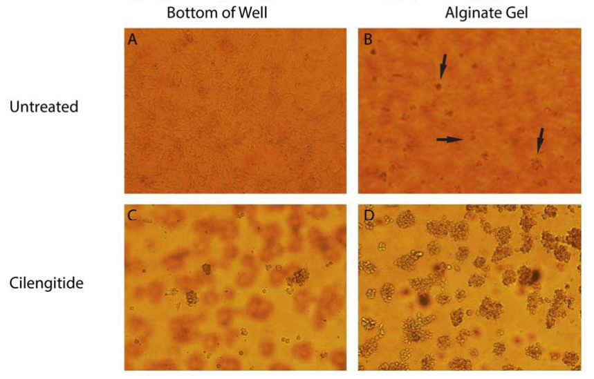
Cilengitide purchased from MedChemExpress. Usage Cited in: Santa Clara University. 2014 Jun 12.
Cilengitide Photos: Pictures of migration through alginate with and without Cilengitide after four days. A) The view of the bottom of the untreated well, showing a healthy monolayer of U87s. B) The view of the cells suspended in alginate in the untreated well. A few of the multiple migrated cells within the image are marked with arrows. C) The view of the bottom of the Cilengitide treated well, showing very few, ill-attached U87s. D). The view of cells suspended in alginate in the Cilengitide-tr


Lösungsmittel & Löslichkeit
In Vitro:
DMSO : ≥ 44 mg/mL (74.75 mM; Hygroscopic DMSO has a significant impact on the solubility of product, please use newly opened DMSO)
H2O : 35 mg/mL (59.46 mM; Need ultrasonic)
* "≥" means soluble, but saturation unknown.
Please refer to the solubility information to select the appropriate solvent. Once prepared, please aliquot and store the solution to prevent product inactivation from repeated freeze-thaw cycles.
Storage method and period of stock solution: -80°C, 1 year; -20°C, 6 months. When stored at -80°C, please use it within 1 year. When stored at -20°C, please use it within 6 months.
* Note: If you choose water as the stock solution, please dilute it to the working solution, then filter and sterilize it with a 0.22 μm filter before use.
Please refer to the solubility information to select the appropriate solvent. Once prepared, please aliquot and store the solution to prevent product inactivation from repeated freeze-thaw cycles.
Storage method and period of stock solution: -80°C, 1 year; -20°C, 6 months. When stored at -80°C, please use it within 1 year. When stored at -20°C, please use it within 6 months.
* Note: If you choose water as the stock solution, please dilute it to the working solution, then filter and sterilize it with a 0.22 μm filter before use.
Konzentration (Stammlösung) × Volumen (Stammlösung) = Konzentration (Ziellösung) × Volumen (Ziellösung)
In Vivo:
For the following dissolution methods, please prepare the working solution directly:
It is recommended to prepare fresh solutions and use them promptly within a short period of time.
The percentages shown for the solvents indicate their volumetric ratio in the final prepared solution. If precipitation or phase separation occurs during preparation, heat and/or sonication can be used to aid dissolution.
Add each solvent one by one: PBS
Solubility: 100 mg/mL (169.88 mM); Clear solution; Need ultrasonic


Reinheit & Dokumentation
-
Data Sheet (304 KB)
-
SDS (396 KB)
- English - EN (396 KB)
- Français - FR (396 KB)
- Deutsch - DE (396 KB)
- Norwegian - NO (396 KB)
- Español - ES (396 KB)
- Swedish - SV (396 KB)
- Italian - IT (396 KB)
- Korean - KR (396 KB)
- Portuguese - PT (396 KB)
-
Handling Instructions (2659 KB)
Verweise
[1]. Kapp TG, et al. A Comprehensive Evaluation of the Activity and Selectivity Profile of Ligands for RGD-binding Integrins. Scientific reports. 2017 Jan 11;7:39805. [Content Brief]
[2]. Pan X, et al. Cilengitide, an αvβ3-integrin inhibitor, enhances the efficacy of anti-programmed cell death-1 therapy in a murine melanoma model. Bioengineered. 2022 Feb;13(2):4557-4572. [Content Brief]
[3]. Hariharan S, et al. Assessment of the biological and pharmacological effects of the alpha nu beta3 and alpha nu beta5 integrin receptor antagonist, cilengitide (EMD 121974), in patients with advanced solid tumors. Ann Oncol. 2007 Aug;18(8):1400-7. [Content Brief]
[4]. MacDonald TJ, et al. Phase I clinical trial of cilengitide in children with refractory brain tumors: Pediatric Brain Tumor Consortium Study PBTC-012. Journal of clinical oncology : official journal of the American Society of Clinical Oncology. 2008 Feb 20;26(6):919-24. [Content Brief]


Complete Stock Solution Preparation Table
Please refer to the solubility information to select the appropriate solvent. Once prepared, please aliquot and store the solution to prevent product inactivation from repeated freeze-thaw cycles.
Storage method and period of stock solution: -80°C, 1 year; -20°C, 6 months. When stored at -80°C, please use it within 1 year. When stored at -20°C, please use it within 6 months.
| Optional Solvent | Concentration Solvent Mass | 1 mg | 5 mg | 10 mg | 25 mg |
|---|---|---|---|---|---|
| H2O / DMSO | 1 mM | 1.6988 mL | 8.4939 mL | 16.9877 mL | 42.4693 mL |
| 5 mM | 0.3398 mL | 1.6988 mL | 3.3975 mL | 8.4939 mL | |
| 10 mM | 0.1699 mL | 0.8494 mL | 1.6988 mL | 4.2469 mL | |
| 15 mM | 0.1133 mL | 0.5663 mL | 1.1325 mL | 2.8313 mL | |
| 20 mM | 0.0849 mL | 0.4247 mL | 0.8494 mL | 2.1235 mL | |
| 25 mM | 0.0680 mL | 0.3398 mL | 0.6795 mL | 1.6988 mL | |
| 30 mM | 0.0566 mL | 0.2831 mL | 0.5663 mL | 1.4156 mL | |
| 40 mM | 0.0425 mL | 0.2123 mL | 0.4247 mL | 1.0617 mL | |
| 50 mM | 0.0340 mL | 0.1699 mL | 0.3398 mL | 0.8494 mL | |
| DMSO | 60 mM | 0.0283 mL | 0.1416 mL | 0.2831 mL | 0.7078 mL |
* Note: If you choose water as the stock solution, please dilute it to the working solution, then filter and sterilize it with a 0.22 μm filter before use.


Cilengitide Related Classifications
HY-16141 Related Classifications
 Powered by Bioz