



BACH1 Antibody (YA7972)
(Synonyms: BACH-1; BTBD24)
Based on 1 Customer Validation
BACH1 Antibody (YA7972) is a Mouse-derived and non-conjugated IgG1 monoclonal antibody, targeting to BACH1.
-
Host:
Mouse
-
Isotype:
IgG1
-
Application:
WB, IHC-P, FC
-
Reactivity :
Human
-
Formulation:
Spplied in PBS (pH 7.3) containing 1% BSA, 50% glycerol and 0.02% sodium azide.
-
Conjugation:
Non-conjugated


To place orders, for customer services and technical support, please contact: MedChemExpress USA
Tel: 609-228-6898 E-mail: [email protected] [email protected]





Applications
| Application |
WB

WB: Western Blot
|
IHC-P
IHC-P: Immunohistochemistry-Paraffin
|
FC
FC: Flow Cytometry
|
|---|---|---|---|
| Dilution Ratio | 1:500-2000 | 1:150-500 | 1:100 |


Product Details
BACH1 Antibody (YA7972) is a Mouse-derived and non-conjugated IgG1 monoclonal antibody, targeting to BACH1.
-
Host Mouse

-
Clonality Monoclonal

-
Species ReactivityHuman
-
Calculated Molecular Weight Predicted band size: 81.8 kDa

Full length human recombinant protein of human BACH1 produced in HEK293T cell.
Endogenous
Affinity purified
Non-conjugated
Unmodified
IgG1


Product Properties
-
Appearance
Liquid
-
Formulation
Spplied in PBS (pH 7.3) containing 1% BSA, 50% glycerol and 0.02% sodium azide.
-
Concentration
Batch-dependent, Please check the COA for the concentration of each lot. Check Lot Concentration
-
Storage & Stability
Stored at -20°C for 1 year. Avoid repeated freeze / thaw cycles.
-
Shipping
Shipping with blue ice.


Verification Images
-
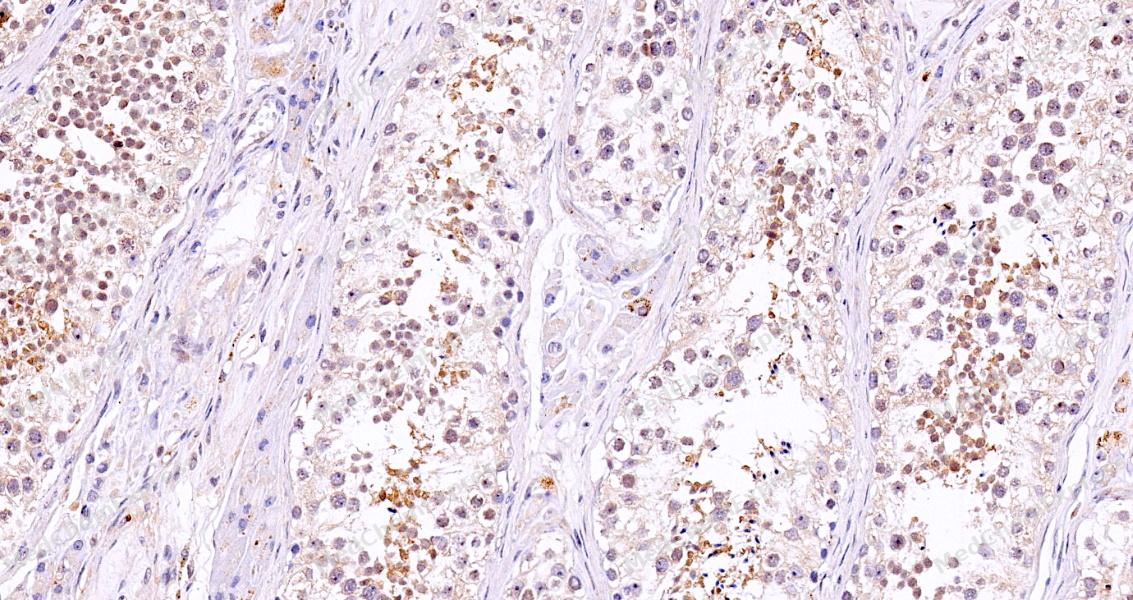 Immunohistochemical analysis of paraffin-embedded human testis tissue using BACH1 Antibody (HY-P88288, 1/500). The section was pretreated using heat mediated antigen retrieval with sodium citrate buffer (pH 6.0) for 8 minutes. The tissues were blocked with quick block buffer for 0.5 hours at room temperature, washed with PBS and PBST, and then incubated with the primary antibody overnight at 4℃. The detection was performed using an HRP conjugated compact polymer system. DAB was used as the chromogen. Tissues were counterstained with hematoxylin and mounted with DPX.
 -
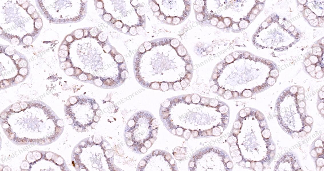 Immunohistochemical analysis of paraffin-embedded human small intestine tissue using BACH1 Antibody (HY-P88288, 1/500). The section was pretreated using heat mediated antigen retrieval with sodium citrate buffer (pH 6.0) for 8 minutes. The tissues were blocked with quick block buffer for 0.5 hours at room temperature, washed with PBS and PBST, and then incubated with the primary antibody overnight at 4℃. The detection was performed using an HRP conjugated compact polymer system. DAB was used as the chromogen. Tissues were counterstained with hematoxylin and mounted with DPX.
 -
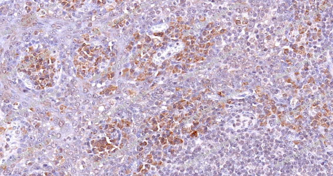 Immunohistochemical analysis of paraffin-embedded human tonsil tissue using BACH1 Antibody (HY-P88288, 1/500). The section was pretreated using heat mediated antigen retrieval with sodium citrate buffer (pH 6.0) for 8 minutes. The tissues were blocked with quick block buffer for 0.5 hours at room temperature, washed with PBS and PBST, and then incubated with the primary antibody overnight at 4℃. The detection was performed using an HRP conjugated compact polymer system. DAB was used as the chromogen. Tissues were counterstained with hematoxylin and mounted with DPX.
 -
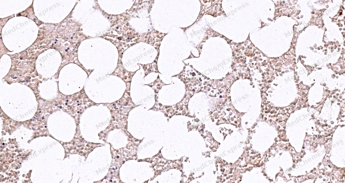 Immunohistochemical analysis of paraffin-embedded human bone marrow tissue using BACH1 Antibody (HY-P88288, 1/500). The section was pretreated using heat mediated antigen retrieval with sodium citrate buffer (pH 6.0) for 8 minutes. The tissues were blocked with quick block buffer for 0.5 hours at room temperature, washed with PBS and PBST, and then incubated with the primary antibody overnight at 4℃. The detection was performed using an HRP conjugated compact polymer system. DAB was used as the chromogen. Tissues were counterstained with hematoxylin and mounted with DPX.
 -
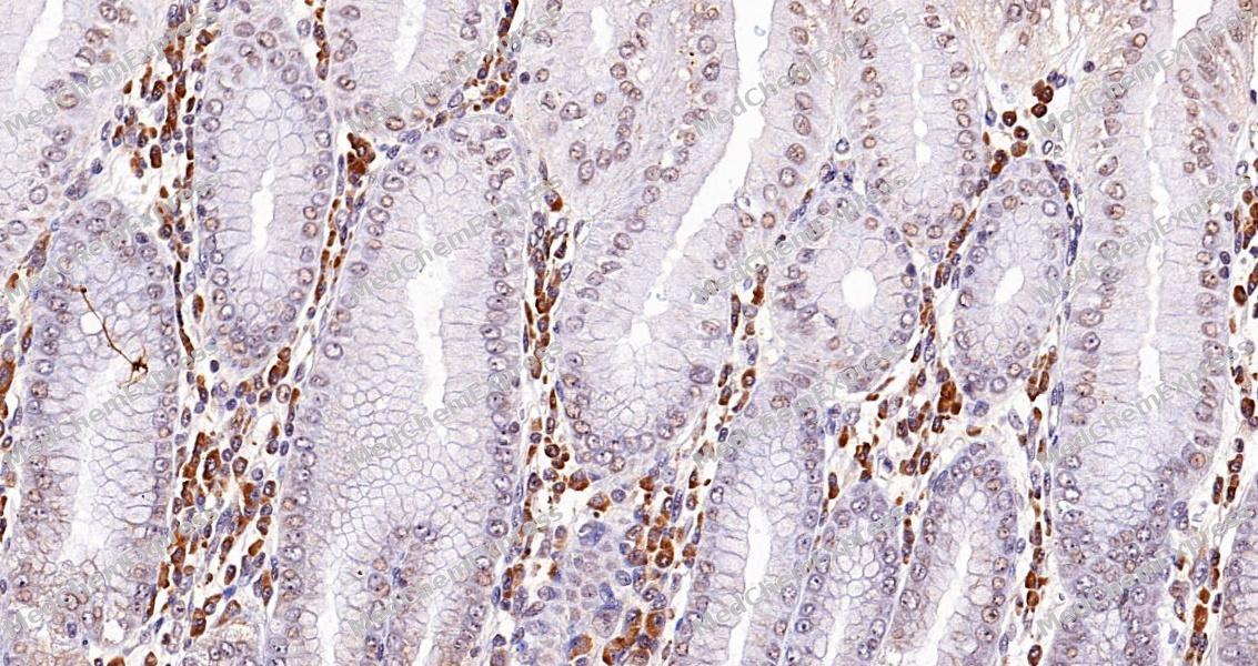 Immunohistochemical analysis of paraffin-embedded human stomach tissue using BACH1 Antibody (HY-P88288, 1/500). The section was pretreated using heat mediated antigen retrieval with sodium citrate buffer (pH 6.0) for 8 minutes. The tissues were blocked with quick block buffer for 0.5 hours at room temperature, washed with PBS and PBST, and then incubated with the primary antibody overnight at 4℃. The detection was performed using an HRP conjugated compact polymer system. DAB was used as the chromogen. Tissues were counterstained with hematoxylin and mounted with DPX.
 -
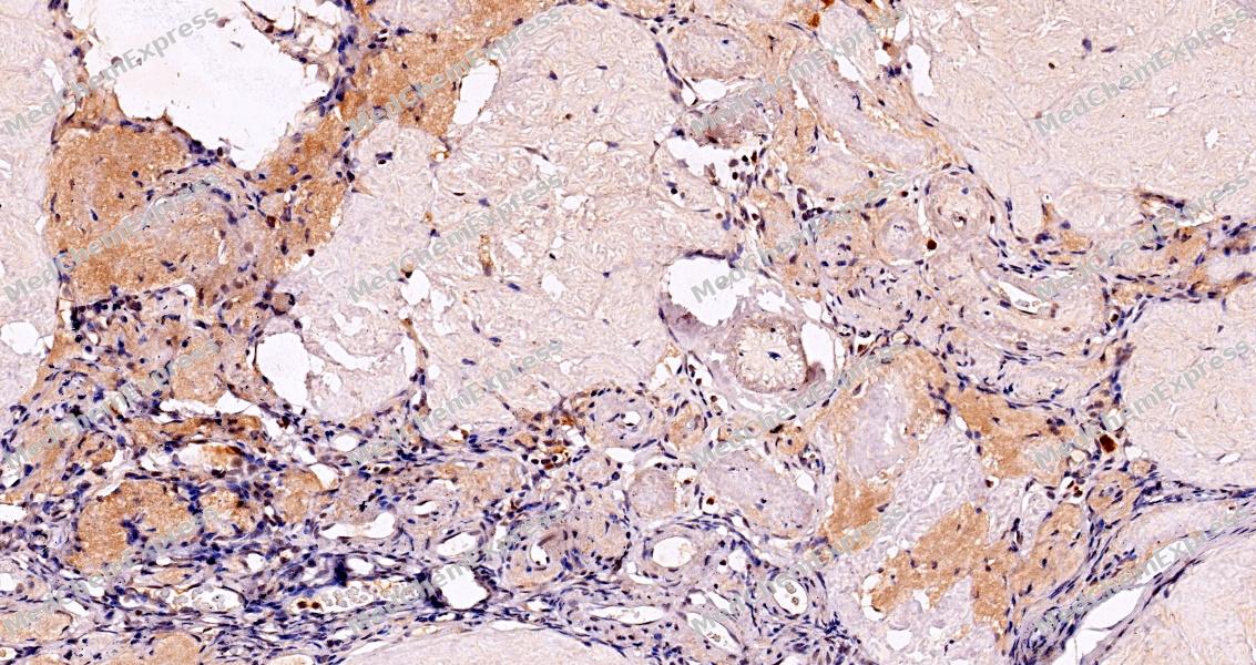 Immunohistochemical analysis of paraffin-embedded human ovary tissue using BACH1 Antibody (HY-P88288, 1/500). The section was pretreated using heat mediated antigen retrieval with sodium citrate buffer (pH 6.0) for 8 minutes. The tissues were blocked with quick block buffer for 0.5 hours at room temperature, washed with PBS and PBST, and then incubated with the primary antibody overnight at 4℃. The detection was performed using an HRP conjugated compact polymer system. DAB was used as the chromogen. Tissues were counterstained with hematoxylin and mounted with DPX.


Background
-
Function
BACH1 transcriptional regulator that acts as a repressor or activator, depending on the context. Binds to NF-E2 DNA binding sites. Plays important roles in coordinating transcription activation and repression by MAFK. Together with MAF, represses the transcription of genes under the control of the NFE2L2 oxidative stress pathway[1].
-
Subcellular Localization
Nucleus
-
Isoforms & Post-Translational Modification
O14867: 736 amino acids, molecular weight 81958 Da.
-
Subunit
Heterodimer of BACH1 and MAFK
-
SwissProt ID
-
Synonyms
BACH-1; BTBD24


Documentation

 Powered by Bioz
