



BDNF Antibody (YA3894)
(Synonyms: ANON2; BULN2)Based on 1 Customer Validation
BDNF Antibody (YA3894) is a Mouse-derived and non-conjugated IgG1 monoclonal antibody, targeting to BDNF.
-
Host:
Mouse
-
Isotype:
IgG1
-
Application:
WB, ICC/IF, ELISA
-
Reactivity :
Human
-
Formulation:
Supplied in PBS with 0.05% sodium azide.
-
Conjugation:
Non-conjugated


To place orders, for customer services and technical support, please contact: MedChemExpress USA
Tel: 609-228-6898 E-mail: [email protected] [email protected]





Applications
| Application |
WB

WB: Western Blot
|
ICC/IF
ICC/IF: Immunocytochemistry/
Immunofluorescence |
ELISA
ELISA: Enzyme Linked Immunosorbent Assay
|
|---|---|---|---|
| Dilution Ratio | 1:500-1:2000 | 1:200-1:1000 | 1:10000 |


Product Details
BDNF Antibody (YA3894) is a Mouse-derived and non-conjugated IgG1 monoclonal antibody, targeting to BDNF.
-
Host Mouse

-
Clonality Monoclonal

-
Species ReactivityHuman
-
Observed Molecular WeightObserved band size: 28 kDa
Note: Due to possible protein modifications or aggregation, the molecular weight should be confirmed by actual measurement, and the predicted value is for reference only.

-
Calculated Molecular Weight Predicted band size: 28 kDa

Purified recombinant fragment of human BDNF (AA: 19-248) expressed in E. Coli.
affinity purified.
Non-conjugated
Unmodified
IgG1


Product Properties
-
Appearance
Liquid
-
Formulation
Supplied in PBS with 0.05% sodium azide.
-
Concentration
Batch-dependent, Please check the COA for the concentration of each lot. Check Lot Concentration
-
Storage & Stability
Stored at -20°C for 1 year. Avoid repeated freeze / thaw cycles.
-
Shipping
Shipping with blue ice.


Verification Images
-
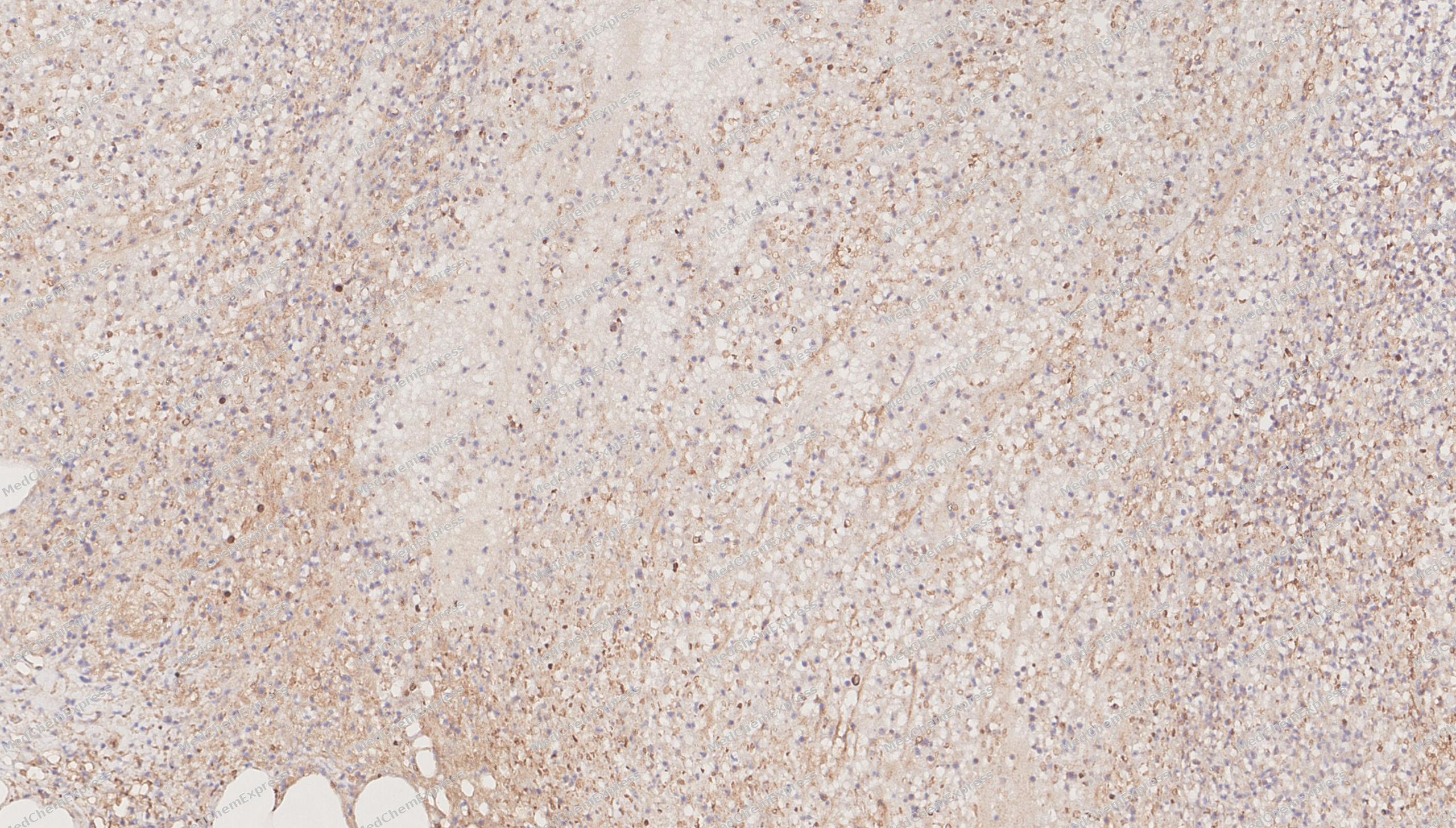 Immunohistochemical analysis of paraffin-embedded human appendix tissue using BDNF antibody was performed. The section was pretreated using high-temperature mediated EDTA antigen retrieval buffer (pH 9.0), for 20 minutes. The tissues were incubated with primary antibody (HY-P84197, 1:500 dilution) at room temperature for 20 minutes. Detection was performed using an HRP conjugated compact polymer system. DAB was used as the chromogen. The tissues were counterstained with hematoxylin and mounted with neutral balsam mounting medium.
 -
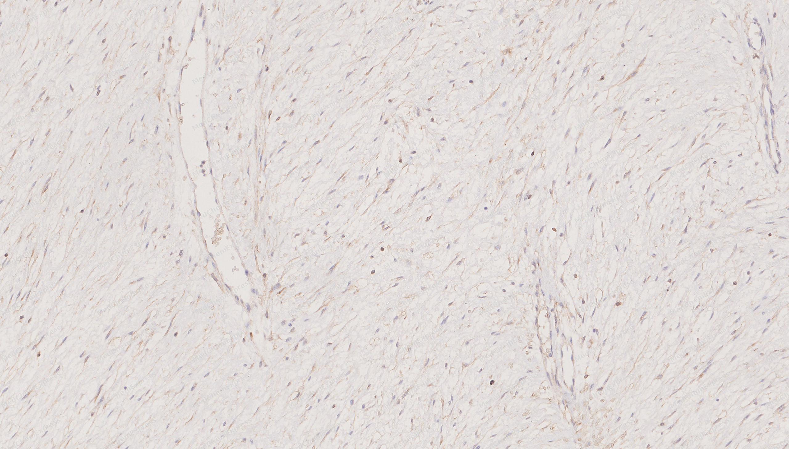 Immunohistochemical analysis of paraffin-embedded human aggressive fibromatosis tissue using BDNF antibody was performed. The section was pretreated using high-temperature mediated EDTA antigen retrieval buffer (pH 9.0), for 20 minutes. The tissues were incubated with primary antibody (HY-P84197, 1:500 dilution) at room temperature for 20 minutes. Detection was performed using an HRP conjugated compact polymer system. DAB was used as the chromogen. The tissues were counterstained with hematoxylin and mounted with neutral balsam mounting medium.
 -
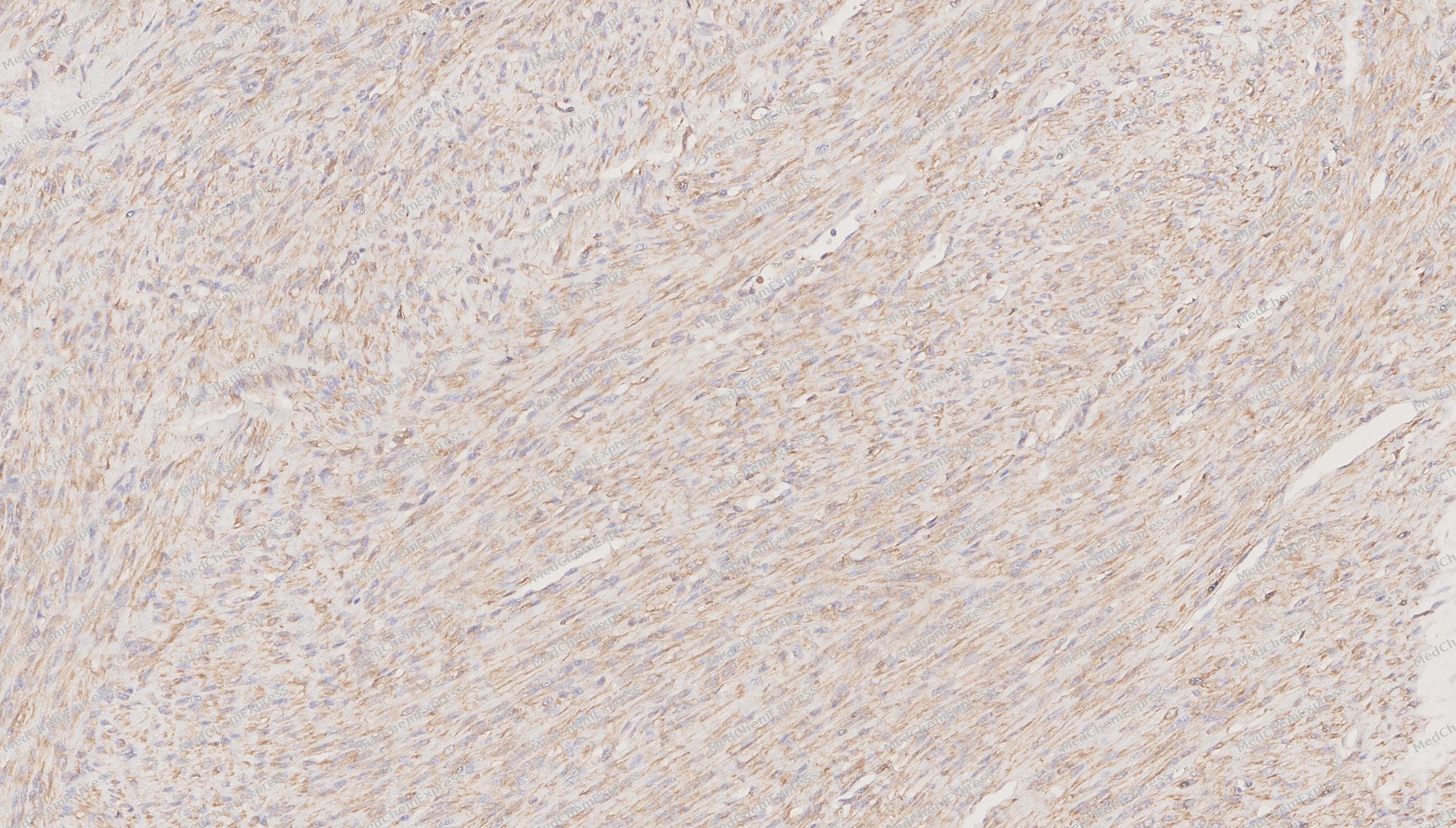 Immunohistochemical analysis of paraffin-embedded human Uterine leiomyoma tissue using BNDF antibody was performed. The section was pretreated using high-temperature mediated EDTA antigen retrieval buffer (pH 9.0), for 20 minutes. The tissues were incubated with primary antibody (HY-P84197, 1:500 dilution) at room temperature for 20 minutes. Detection was performed using an HRP conjugated compact polymer system. DAB was used as the chromogen. The tissues were counterstained with hematoxylin and mounted with neutral balsam mounting medium.
 -
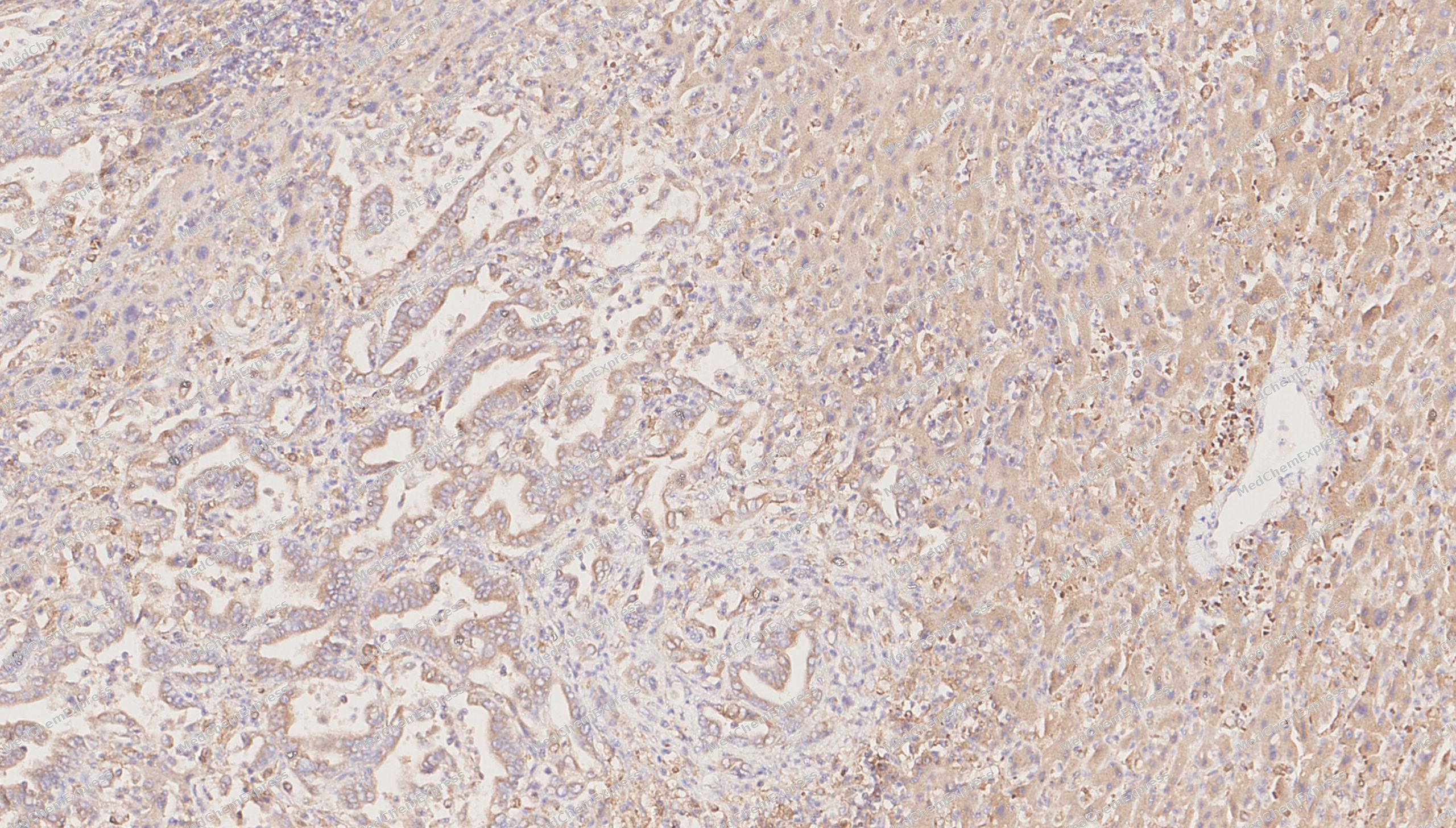 Immunohistochemical analysis of paraffin-embedded human Intrahepatic cholangiocarcinoma tissue using BDNF antibody was performed. The section was pretreated using high-temperature mediated EDTA antigen retrieval buffer (pH 9.0), for 20 minutes. The tissues were incubated with primary antibody (HY-P84197, 1:500 dilution) at room temperature for 20 minutes. Detection was performed using an HRP conjugated compact polymer system. DAB was used as the chromogen. The tissues were counterstained with hematoxylin and mounted with neutral balsam mounting medium.
 -
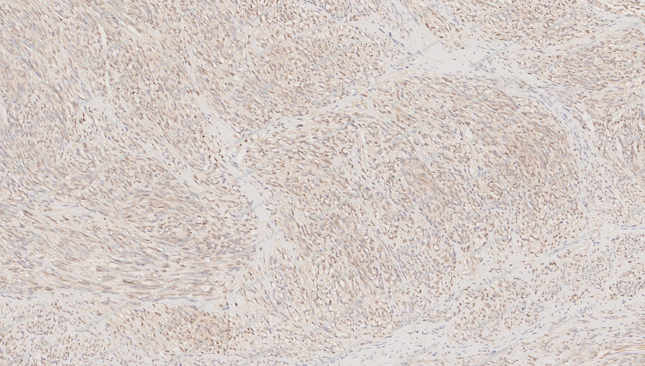 Immunohistochemical analysis of paraffin-embedded human Stromal tumor tissue using BDNF antibody was performed. The section was pretreated using high-temperature mediated EDTA antigen retrieval buffer (pH 9.0), for 20 minutes. The tissues were incubated with primary antibody (HY-P84197, 1:500 dilution) at room temperature for 20 minutes. Detection was performed using an HRP conjugated compact polymer system. DAB was used as the chromogen. The tissues were counterstained with hematoxylin and mounted with neutral balsam mounting medium.
 -
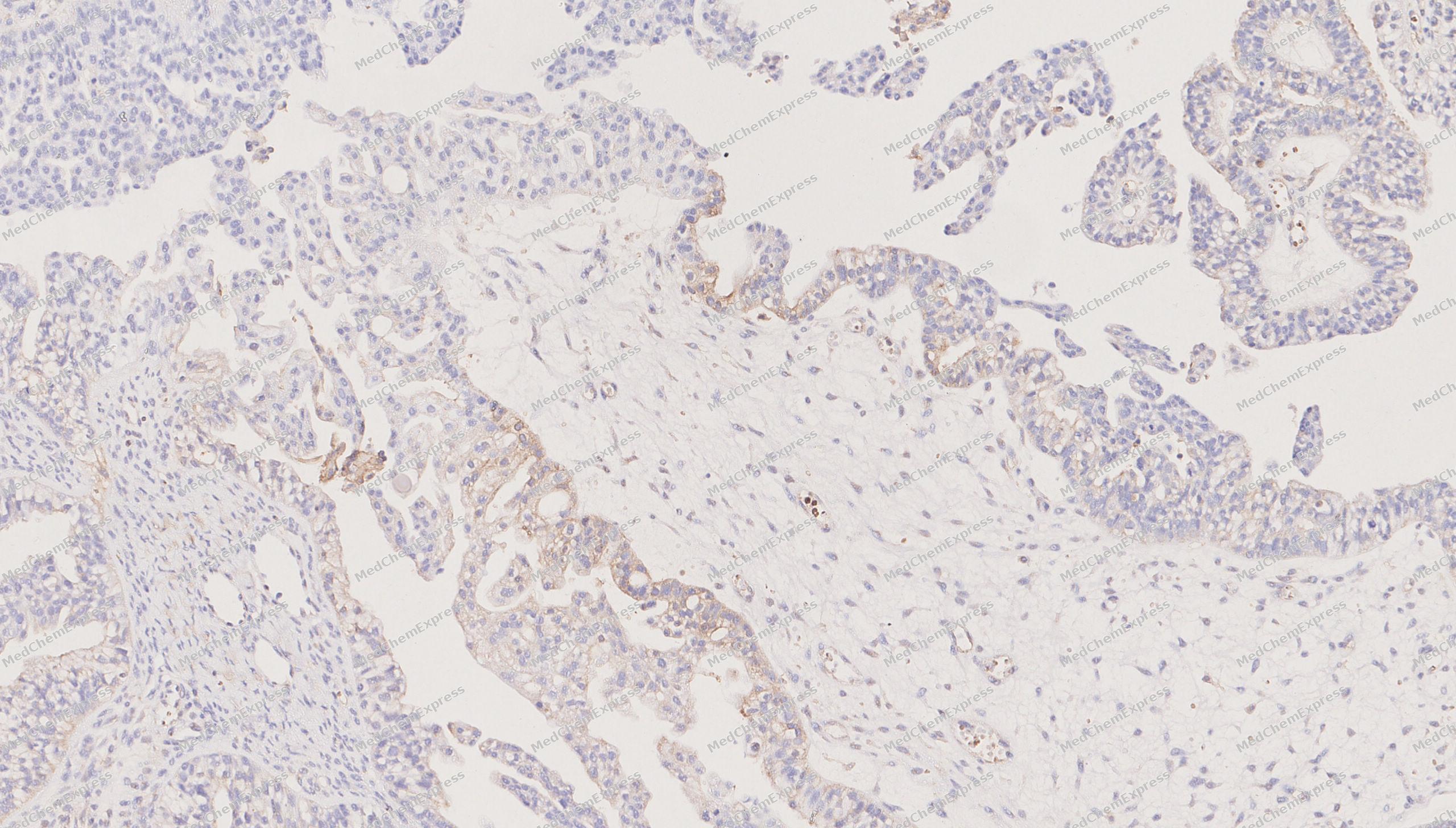 Immunohistochemical analysis of paraffin-embedded human serous ovarian cancer tissue using BDNF antibody was performed. The section was pretreated using high-temperature mediated EDTA antigen retrieval buffer (pH 9.0), for 20 minutes. The tissues were incubated with primary antibody (HY-P84197, 1:500 dilution) at room temperature for 20 minutes. Detection was performed using an HRP conjugated compact polymer system. DAB was used as the chromogen. The tissues were counterstained with hematoxylin and mounted with neutral balsam mounting medium.
 -
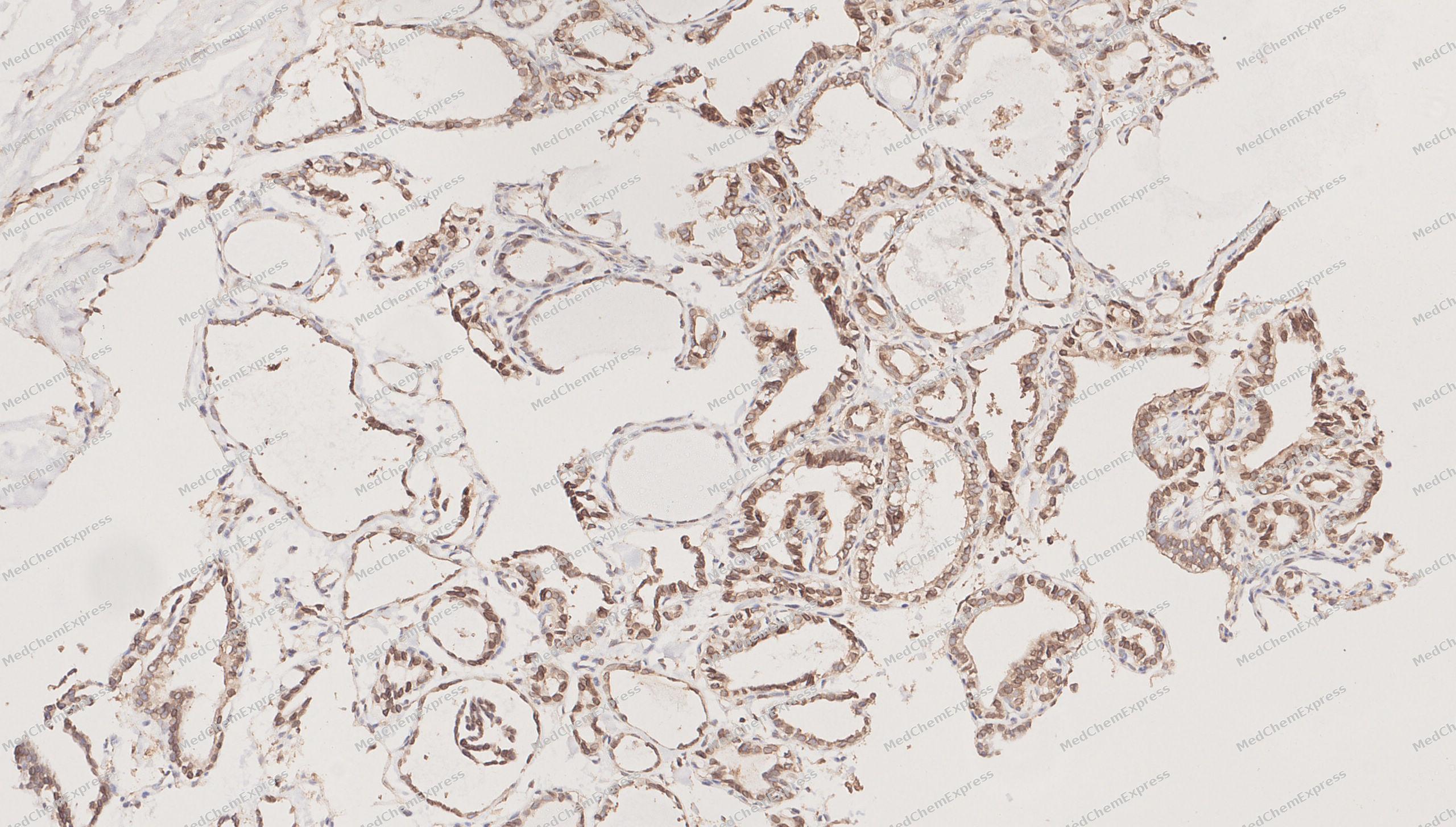 Immunohistochemical analysis of paraffin-embedded human Thyroid carcinoma tissue using BDNF antibody was performed. The section was pretreated using high-temperature mediated EDTA antigen retrieval buffer (pH 9.0), for 20 minutes. The tissues were incubated with primary antibody (HY-P84197, 1:500 dilution) at room temperature for 20 minutes. Detection was performed using an HRP conjugated compact polymer system. DAB was used as the chromogen. The tissues were counterstained with hematoxylin and mounted with neutral balsam mounting medium.
 -
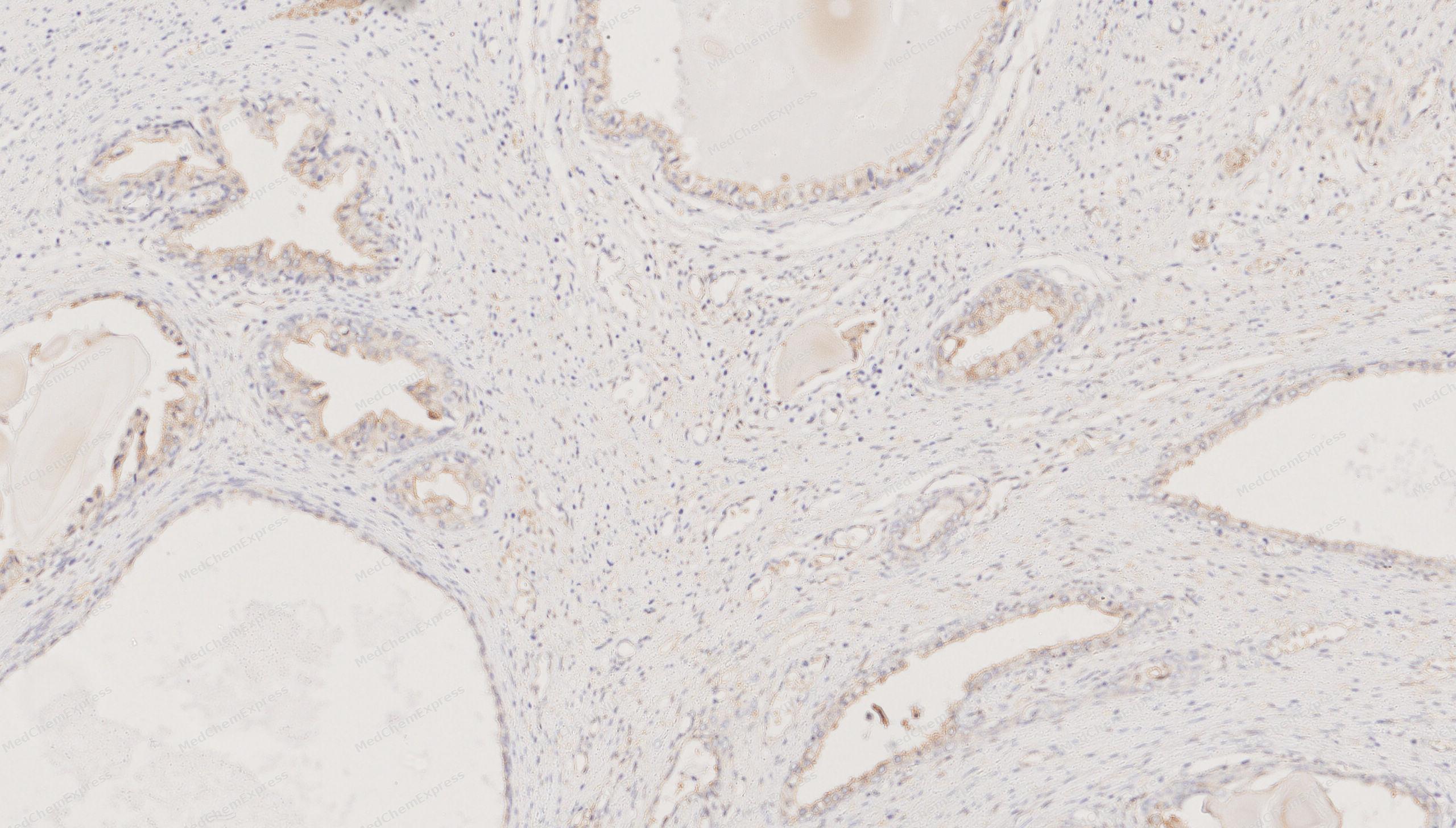 Immunohistochemical analysis of paraffin-embedded human Prostate cancer tissue using BDNF antibody was performed. The section was pretreated using high-temperature mediated EDTA antigen retrieval buffer (pH 9.0), for 20 minutes. The tissues were incubated with primary antibody (HY-P84197, 1:500 dilution) at room temperature for 20 minutes. Detection was performed using an HRP conjugated compact polymer system. DAB was used as the chromogen. The tissues were counterstained with hematoxylin and mounted with neutral balsam mounting medium.
 -
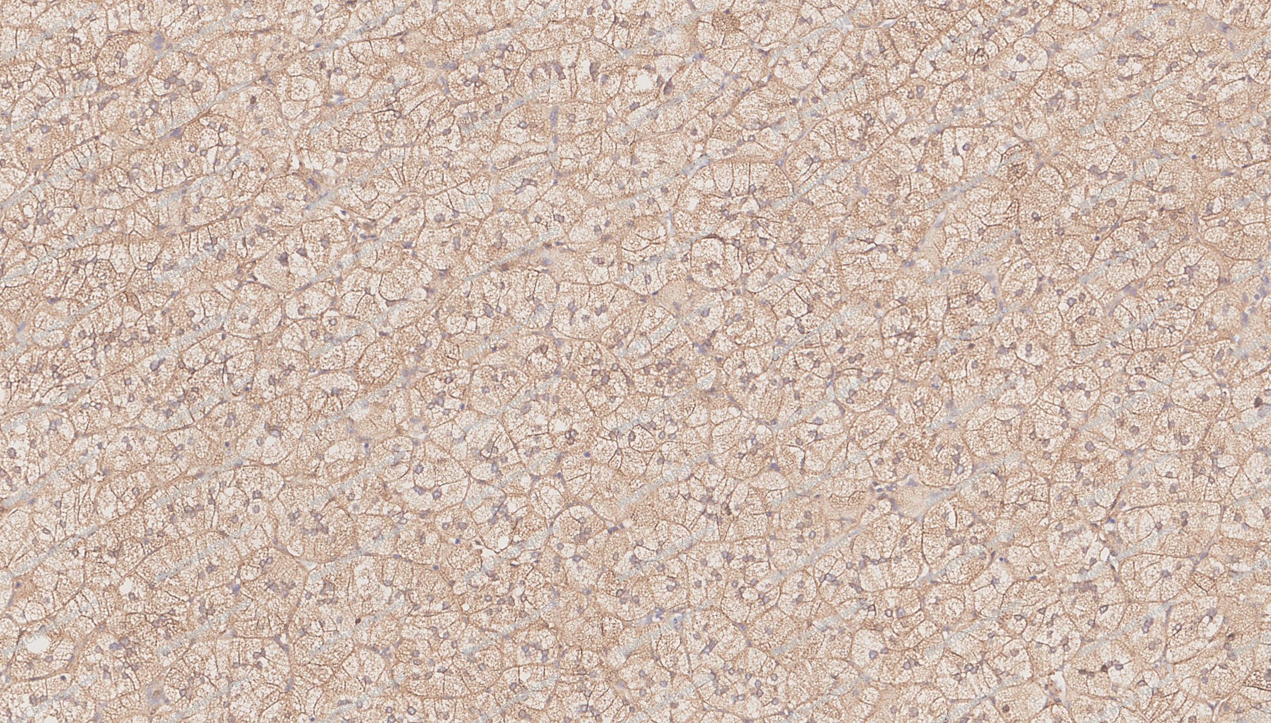 Immunohistochemical analysis of paraffin-embedded human adrenocortical adenoma tissue using BDNF antibody was performed. The section was pretreated using high-temperature mediated EDTA antigen retrieval buffer (pH 9.0), for 20 minutes. The tissues were incubated with primary antibody (HY-P84197, 1:500 dilution) at room temperature for 20 minutes. Detection was performed using an HRP conjugated compact polymer system. DAB was used as the chromogen. The tissues were counterstained with hematoxylin and mounted with neutral balsam mounting medium.


Background
-
Function
BDNF is important signaling molecule that activates signaling cascades downstream of NTRK2. During development, promotes the survival and differentiation of selected neuronal populations of the peripheral and central nervous systems. Participates in axonal growth, pathfinding and in the modulation of dendritic growth and morphology. Major regulator of synaptic transmission and plasticity at adult synapses in many regions of the CNS. The versatility of BDNF is emphasized by its contribution to a range of adaptive neuronal responses including long-term potentiation (LTP), long-term depression (LTD), certain forms of short-term synaptic plasticity, as well as homeostatic regulation of intrinsic neuronal excitability; Important signaling molecule that activates signaling cascades downstream of NTRK2. Activates signaling cascades via the heterodimeric receptor formed by NGFR and SORCS2. Signaling via NGFR and SORCS2 plays a role in synaptic plasticity and long-term depression (LTD). Binding to NGFR and SORCS2 promotes neuronal apoptosis. Promotes neuronal growth cone collapse[1][2][3].
-
Subcellular Localization
Secreted; Secreted
-
Expression
Tissue_specificity:Protein levels were detected in both plasma and saliva (PubMed: 11152678, PubMed: 19467646) . High expression was observed in the hippocampus, amygdala, cerebral cortex, and cerebellum of brain tissue. It was also expressed in the heart, lungs, skeletal muscle, testes, prostate, and placenta. -
Isoforms & Post-Translational Modification
P23560 has 5 isomers: P23560-1: 27818 Da (predicted); P23560-2: 28872 Da (predicted); P23560-3: 29817 Da (predicted); P23560-4: 37120 Da (predicted); P23560-5: 31116 Da (predicted).
N-glycosylated and glycosulfated, contrary to mature BDNF;Mature BDNF is produced by proteolytic removal of the propeptide, catalyzed by a FURIN family member. In addition, the precursor form is proteolytically cleaved within the propeptide, but this is not an obligatory intermediate for the production of mature BDNF (PubMed:11152678). Can be converted into mature BDNF by plasmin (PLG) (PubMed:19467646) -
Subunit
Monomers and homodimers. Binds to NTRK2/TRKB (PubMed:11152678, PubMed:8527932). Can form heterodimers with other neurotrophin family members, such as NTF3 and NTF4 (in vitro), but the physiological relevance of this is not clear (PubMed:10631974, PubMed:7703225). BDNF precursor form: interacts with the heterodimer formed by NGFR and SORCS2 (PubMed:24908487)
-
SwissProt ID
-
Synonyms
ANON2; BULN2


Documentation
-
Data Sheet (234 KB)
-
SDS (251 KB)
- English - EN (251 KB)
- Français - FR (251 KB)
- Deutsch - DE (251 KB)
- Norwegian - NO (251 KB)
- Español - ES (251 KB)
- Swedish - SV (251 KB)
- Italian - IT (251 KB)
- Korean - KR (251 KB)
- Portuguese - PT (251 KB)
-
User Guide for Antibodies (1077 KB)

 Powered by Bioz
