



Caldesmon Antibody (YA2332)
(Synonyms: CDM; HCAD; LCAD; H-CAD; L-CAD; NAG22)
Based on 1 Customer Validation
Caldesmon Antibody (YA2332) is a Rabbit-derived and non-conjugated IgG monoclonal antibody, targeting to Caldesmon.
-
Host:
Rabbit
-
Isotype:
IgG
-
Application:
WB, IHC-P, IP
-
Reactivity :
Human, Mouse, Rat
-
Formulation:
Supplied in 50mM Tris-Glycine(pH 7.4), 0.15M NaCl, 40% Glycerol, 0.01% Sodium azide and 0.05% BSA
-
Conjugation:
Non-conjugated


To place orders, for customer services and technical support, please contact: MedChemExpress USA
Tel: 609-228-6898 E-mail: [email protected] [email protected]





Applications
| Application |
WB

WB: Western Blot
|
IHC-P
IHC-P: Immunohistochemistry-Paraffin
|
IP
IP: Immunoprecipitation
|
|---|---|---|---|
| Dilution Ratio | 1:500-1:1000 | 1:50-1:100 | 1:20 |


Product Details
Caldesmon Antibody (YA2332) is a Rabbit-derived and non-conjugated IgG monoclonal antibody, targeting to Caldesmon.
-
Host Rabbit

-
Clonality Recombinant,Monoclonal

-
Species ReactivityHuman, Mouse, Rat
-
Observed Molecular WeightObserved band size: 70-80 kDa
Note: Due to possible protein modifications or aggregation, the molecular weight should be confirmed by actual measurement, and the predicted value is for reference only.

-
Calculated Molecular Weight Predicted band size: 93 kDa

Entrez Gene: 800 Human ; 109624 Mouse ; 25687 Rat
SwissProt: Q05682 Human ; Q62736 Rat
OMIM: 114213 Human
A synthetic peptide of human Caldesmon aa782-793.
Endogenous
Affinity Purified
Non-conjugated
Unmodified
IgG


Product Properties
-
Appearance
Solution
-
Formulation
Supplied in 50mM Tris-Glycine(pH 7.4), 0.15M NaCl, 40% Glycerol, 0.01% Sodium azide and 0.05% BSA
-
Concentration
Batch-dependent, Please check the COA for the concentration of each lot. Check Lot Concentration
-
Storage & Stability
Stored at -20°C for 1 year. Avoid repeated freeze / thaw cycles.
-
Shipping
Shipping with blue ice.


Verification Images
-
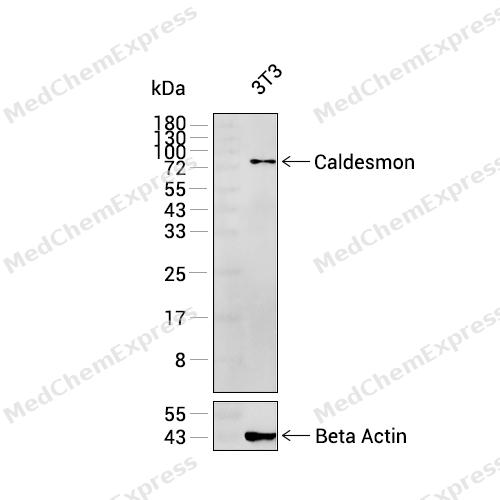 Western blot analysis was performed on extracts from 3T3 (lane 1, 15 μg) using Caldesmon Rabbit mAb.Proteins were transferred to a PVDF membrane and blocked with 5% non-fat milk in TBST at 4°C overnight.The primary antibody (1:1000 dilution) and the loading control antibody (beta-Actin, HY-P83730, 1:20000 dilution) were incubated in 5% non-fat milk in TBST for 1 hour at 37°C.Goat Anti-Rabbit IgG-HRP Secondary Antibody (1:20000 dilution) was then applied for 40 minutes at 37°C.
 -
 Immunohistochemical analysis of paraffin-embedded mouse lung tissue using Caldesmon Antibody (HY-P82587, 1/100). The section was pretreated using heat mediated antigen retrieval with sodium citrate buffer (pH 6.0) for 8 minutes. The tissues were blocked with quick block buffer for 0.5 hours at room temperature, washed with PBS and PBST, and then incubated with the primary antibody overnight at 4℃. The detection was performed using an HRP conjugated compact polymer system. DAB was used as the chromogen. Tissues were counterstained with hematoxylin and mounted with DPX.
 -
 Immunohistochemical analysis of paraffin-embedded mouse stomach tissue using Caldesmon Antibody (HY-P82587, 1/100). The section was pretreated using heat mediated antigen retrieval with sodium citrate buffer (pH 6.0) for 8 minutes. The tissues were blocked with quick block buffer for 0.5 hours at room temperature, washed with PBS and PBST, and then incubated with the primary antibody overnight at 4℃. The detection was performed using an HRP conjugated compact polymer system. DAB was used as the chromogen. Tissues were counterstained with hematoxylin and mounted with DPX.
 -
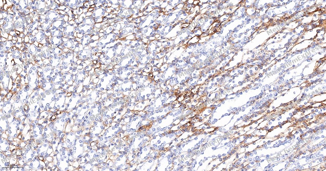 Immunohistochemical analysis of paraffin-embedded mouse kidney tissue using Caldesmon Antibody (HY-P82587, 1/100). The section was pretreated using heat mediated antigen retrieval with sodium citrate buffer (pH 6.0) for 8 minutes. The tissues were blocked with quick block buffer for 0.5 hours at room temperature, washed with PBS and PBST, and then incubated with the primary antibody overnight at 4℃. The detection was performed using an HRP conjugated compact polymer system. DAB was used as the chromogen. Tissues were counterstained with hematoxylin and mounted with DPX.
 -
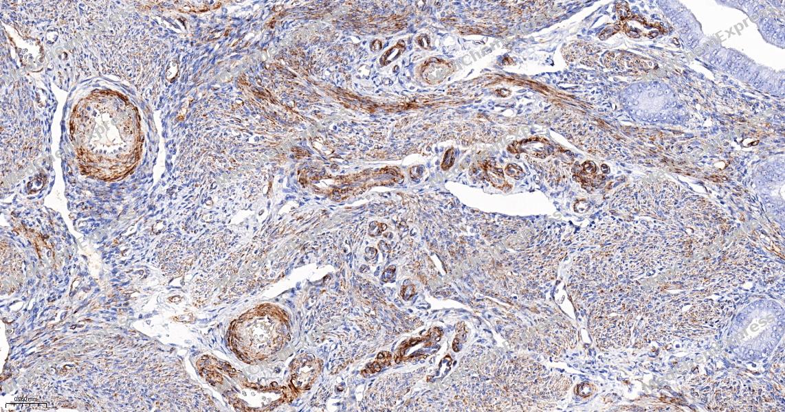 Immunohistochemical analysis of paraffin-embedded human endometrium tissue using Caldesmon Antibody (HY-P82587, 1/100). The section was pretreated using heat mediated antigen retrieval with sodium citrate buffer (pH 6.0) for 8 minutes. The tissues were blocked with quick block buffer for 0.5 hours at room temperature, washed with PBS and PBST, and then incubated with the primary antibody overnight at 4℃. The detection was performed using an HRP conjugated compact polymer system. DAB was used as the chromogen. Tissues were counterstained with hematoxylin and mounted with DPX.
 -
 Immunohistochemical analysis of paraffin-embedded human breast tissue using Caldesmon Antibody (HY-P82587, 1/100). The section was pretreated using heat mediated antigen retrieval with sodium citrate buffer (pH 6.0) for 8 minutes. The tissues were blocked with quick block buffer for 0.5 hours at room temperature, washed with PBS and PBST, and then incubated with the primary antibody overnight at 4℃. The detection was performed using an HRP conjugated compact polymer system. DAB was used as the chromogen. Tissues were counterstained with hematoxylin and mounted with DPX.
 -
 Immunohistochemical analysis of paraffin-embedded human tonsil tissue using Caldesmon Antibody (HY-P82587, 1/100). The section was pretreated using heat mediated antigen retrieval with sodium citrate buffer (pH 6.0) for 8 minutes. The tissues were blocked with quick block buffer for 0.5 hours at room temperature, washed with PBS and PBST, and then incubated with the primary antibody overnight at 4℃. The detection was performed using an HRP conjugated compact polymer system. DAB was used as the chromogen. Tissues were counterstained with hematoxylin and mounted with DPX.


Background
-
Function
Caldesmon is an Actin- and myosin-binding protein implicated in the regulation of actomyosin interactions in smooth muscle and nonmuscle cells (could act as a bridge between myosin and actin filaments). Stimulates actin binding of tropomyosin which increases the stabilization of actin filament structure. In muscle tissues, inhibits the actomyosin ATPase by binding to F-actin. This inhibition is attenuated by calcium-calmodulin and is potentiated by tropomyosin. Interacts with actin, myosin, two molecules of tropomyosin and with calmodulin. Also plays an essential role during cellular mitosis and receptor capping. Involved in Schwann cell migration during peripheral nerve regeneration
-
Subcellular Localization
Cytoplasm, cytoskeleton; Cytoplasm, myofibril; Cytoplasm, cytoskeleton, stress fiber
-
Expression
Tissue_specificity:High molecular weight calmodulin (isoform 1) is primarily expressed in smooth muscle, while low molecular weight calmodulin (isoforms 2, 3, 4, and 5) are widely distributed in non-muscle tissues and cells. It is not expressed in skeletal muscle or the heart. -
Isoforms & Post-Translational Modification
Q05682 has 6 isomers: Q05682-1: 93231 Da (predicted); Q05682-2: 65707 Da (predicted); Q05682-3: 64256 Da (predicted); Q05682-4: 62663 Da (predicted); Q05682-5: 61213 Da (predicted); Q05682-6: 65620 Da (predicted).
In non-muscle cells, phosphorylation by CDK1 during mitosis causes caldesmon to dissociate from microfilaments. Phosphorylation reduces caldesmon binding to actin, myosin, and calmodulin as well as its inhibition of actomyosin ATPase activity. Phosphorylation also occurs in both quiescent and dividing smooth muscle cells with similar effects on the interaction with actin and calmodulin and on microfilaments reorganization. CDK1-mediated phosphorylation promotes Schwann cell migration during peripheral nerve regeneration (By similarity) -
SwissProt ID
-
Synonyms
CDM; HCAD; LCAD; H-CAD; L-CAD; NAG22
-
Research Field
Cell Biology


Documentation
-
Data Sheet (234 KB)
-
SDS (251 KB)
- English - EN (251 KB)
- Français - FR (251 KB)
- Deutsch - DE (251 KB)
- Norwegian - NO (251 KB)
- Español - ES (251 KB)
- Swedish - SV (251 KB)
- Italian - IT (251 KB)
- Korean - KR (251 KB)
- Portuguese - PT (251 KB)
-
User Guide for Antibodies (1077 KB)

 Powered by Bioz
