



CD147 Antibody (YA3974)
(Synonyms: BSG; OK; 5F7; TCSF; EMMPRIN)Based on 1 Customer Validation
CD147 Antibody (YA3974) is a Mouse-derived and non-conjugated IgG1 monoclonal antibody, targeting to CD147.
-
Host:
Mouse
-
Isotype:
IgG1
-
Application:
IHC-P, ICC/IF, FC, ELISA
-
Reactivity :
Human
-
Formulation:
Supplied in PBS with 0.05% sodium azide
-
Conjugation:
Non-conjugated


To place orders, for customer services and technical support, please contact: MedChemExpress USA
Tel: 609-228-6898 E-mail: [email protected] [email protected]





Applications
| Application |
IHC-P

IHC-P: Immunohistochemistry-Paraffin
|
ICC/IF
ICC/IF: Immunocytochemistry/
Immunofluorescence |
FC
FC: Flow Cytometry
|
ELISA
ELISA: Enzyme Linked Immunosorbent Assay
|
|---|---|---|---|---|
| Dilution Ratio | 1:200-1:1000 | 1:50-1:200 | 1:200-1:400 | 1:10000 |


Product Details
CD147 Antibody (YA3974) is a Mouse-derived and non-conjugated IgG1 monoclonal antibody, targeting to CD147.
-
Host Mouse

-
Clonality Monoclonal

-
Species ReactivityHuman
-
Observed Molecular WeightObserved band size: 60 kDa
Note: Due to possible protein modifications or aggregation, the molecular weight should be confirmed by actual measurement, and the predicted value is for reference only.

-
Calculated Molecular Weight Predicted band size: 42 kDa

Purified recombinant fragment of human CD147 (AA: extra 138-323) expressed in E. Coli.
affinity purified.
Non-conjugated
Unmodified
IgG1


Product Properties
-
Appearance
Liquid
-
Formulation
Supplied in PBS with 0.05% sodium azide
-
Concentration
Batch-dependent, Please check the COA for the concentration of each lot. Check Lot Concentration
-
Storage & Stability
Stored at -20°C for 1 year. Avoid repeated freeze / thaw cycles.
-
Shipping
Shipping with blue ice.


Verification Images
-
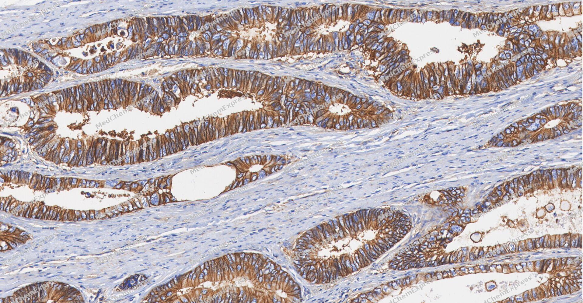 Immunohistochemical analysis of paraffin-embedded human Colon cancer tissue using CD147 antibody was performed. The section was pretreated using high-temperature and high-pressure mediated EDTA antigen retrieval buffer (pH 9.0), for 5 minutes. The tissues were incubated with primary antibody (HY-P84277, 1:400 dilution) at room temperature for 60 minutes. Detection was performed using an HRP conjugated compact polymer system. DAB was used as the chromogen. The tissues were counterstained with hematoxylin and mounted with neutral balsam mounting medium.
 -
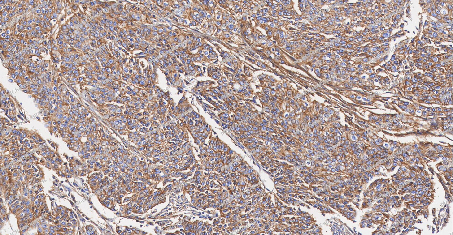 Immunohistochemical analysis of paraffin-embedded human Gastric Cancer tissue using CD147 antibody was performed. The section was pretreated using high-temperature and high-pressure mediated EDTA antigen retrieval buffer (pH 9.0), for 5 minutes. The tissues were incubated with primary antibody (HY-P84277, 1:400 dilution) at room temperature for 60 minutes. Detection was performed using an HRP conjugated compact polymer system. DAB was used as the chromogen. The tissues were counterstained with hematoxylin and mounted with neutral balsam mounting medium.
 -
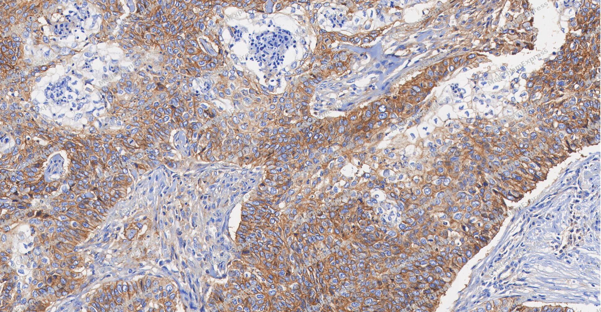 Immunohistochemical analysis of paraffin-embedded human Esophageal Carcinoma tissue using CD147 antibody was performed. The section was pretreated using high-temperature and high-pressure mediated EDTA antigen retrieval buffer (pH 9.0), for 5 minutes. The tissues were incubated with primary antibody (HY-P84277, 1:400 dilution) at room temperature for 60 minutes. Detection was performed using an HRP conjugated compact polymer system. DAB was used as the chromogen. The tissues were counterstained with hematoxylin and mounted with neutral balsam mounting medium.
 -
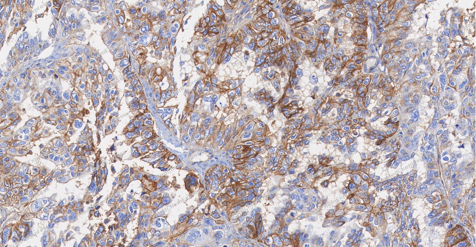 Immunohistochemical analysis of paraffin-embedded human ovarian carcinoma tissue using CD147 antibody was performed. The section was pretreated using high-temperature and high-pressure mediated EDTA antigen retrieval buffer (pH 9.0), for 5 minutes. The tissues were incubated with primary antibody (HY-P84277, 1:400 dilution) at room temperature for 60 minutes. Detection was performed using an HRP conjugated compact polymer system. DAB was used as the chromogen. The tissues were counterstained with hematoxylin and mounted with neutral balsam mounting medium.
 -
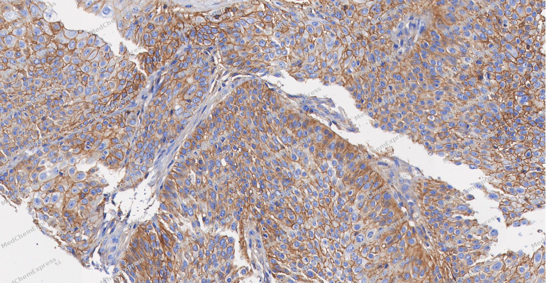 Immunohistochemical analysis of paraffin-embedded human Cervical cancer tissue using CD147 antibody was performed. The section was pretreated using high-temperature and high-pressure mediated EDTA antigen retrieval buffer (pH 9.0), for 5 minutes. The tissues were incubated with primary antibody (HY-P84277, 1:400 dilution) at room temperature for 60 minutes. Detection was performed using an HRP conjugated compact polymer system. DAB was used as the chromogen. The tissues were counterstained with hematoxylin and mounted with neutral balsam mounting medium.
 -
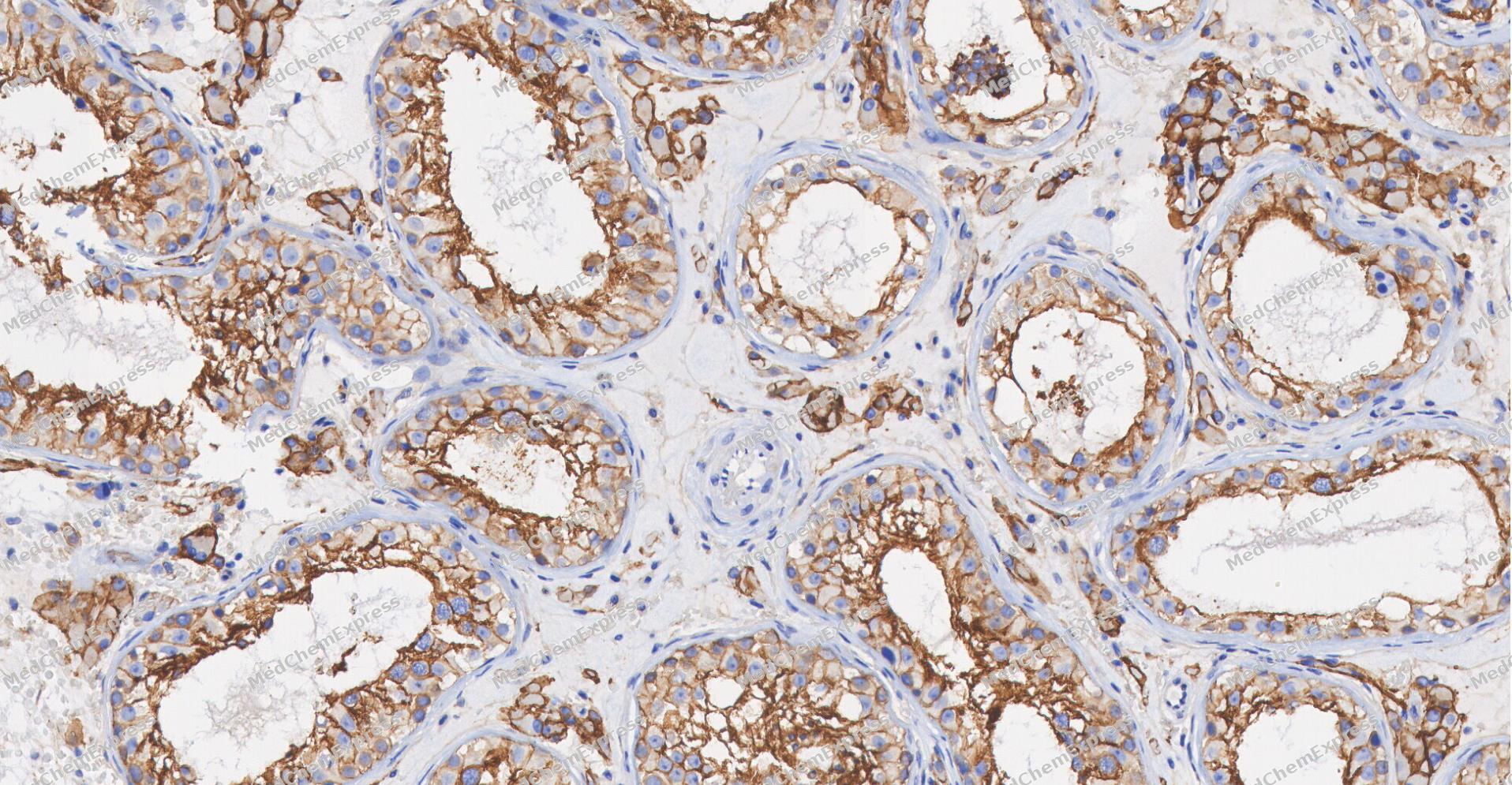 Immunohistochemical analysis of paraffin-embedded human Testis tissue using CD147 antibody was performed. The section was pretreated using high-temperature and high-pressure mediated EDTA antigen retrieval buffer (pH 9.0), for 5 minutes. The tissues were incubated with primary antibody (HY-P84277, 1:400 dilution) at room temperature for 60 minutes. Detection was performed using an HRP conjugated compact polymer system. DAB was used as the chromogen. The tissues were counterstained with hematoxylin and mounted with neutral balsam mounting medium.


Background
-
Function
CD147 is essential for normal retinal maturation and development. Acts as a retinal cell surface receptor for NXNL1 and plays an important role in NXNL1-mediated survival of retinal cone photoreceptors. In association with glucose transporter SLC16A1/GLUT1 and NXNL1, promotes retinal cone survival by enhancing aerobic glycolysis and accelerating the entry of glucose into photoreceptors. May act as a potent stimulator of IL6 secretion in multiple cell lines that include monocytes; (Microbial infection) Erythrocyte receptor for P.falciparum RH5 which is essential for erythrocyte invasion by the merozoite stage of P.falciparum isolates 3D7 and Dd2; Signaling receptor for cyclophilins, essential for PPIA/CYPA and PPIB/CYPB-dependent signaling related to chemotaxis and adhesion of immune cells. Plays an important role in targeting monocarboxylate transporters SLC16A1/GLUT1, SLC16A11 and SLC16A12 to the plasma membrane. Acts as a coreceptor for vascular endothelial growth factor receptor 2 (KDR/VEGFR2) in endothelial cells enhancing its VEGFA-mediated activation and downstream signaling. Promotes angiogenesis through EPAS1/HIF2A-mediated up-regulation of VEGFA (isoform VEGF-165 and VEGF-121) and KDR/VEGFR2 in endothelial cells. Plays a key role in regulating tumor growth, invasion, metastasis and neoangiogenesis by stimulating the production and release of extracellular matrix metalloproteinases and KDR/VEGFR2 by both tumor cells and stromal cells (fibroblasts and endothelial cells); (Microbial infection) Erythrocyte receptor for P.falciparum RH5 which is essential for erythrocyte invasion by the merozoite stage of P.falciparum isolates 3D7, Dd2, 7G8 and HB3. Binding of P.falciparum RH5 results in BSG dimerization which triggers an increase in intracellular Ca(2+) in the erythrocyte. This essential step leads to a rearrangement of the erythrocyte cytoskeleton required for the merozoite invasion; (Microbial infection) Can facilitate human SARS coronavirus (SARS-CoV-1) infection via its interaction with virus-associated PPIA/CYPA; (Microbial infection) Can facilitate HIV-1 infection via its interaction with virus-associated PPIA/CYPA; (Microbial infection) First described as a receptor for severe acute respiratory syndrome coronavirus 2 (SARS-CoV-2), it is not required for SARS-CoV-2 infection; (Microbial infection) Acts as a receptor for measles virus; (Microbial infection) Promotes entry of pentamer-expressing human cytomegalovirus (HCMV) into epithelial and endothelial cells[1][2][3][4][5][6][7][8][9][10][11][12][13][14][15].
-
Subcellular Localization
Melanosome; Cell membrane; Single-pass type I membrane protein; Photoreceptor inner segment; Cell projection, cilium, photoreceptor outer segment; Cell membrane; Single-pass type I membrane protein; Endosome; Endoplasmic reticulum membrane; Single-pass type I membrane protein; Basolateral cell membrane; Single-pass type I membrane protein; Cell membrane; Single-pass type I membrane protein; Cell membrane; Single-pass type I membrane protein
-
Expression
Tissue_specificity:Retinal-specific expression (PubMed: 25957687) . Expression in retinal cone photoreceptor cells (protein level) (PubMed: 25957687) ; expression in erythrocytes (protein level) (PubMed: 26195724, PubMed: 28409866) . High expression in melanoma cell lines (protein level) (PubMed: 11992541) . High expression in heart, kidney, skeletal muscle, and testis (PubMed: 21536654) ; high expression in bone marrow, fetal liver, lung, testis, and thymus. -
Isoforms & Post-Translational Modification
P35613 has 4 isomers: P35613-1: 42200 Da (predicted); P35613-2: 29221 Da (predicted); P35613-3: 19438 Da (predicted); P35613-4: 22764 Da (predicted).
N-glycosylated;N-glycosylated;N-glycosylated -
Subunit
Homooligomer (PubMed:19768682). Interacts with NXNL1 (PubMed:25957687). Interacts with SLC2A1 and SLC16A1/GLUT1 (By similarity). Interacts with XKR8; promoting its localization at the cell membrane (PubMed:27503893)
-
SwissProt ID
-
Synonyms
BSG; OK; 5F7; TCSF; EMMPRIN


Documentation
-
Data Sheet (235 KB)
-
SDS (251 KB)
- English - EN (251 KB)
- Français - FR (251 KB)
- Deutsch - DE (251 KB)
- Norwegian - NO (251 KB)
- Español - ES (251 KB)
- Swedish - SV (251 KB)
- Italian - IT (251 KB)
- Korean - KR (251 KB)
- Portuguese - PT (251 KB)
-
User Guide for Antibodies (1077 KB)

 Powered by Bioz
