



CD80 Antibody (YA3963)
(Synonyms: B7; BB1; B7-1; B7.1; LAB7; CD28LG; CD28LG1)Based on 1 publication(s) in Google Scholar
CD80 Antibody (YA3963) is a Mouse-derived and non-conjugated IgG1 monoclonal antibody, targeting to CD80.
-
Host:
Mouse
-
Isotype:
IgG
-
Application:
IHC-P, FC, ELISA
-
Reactivity :
Human, Mouse, Rat, Rabbit
-
Formulation:
Supplied in PBS with 0.05% sodium azide
-
Conjugation:
Non-conjugated


To place orders, for customer services and technical support, please contact: MedChemExpress USA
Tel: 609-228-6898 E-mail: [email protected] [email protected]



Publications Citing Use of MedChemExpress (MCE) CD80 Antibody (YA3963)
More

Applications
| Application |
IHC-P

IHC-P: Immunohistochemistry-Paraffin
|
FC
FC: Flow Cytometry
|
ELISA
ELISA: Enzyme Linked Immunosorbent Assay
|
|---|---|---|---|
| Dilution Ratio | 1:100-1:500 | 1:200-1:400 | 1:10000 |


Product Details
CD80 Antibody (YA3963) is a Mouse-derived and non-conjugated IgG1 monoclonal antibody, targeting to CD80.
-
Host Mouse

-
Clonality Monoclonal

-
Species ReactivityHuman, Mouse, Rat, Rabbit
-
Observed Molecular WeightObserved band size: 50-75 kDa
Note: Due to possible protein modifications or aggregation, the molecular weight should be confirmed by actual measurement, and the predicted value is for reference only.

-
Calculated Molecular Weight Predicted band size: 33 kDa

Purified recombinant fragment of human CD80 (AA: 35-288) expressed in E. Coli.
affinity purified.
Non-conjugated
Unmodified
IgG


Product Properties
-
Appearance
Solution
-
Formulation
Supplied in PBS with 0.05% sodium azide
-
Concentration
Batch-dependent, Please check the COA for the concentration of each lot. Check Lot Concentration
-
Storage & Stability
Stored at -20°C for 1 year. Avoid repeated freeze / thaw cycles.
-
Shipping
Shipping with blue ice.


Publications (1)
-
Journal Impact Factor
-
Most Recent
-
Biochem Biophys Res Commun
Non-fused ring A-D-A-type dyes for near-infrared II fluorescence imaging guided phototherapy and immune activation of tumour. [Abstract]2026 Jun 16:829:154158. PMID: 42361740


Verification Images
-
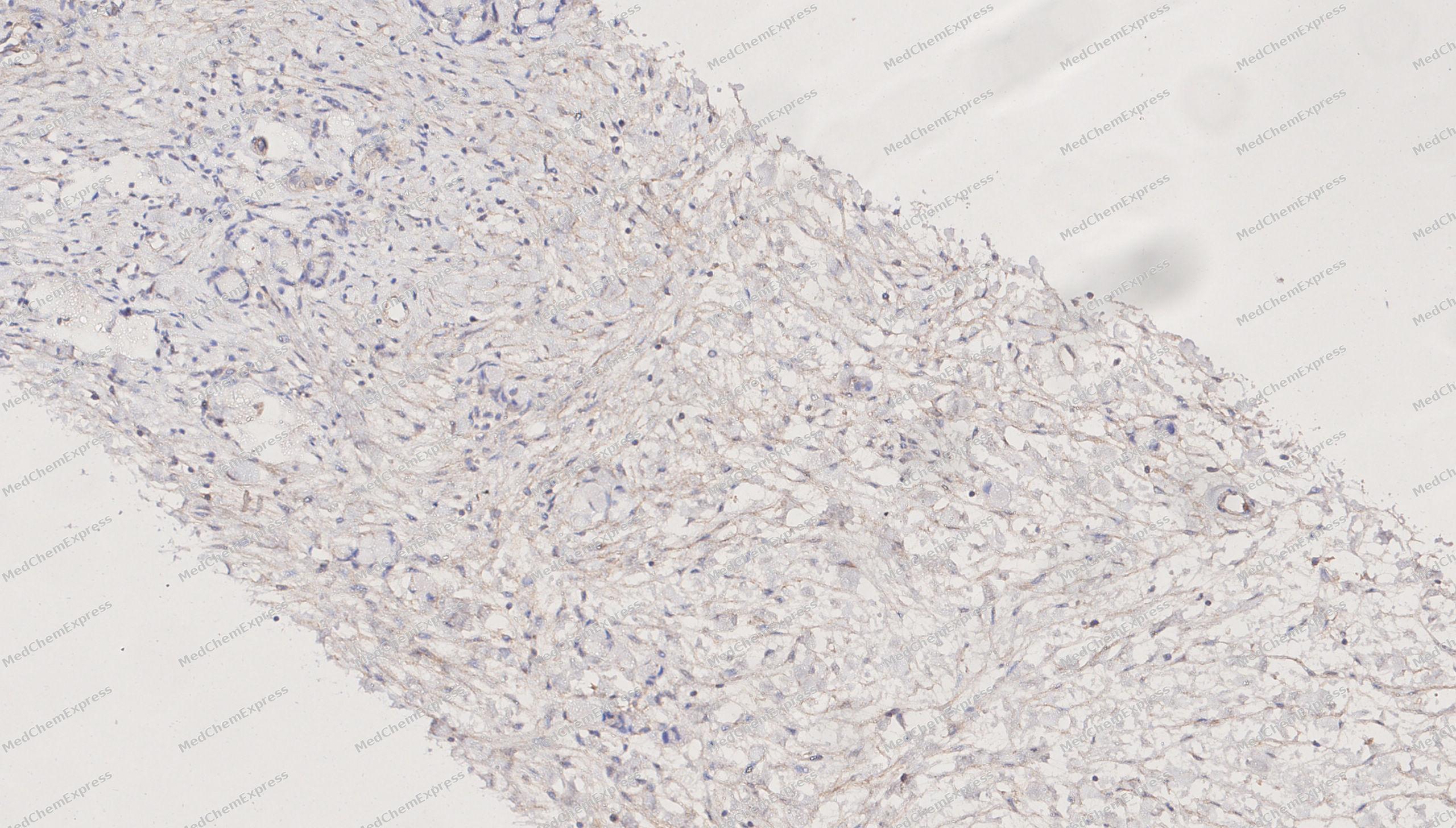 Immunohistochemical analysis of paraffin-embedded human uterine appendages tissue using CD80 antibody was performed. The section was pretreated using high-temperature mediated EDTA antigen retrieval buffer (pH 9.0), for 20 minutes. The tissues were incubated with primary antibody (HY-P84266, 1:600 dilution) at room temperature for 20 minutes. Detection was performed using an HRP conjugated compact polymer system. DAB was used as the chromogen. The tissues were counterstained with hematoxylin and mounted with neutral balsam mounting medium.
 -
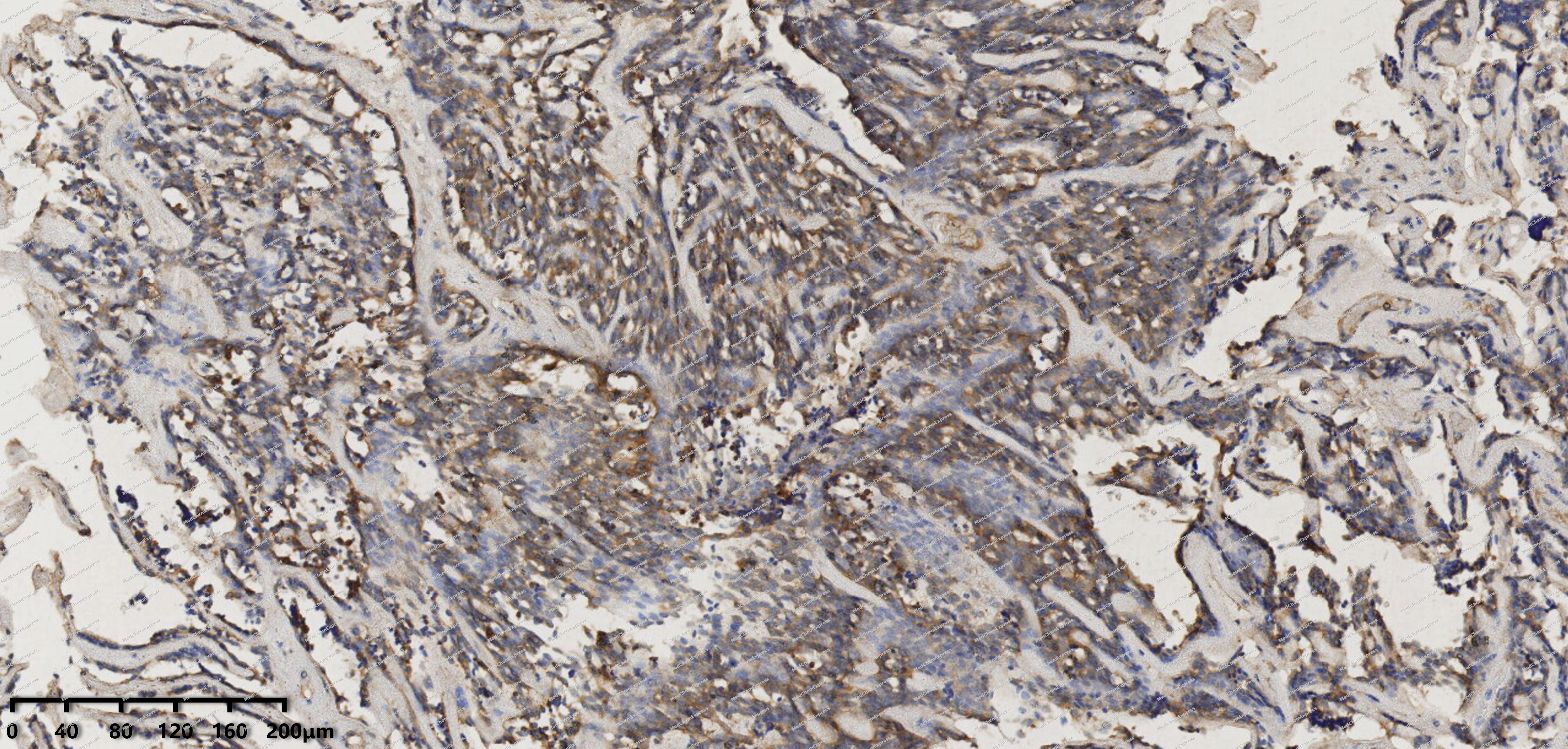 Immunohistochemical analysis of paraffin-embedded human mesothelioma tissue using CD80 antibody was performed. The section was pretreated using high-temperature mediated EDTA antigen retrieval buffer (pH 9.0), for 20 minutes. The tissues were incubated with primary antibody (HY-P84266, 1:600 dilution) at room temperature for 20 minutes. Detection was performed using an HRP conjugated compact polymer system. DAB was used as the chromogen. The tissues were counterstained with hematoxylin and mounted with neutral balsam mounting medium.
 -
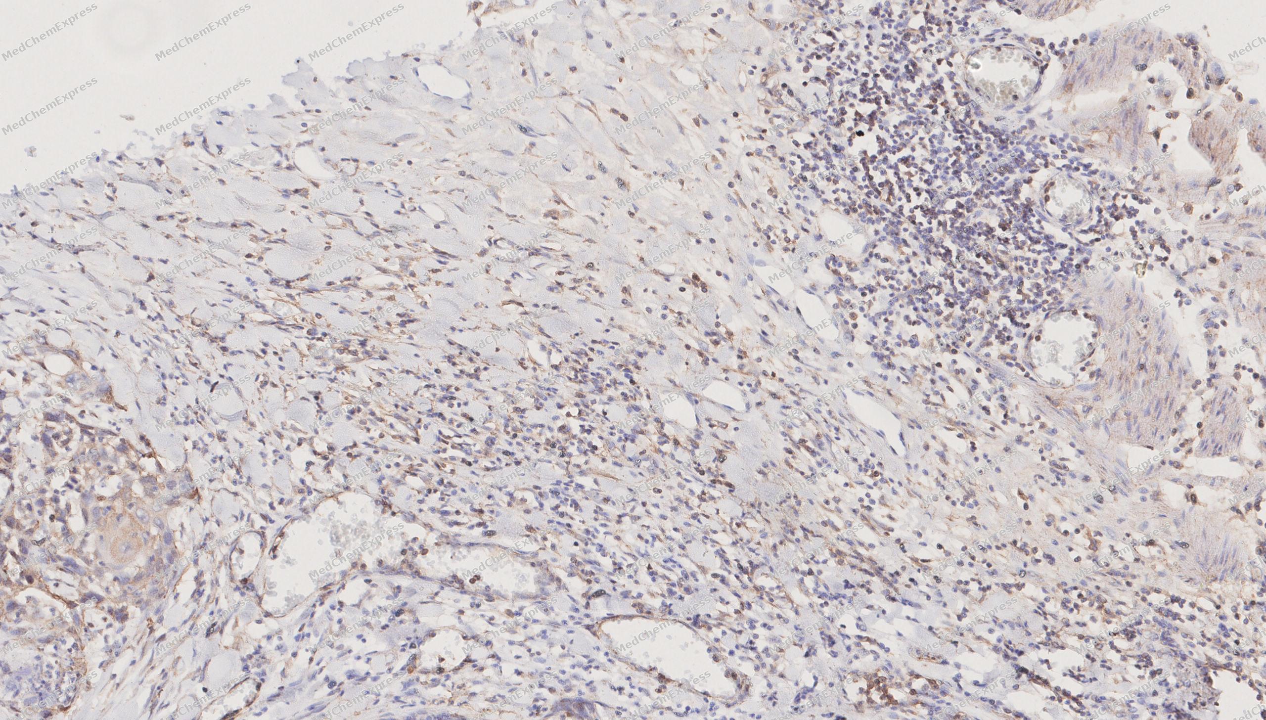 Immunohistochemical analysis of paraffin-embedded human esophagus tissue using CD80 antibody was performed. The section was pretreated using high-temperature mediated EDTA antigen retrieval buffer (pH 9.0), for 20 minutes. The tissues were incubated with primary antibody (HY-P84266, 1:600 dilution) at room temperature for 20 minutes. Detection was performed using an HRP conjugated compact polymer system. DAB was used as the chromogen. The tissues were counterstained with hematoxylin and mounted with neutral balsam mounting medium.
 -
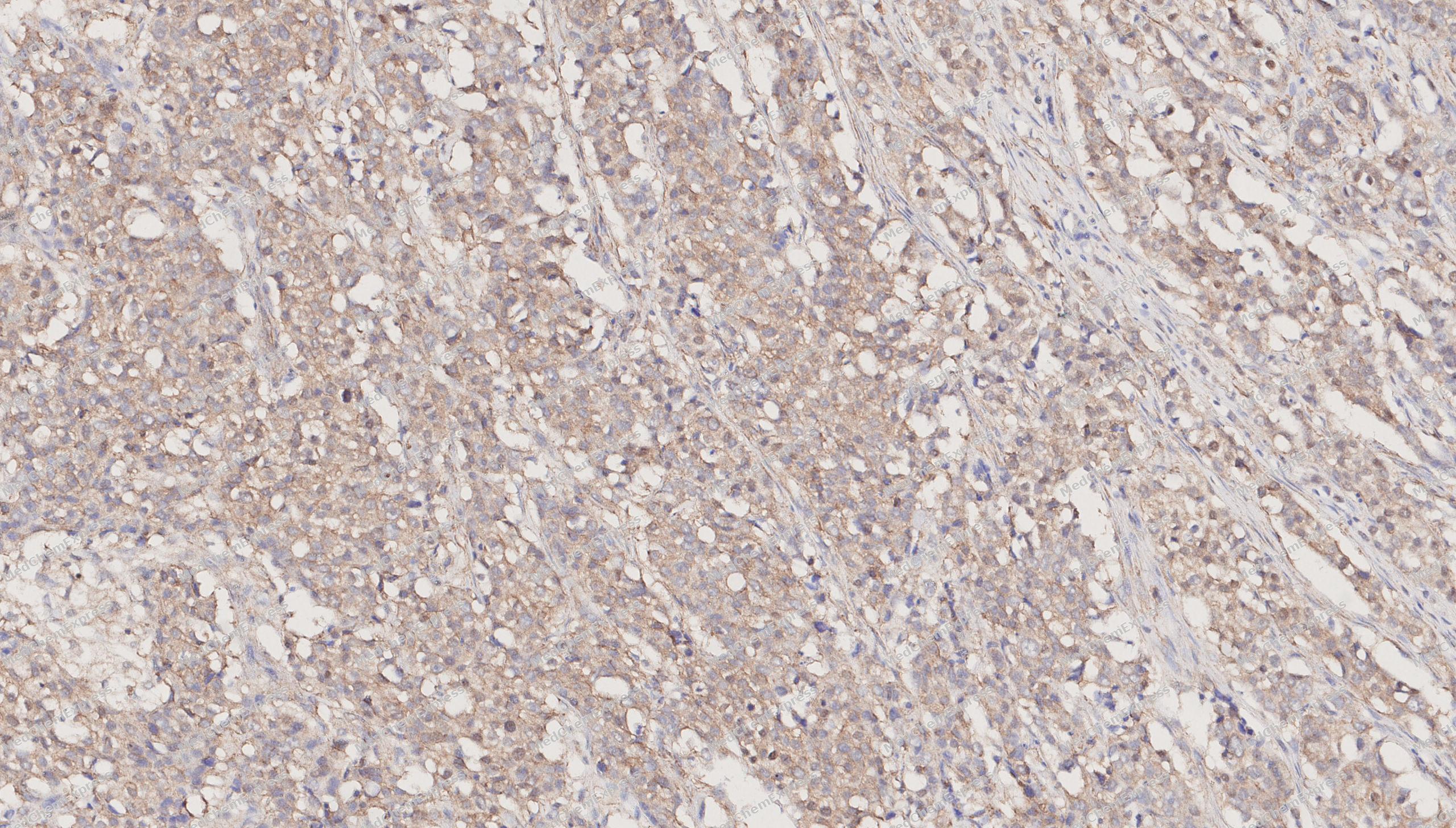 Immunohistochemical analysis of paraffin-embedded human breast cancer tissue using CD80 antibody was performed. The section was pretreated using high-temperature mediated EDTA antigen retrieval buffer (pH 9.0), for 20 minutes. The tissues were incubated with primary antibody (HY-P84266, 1:600 dilution) at room temperature for 20 minutes. Detection was performed using an HRP conjugated compact polymer system. DAB was used as the chromogen. The tissues were counterstained with hematoxylin and mounted with neutral balsam mounting medium.
 -
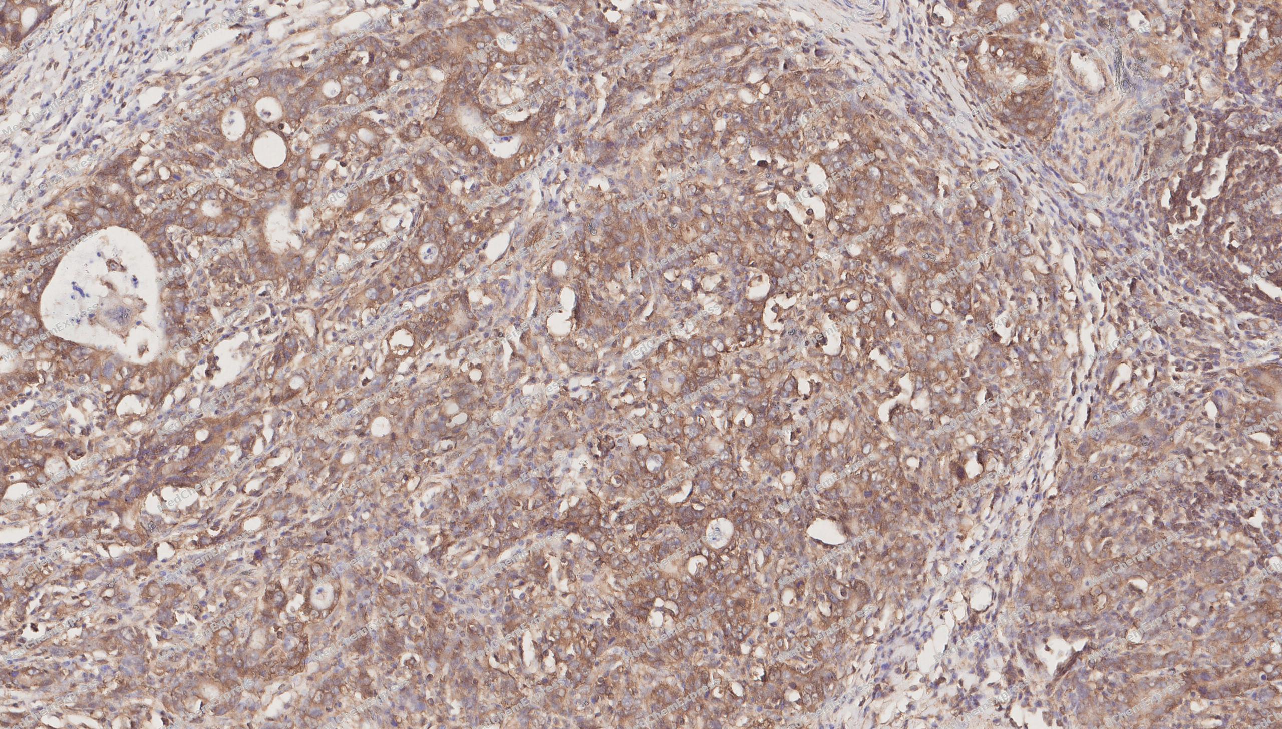 Immunohistochemical analysis of paraffin-embedded human gastric cancer tissue using CD80 antibody was performed. The section was pretreated using high-temperature mediated EDTA antigen retrieval buffer (pH 9.0), for 20 minutes. The tissues were incubated with primary antibody (HY-P84266, 1:600 dilution) at room temperature for 20 minutes. Detection was performed using an HRP conjugated compact polymer system. DAB was used as the chromogen. The tissues were counterstained with hematoxylin and mounted with neutral balsam mounting medium.
 -
 Flow cytometric analysis of 1X10⁶ C2C12 cells labeling CD80 Antibody (HY-P84266, red). Cells were stained with the primary antibody at 1/400 dilution for an hour at 4℃. AF488-conjugated Goat Anti-Mouse IgG H&L (HY-P8005) was used as the secondary antibody at 1/1,000 dilution for 30 minutes at 4℃. Mouse IgG Isotype Control (HY-P80757, blue) was used as the isotype control, cells without incubation with primary antibody were used as the unlabeled control (black).


Background
-
Function
CD28/CD80 is a co-stimulatory molecule belonging to the immunoglobulin superfamily that plays a crucial role in T-lymphocyte activation. CD80 interacts with the CD28 receptor-constitutively expressed on the surface of T cells-to serve as a primary co-stimulatory signal that enhances MHC/TCR signaling in naive T cells. This triggers the activation of various signaling pathways, such as NF-κB or MAPK, ultimately leading to the production of multiple cytokines. Furthermore, the CD28/CD80 co-stimulatory signal promotes T-cell glucose metabolism and ATP synthesis by activating the PI3K/Akt signaling pathway. CD80 also acts as a regulator of the PD-L1/PDCD1 interaction, limiting the excessive activation of PD-L1 and its inhibitory effects during immune responses. Expressed on the surface of B cells, CD80 plays a pivotal regulatory role in the interactions between B cells and T cells during both early and late germinal center reactions.
-
Subcellular Localization
Cell membrane; Single-pass type I membrane protein
-
Expression
Tissue_specificity:It is expressed on activated B cells, macrophages, and dendritic cells. -
Isoforms & Post-Translational Modification
CD80 has 3 isomers: P33681-1: 33048 Da (predicted); P33681-2: 29413 Da (predicted); P33681-3: 18761 Da (predicted).
Palmitoylated by ZDHHC20; palmitoylation protects CD80 from ubiquitin-mediated degradation, regulating the protein stability, and ensures its accurate plasma membrane localization -
Subunit
Homodimer. Interacts with CTLA4; this interaction inhibits T-cell activation. Interacts with PDL1/CD274; this interaction blocks PDL1/PDCD1 binding and thus PDL1/CD274 inhibitory function. Interacts with CD28
-
SwissProt ID
-
Synonyms
B7; BB1; B7-1; B7.1; LAB7; CD28LG; CD28LG1
Documentation
-
Data Sheet (261 KB)
-
SDS (251 KB)
- English - EN (251 KB)
- Français - FR (251 KB)
- Deutsch - DE (251 KB)
- Norwegian - NO (251 KB)
- Español - ES (251 KB)
- Swedish - SV (251 KB)
- Italian - IT (251 KB)
- Korean - KR (251 KB)
- Portuguese - PT (251 KB)
-
User Guide for Antibodies (1077 KB)
References
[1]. Lu B, et al. zDHHC20-driven S-palmitoylation of CD80 is required for its costimulatory function. Acta Pharmacol Sin. 2024 Jun;45(6):1214-1223. [Content Brief]
[2]. Collins AV, et al. The interaction properties of costimulatory molecules revisited. Immunity. 2002 Aug;17(2):201-10. [Content Brief]
[3]. Boulougouris G, et al. IL-2-independent activation and proliferation in human T cells induced by CD28. J Immunol. 1999 Aug 15;163(4):1809-16. [Content Brief]
[4]. Frauwirth KA, et al. The CD28 signaling pathway regulates glucose metabolism. Immunity. 2002 Jun;16(6):769-77. [Content Brief]
[5]. Kennedy A, et al. The CTLA-4 immune checkpoint protein regulates PD-L1:PD-1 interaction via transendocytosis of its ligand CD80. EMBO J. 2023 Mar 1;42(5):e111556. [Content Brief]

 Powered by Bioz
