



Chk1 Antibody
(Synonyms: CHEK1; CHK1; Serine/threonine-protein kinase Chk1; CHK1 checkpoint homolog; Cell cycle checkpoint kinase; Checkpoint kinase-1)
Based on 1 Customer Validation
Chk1 Antibody is a Rabbit-derived and non-conjugated IgG polyclonal antibody, targeting to Chk1.
-
Host:
Rabbit
-
Isotype:
IgG
-
Application:
WB, ICC/IF, IP
-
Reactivity :
Human, Mouse, Rat
-
Formulation:
Supplied in Rabbit IgG in PBS(pH 7.4), 150mM sodium chloride, 0.05% BSA, 0.02% sodium azide and 50% glycerol.
-
Conjugation:
Non-conjugated


To place orders, for customer services and technical support, please contact: MedChemExpress USA
Tel: 609-228-6898 E-mail: [email protected] [email protected]





Applications
| Application |
WB

WB: Western Blot
|
ICC/IF
ICC/IF: Immunocytochemistry/
Immunofluorescence |
IP
IP: Immunoprecipitation
|
|---|---|---|---|
| Dilution Ratio | 1:1000-1:2000 | 1:50-1:200 | 1:20 |


Product Details
Chk1 Antibody is a Rabbit-derived and non-conjugated IgG polyclonal antibody, targeting to Chk1.
-
Host Rabbit

-
Clonality Polyclonal

-
Species ReactivityHuman, Mouse, Rat
-
Observed Molecular WeightObserved band size: 54 kDa
Note: Due to possible protein modifications or aggregation, the molecular weight should be confirmed by actual measurement, and the predicted value is for reference only.

-
Calculated Molecular Weight Predicted band size: 54 kDa

Entrez Gene: 1111 Human ; 12649 Mouse ; 140583 Rat
SwissProt: O14757 Human ; O35280 Mouse ; Q91ZN7 Rat
OMIM: 620610 Human
Synthetic peptide corresponding to Human Chk1 aa1-67.
Endogenous
affinity purified
Non-conjugated
Unmodified
IgG


Product Properties
-
Appearance
Solution
-
Formulation
Supplied in Rabbit IgG in PBS(pH 7.4), 150mM sodium chloride, 0.05% BSA, 0.02% sodium azide and 50% glycerol.
-
Concentration
Batch-dependent, Please check the COA for the concentration of each lot. Check Lot Concentration
-
Storage & Stability
Stored at -20°C for 1 year. Avoid repeated freeze / thaw cycles.
-
Shipping
Shipping with blue ice.


Verification Images
-
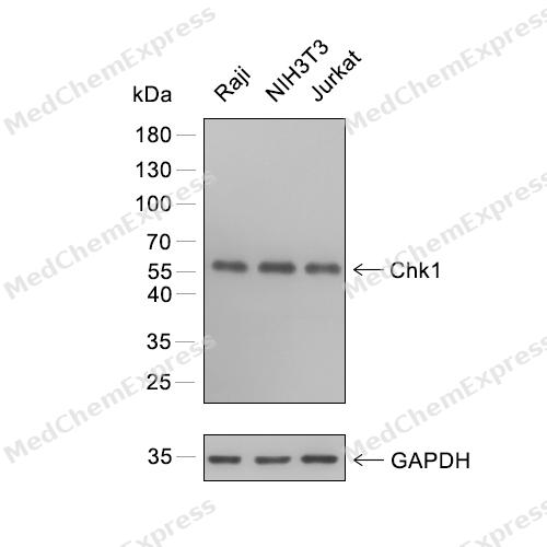 Western blot analysis of extracts from Raji (lane 1) and NIH3T3 (lane 2) and Jurkat (lane 3) using Chk1 antibody. Proteins were transferred to a PVDF membrane and blocked with 5% nonfat powdered milk in PBST for 2 hour at room temperature. The primary antibody (1/1000) and Loading control antibody (GAPDH, 1/3000) was diluted with 5% nonfat powdered milk in PBST at 4°C overnight. Goat Anti-Rabbit IgG-HRP Secondary Antibody (1/8,000) was incubated for 45min at room temperature.
 -
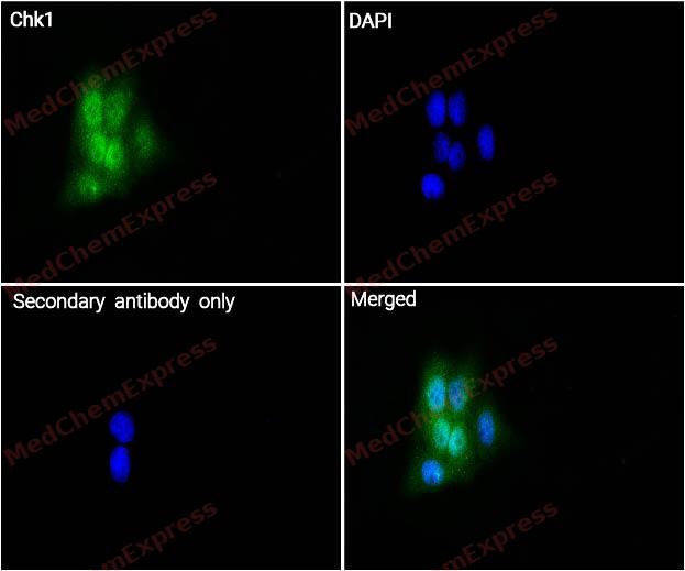 Immunocytochemistry analysis of Hela cells labeling Chk1 with Chk1 Antibody (HY-P80617) at 1/100 dilution. Cells were fixed in 4% paraformaldehyde for 15 minutes at room temperature, permeabilized with 0.1% Triton X-100 in PBS for 15 minutes at room temperature, then blocked with quick block buffer for 10 minutes at room temperature. Cells were then incubated with Chk1 Antibody (HY-P80617) at 1/100 dilution in quick block buffer overnight at 4 ℃. AF488-conjugated Goat Anti-Rabbit IgG H&L(HY-P8002, Green) was used as the secondary antibody at 1/1,000 dilution. PBS instead of the primary antibody was used as the secondary antibody only control. The Nuclear counterstain was DAPI (Blue).
 -
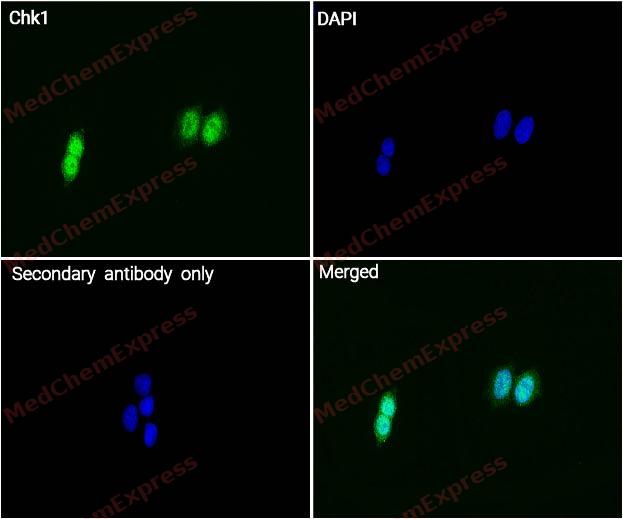 Immunocytochemistry analysis of MCF-7 cells labeling Chk1 with Chk1 Antibody (HY-P80617) at 1/100 dilution. Cells were fixed in 4% paraformaldehyde for 15 minutes at room temperature, permeabilized with 0.1% Triton X-100 in PBS for 15 minutes at room temperature, then blocked with quick block buffer for 10 minutes at room temperature. Cells were then incubated with Chk1 Antibody (HY-P80617) at 1/100 dilution in quick block buffer overnight at 4 ℃. AF488-conjugated Goat Anti-Rabbit IgG H&L(HY-P8002, Green) was used as the secondary antibody at 1/1,000 dilution. PBS instead of the primary antibody was used as the secondary antibody only control. The Nuclear counterstain was DAPI (Blue).


Background
-
Function
Chk1 (checkpoint kinase 1) mediates the DNA damage response and normal cell-cycle regulation through checkpoint activation, cell-cycle arrest, DNA repair, and cell death control[1]. Mechanistically, Chk1 functions as a key transducer in genome-integrity checkpoints, especially within ATR-linked responses to stalled replication and genotoxic stress[2][3]. In cancer research, this role makes Chk1 relevant to replication stress, BRCA wild-type ovarian cancer models, and studies combining checkpoint inhibition with DNA-damaging therapy[4][5]. Compared with Chk2, Chk1 shows distinct activation biology and experimentally separable checkpoint functions, while Chk1-S acts as an endogenous splice-variant inhibitor that regulates Chk1-dependent checkpoints[2][6]. For experimental applications, Chk1 inhibitors such as prexasertib/LY2606368 induce replication catastrophe and have been tested in tumor models and recurrent high-grade serous ovarian cancer[7][8].
-
Subcellular Localization
Nucleus; Chromosome; Cytoplasm; Cytoplasm, cytoskeleton, microtubule organizing center, centrosome
-
Expression
Tissue_specificity:This gene is widely expressed in various tissues, with the highest expression in the thymus, testes, small intestine, and colon. -
Isoforms & Post-Translational Modification
O14757 has 3 isomers: O14757-1: 54434 Da (predicted); O14757-2: 43703 Da (predicted); O14757-3: 50415 Da (predicted).
Phosphorylated by ATR in a RAD17-dependent manner in response to ultraviolet irradiation and inhibition of DNA replication (PubMed:10859164, PubMed:11390642, PubMed:12446774, PubMed:12588868, PubMed:12676583, PubMed:12676925, PubMed:12676962, PubMed:14681223, PubMed:14988723, PubMed:15650047, PubMed:15707391, PubMed:15870257, PubMed:25083873, PubMed:31316063). Phosphorylated by ATM in response to ionizing irradiation (PubMed:12588868, PubMed:12676583). ATM and ATR can both phosphorylate Ser-317 and Ser-345 and this results in enhanced kinase activity (PubMed:11390642, PubMed:12446774, PubMed:12588868, PubMed:12660173, PubMed:12676583, PubMed:12676962, PubMed:14657349, PubMed:15665856, PubMed:15707391, PubMed:15870257, PubMed:25083873). Phosphorylation at Ser-345 induces a change in the conformation of the protein, activates the kinase activity and is a prerequisite for interaction with FBXO6 and subsequent ubiquitination at Lys-436 (PubMed:19716789). Phosphorylation at Ser-345 also increases binding to 14-3-3 proteins and promotes nuclear retention (PubMed:12676962). Conversely, dephosphorylation at Ser-345 by PPM1D may contribute to exit from checkpoint mediated cell cycle arrest (PubMed:15870257). Phosphorylation at Ser-280 by AKT1/PKB, may promote mono and/or diubiquitination. Also phosphorylated at undefined residues during mitotic arrest, resulting in decreased activity (By similarity);Ubiquitinated. Mono or diubiquitination promotes nuclear exclusion (By similarity). The activated form (phosphorylated on Ser-345) is polyubiquitinated at Lys-436 by some SCF-type E3 ubiquitin ligase complex containing FBXO6 promoting its degradation. Ubiquitination and degradation are required to terminate the checkpoint and ensure that activated CHEK1 does not accumulate as cells progress through S phase, when replication forks encounter transient impediments during normal DNA replication. 'Lys-63'-mediated ubiquitination by TRAF4 at Lys-132 activates cell cycle arrest and activation of DNA repair (PubMed:32357935);Proteolytically cleaved at the C-terminus by SPRTN during normal DNA replication, thereby promoting CHEK1 removal from chromatin and activating the protein kinase activity -
Subunit
Interacts (phosphorylated by ATR) with RAD51 (PubMed:15665856). Interacts with and phosphorylates CLSPN, an adapter protein that regulates the ATR-dependent phosphorylation of CHEK1 (PubMed:16963448). Interacts with BRCA1 (PubMed:11836499). Interacts with and phosphorylates CDC25A, CDC25B and CDC25C (PubMed:9278511). Interacts with FBXO6, which regulates CHEK1 (PubMed:19716789). Interacts with PPM1D, which regulates CHEK1 through dephosphorylation (PubMed:15870257). Interacts with TIMELESS; DNA damage-dependent (PubMed:15798197). Interacts with FEM1B; activates CHEK1 in response to stress (PubMed:19330022). Interacts with TLK1 (PubMed:12660173). Interacts with XPO1 and YWHAZ (PubMed:12676962). Interacts with CDK5RAP3; antagonizes CHEK1 (PubMed:19223857)
-
SwissProt ID
-
Synonyms
CHEK1; CHK1; Serine/threonine-protein kinase Chk1; CHK1 checkpoint homolog; Cell cycle checkpoint kinase; Checkpoint kinase-1
-
Research Field
Epigenetics and Nuclear Signaling


Documentation
-
Data Sheet (261 KB)
-
SDS (252 KB)
- English - EN (252 KB)
- Français - FR (252 KB)
- Deutsch - DE (252 KB)
- Norwegian - NO (252 KB)
- Español - ES (252 KB)
- Swedish - SV (252 KB)
- Italian - IT (252 KB)
- Korean - KR (252 KB)
- Portuguese - PT (252 KB)
-
User Guide for Antibodies (1077 KB)
References
[1]. Patil M, et al. Checkpoint kinase 1 in DNA damage response and cell cycle regulation. Cell Mol Life Sci. 2013 Nov;70(21):4009-21. [Content Brief]
[2]. Bartek J, et al. Chk1 and Chk2 kinases in checkpoint control and cancer. Cancer Cell. 2003 May;3(5):421-9. [Content Brief]
[3]. Dai Y, et al. New insights into checkpoint kinase 1 in the DNA damage response signaling network. Clin Cancer Res. 2010 Jan 15;16(2):376-83. [Content Brief]
[4]. Rabezanahary H, et al. Live virus neutralizing antibodies against pre and post Omicron strains in food and retail workers in Québec, Canada. Heliyon. 2024 May 21;10(10):e31026. [Content Brief]
[5]. Nair J, et al. Resistance to the CHK1 inhibitor prexasertib involves functionally distinct CHK1 activities in BRCA wild-type ovarian cancer. Oncogene. 2020 Aug;39(33):5520-5535. [Content Brief]
[6]. Pabla N, et al. Checkpoint kinase 1 (Chk1)-short is a splice variant and endogenous inhibitor of Chk1 that regulates cell cycle and DNA damage checkpoints. Proc Natl Acad Sci U S A. 2012 Jan 3;109(1):197-202. [Content Brief]
[7]. King C, et al. LY2606368 Causes Replication Catastrophe and Antitumor Effects through CHK1-Dependent Mechanisms. Mol Cancer Ther. 2015 Sep;14(9):2004-13. [Content Brief]
[8]. Lee JM, et al. Prexasertib, a cell cycle checkpoint kinase 1 and 2 inhibitor, in BRCA wild-type recurrent high-grade serous ovarian cancer: a first-in-class proof-of-concept phase 2 study. Lancet Oncol. 2018 Feb;19(2):207-215. [Content Brief]

 Powered by Bioz
