



cIAP1 Antibody (YA2500)
(Synonyms: cIAP1; C-IAP1; IAP homolog B; Inhibitor of apoptosis protein 2; IAP-2; hIAP-2; hIAP2; API1; IAP2; MIHB; RNF48)Based on 1 Customer Validation
cIAP1 Antibody (YA2500) is a Rabbit-derived and non-conjugated IgG monoclonal antibody, targeting to cIAP1.
-
Host:
Rabbit
-
Isotype:
IgG
-
Application:
WB, ICC/IF
-
Reactivity :
Human
-
Formulation:
Supplied in 50mM Tris-Glycine(pH 7.4), 0.15M NaCl, 40% Glycerol, 0.01% Sodium azide and 0.05% BSA
-
Conjugation:
Non-conjugated


To place orders, for customer services and technical support, please contact: MedChemExpress USA
Tel: 609-228-6898 E-mail: [email protected] [email protected]





Applications
| Application |
WB

WB: Western Blot
|
ICC/IF
ICC/IF: Immunocytochemistry/
Immunofluorescence |
|---|---|---|
| Dilution Ratio | 1:500-1:1000 | 1:50-1:200 |


Product Details
cIAP1 Antibody (YA2500) is a Rabbit-derived and non-conjugated IgG monoclonal antibody, targeting to cIAP1.
-
Host Rabbit

-
Clonality Recombinant,Monoclonal

-
Species ReactivityHuman
-
Observed Molecular WeightObserved band size: 70 kDa
Note: Due to possible protein modifications or aggregation, the molecular weight should be confirmed by actual measurement, and the predicted value is for reference only.

-
Calculated Molecular Weight Predicted band size: 70 kDa

A synthetic peptide of human cIAP1 aa29-42.
Endogenous
Affinity Purified
Non-conjugated
Unmodified
IgG


Product Properties
-
Appearance
Solution
-
Formulation
Supplied in 50mM Tris-Glycine(pH 7.4), 0.15M NaCl, 40% Glycerol, 0.01% Sodium azide and 0.05% BSA
-
Concentration
Batch-dependent, Please check the COA for the concentration of each lot. Check Lot Concentration
-
Storage & Stability
Stored at -20°C for 1 year. Avoid repeated freeze / thaw cycles.
-
Shipping
Shipping with blue ice.


Verification Images
-
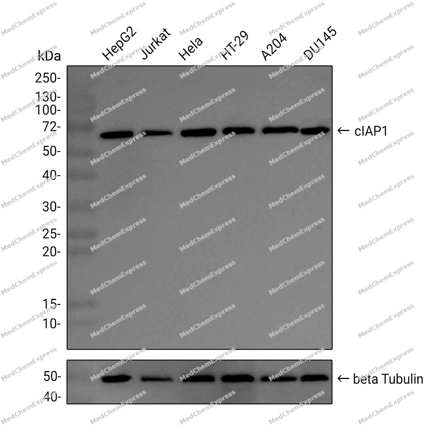
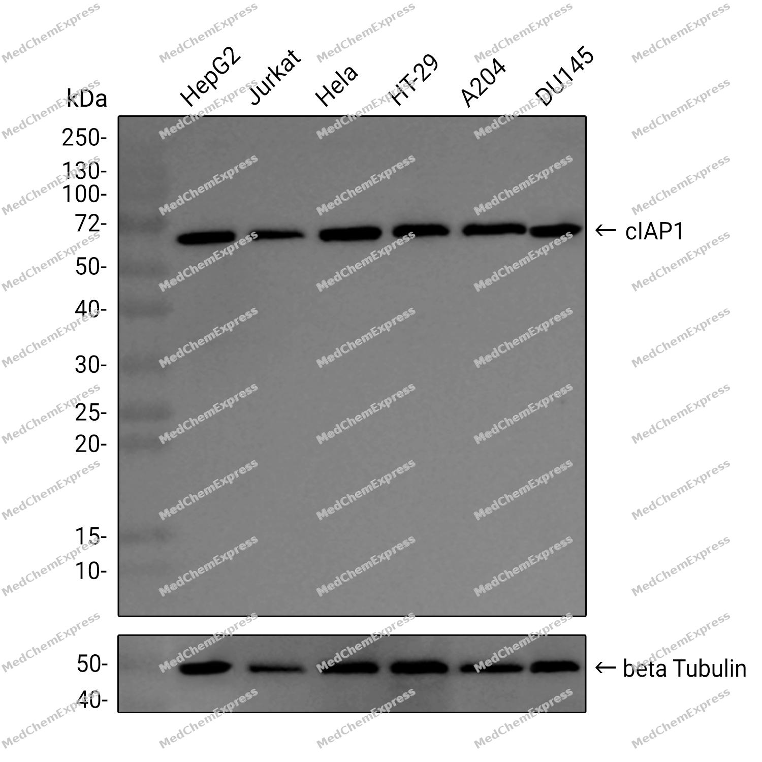 Western blot analysis was performed on extracts from HepG2 (lane 1, 15 μg), Jurkat (lane 2, 15 μg), Hela (lane 3, 15 μg), HT-29 (lane 4, 15 μg), A204 (lane 5, 15 μg), DU145 (lane 6, 15 μg) using cIAP1 Rabbit mAb.Proteins were transferred to a PVDF membrane and blocked with 5% non-fat milk in TBST at 4°C overnight.The primary antibody (1:1000 dilution) and the loading control antibody (beta-Tubulin, HY-P80955, 1:5000 dilution) were incubated in 5% non-fat milk in TBST for 1 hour at 37°C.Goat Anti-Rabbit IgG-HRP Secondary Antibody (1:20000 dilution) was then applied for 40 minutes at 37°C.


Background
-
Function
Cellular inhibitor of apoptosis protein 1 (cIAP1) is a multifunctional E3 ubiquitin ligase that regulates apoptosis, NF-κB signaling, and cell proliferation in both cytoplasmic and nuclear compartments[1][4]. Mechanistically, cIAP1 modulates tumor necrosis factor receptor (TNFR) and pattern recognition receptor (PRR) signaling, thereby influencing innate immunity, inflammasome activation, and transcriptional control via E2F1[1][4][5][6]. Compared with cIAP2, cIAP1 exhibits distinct nuclear functions, including direct binding to E2F1 and enhancement of CCNE and CCNA gene transcription, which promotes cell cycle progression[4]. In disease models, cIAP1 contributes to tumor growth through interactions with TRAF2, activating the canonical NF-κB and JAK/STAT3 pathways, and sustaining survival in gallbladder cancer, non-small cell lung cancer, and skeletal muscle atrophy models[9][6][2][7]. Furthermore, cIAP1 is required for T cell costimulation through CD137 signaling, mediating NF-κB and MAPK activation, which is critical for antitumor immune responses[3]. Pharmacologically, cIAP1 degradation by SMAC mimetics or selective inhibitors potentiates apoptosis by releasing RIP1 and activating caspase complexes, highlighting its potential as a therapeutic target in cancer and immune-related diseases[8][3]. Overall, the unique nuclear and cytoplasmic functions of cIAP1 distinguish it from cIAP2, enabling diverse applications in experimental oncology, immunology, and tissue regeneration studies.
-
Subcellular Localization
Cytoplasm; Nucleus
-
Expression
Tissue_specificity:Present in many human tissues. Mainly expressed in human skeletal muscle, thymus, testis, ovary, and pancreas, low or absent in brain and peripheral blood leukocytes -
Isoforms & Post-Translational Modification
Q13490 has 2 isomers: Q13490-1: 69900 Da (predicted); Q13490-2: 64098 Da (predicted).
Auto-ubiquitinated and degraded by the proteasome in apoptotic cells;Upon stimulation of death receptors, including TNFRSF10B, recruited to receptors and cleaved by caspases. Proteolytic fragments remain associated with the receptors. This cleavage presumably inactivates the protein -
Subunit
Interacts with DIABLO/SMAC and with PRSS25; these interactions inhibit apoptotic suppressor activity. Interacts with CASP9. Interacts (via BIR domains) with TRAF2; the interaction is required for IKBKE ubiquitination. Interacts with E2F1, RIPK1, RIPK2, RIPK3, RIPK4, BIRC5/survivin and USP19. HSP90AB1 (PubMed:25486457). Interacts with UBXN1 (PubMed:25681446). Interacts with GSK3B (PubMed:18846110). Interacts with several death receptors, inclusing FAS, TNFRSF10A and TNFRSF10B (PubMed:18846110). Recruited to TNFRSF10B in the absence of receptor stimulation. When TNFRSF10B is stimulated, further recruited to the receptor and cleaved by caspases. Proteolytic fragments remain associated with TNFRSF10B (PubMed:18846110)
-
SwissProt ID
-
Synonyms
cIAP1; C-IAP1; IAP homolog B; Inhibitor of apoptosis protein 2; IAP-2; hIAP-2; hIAP2; API1; IAP2; MIHB; RNF48
-
Research Field
Cell Biology


Documentation
-
Data Sheet (263 KB)
-
SDS (252 KB)
- English - EN (252 KB)
- Français - FR (252 KB)
- Deutsch - DE (252 KB)
- Norwegian - NO (252 KB)
- Español - ES (252 KB)
- Swedish - SV (252 KB)
- Italian - IT (252 KB)
- Korean - KR (252 KB)
- Portuguese - PT (252 KB)
-
User Guide for Antibodies (1077 KB)
References
[1]. Zadoroznyj A, et al. Cytoplasmic and Nuclear Functions of cIAP1. Biomolecules. 2022 Feb 17;12(2):322. [Content Brief]
[2]. Su W, et al. cIAP1 promotes proliferation and migration and prevents apoptosis in gallbladder cancer in vitro. Biosci Rep. 2019 Apr 12;39(4):BSR20182266. [Content Brief]
[3]. Glez-Vaz J, et al. CD137 (4-1BB) requires physically associated cIAPs for signal transduction and antitumor effects. Sci Adv. 2023 Aug 18;9(33):eadf6692. [Content Brief]
[4]. Cartier J, et al. Cellular inhibitor of apoptosis protein-1 (cIAP1) can regulate E2F1 transcription factor-mediated control of cyclin transcription. J Biol Chem. 2011 Jul 29;286(30):26406-17. [Content Brief]
[7]. Vu NT, et al. Caspase-9b Interacts Directly with cIAP1 to Drive Agonist-Independent Activation of NF-κB and Lung Tumorigenesis. Cancer Res. 2016 May 15;76(10):2977-89. [Content Brief]
[8]. Yang C, et al. LCL161 increases paclitaxel-induced apoptosis by degrading cIAP1 and cIAP2 in NSCLC. J Exp Clin Cancer Res. 2016 Sep 30;35(1):158. [Content Brief]
[9]. Zhang J, et al. Ubiquitin Ligases cIAP1 and cIAP2 Limit Cell Death to Prevent Inflammation. Cell Rep. 2019 May 28;27(9):2679-2689.e3. [Content Brief]
[10]. Dumétier B, et al. IAP-Mediated Protein Ubiquitination in Regulating Cell Signaling. Cells. 2020 Apr 30;9(5):1118. [Content Brief]
[11]. Morrish E, et al. Future Therapeutic Directions for Smac-Mimetics. Cells. 2020 Feb 11;9(2):406. [Content Brief]

 Powered by Bioz
