



EHD1 Antibody (YA1970)
(Synonyms: EHD1; H PAST; hPAST1; PAST; PAST homolog 1; PAST1; Tilin)
Based on 1 Customer Validation
EHD1 Antibody (YA1970) is a Rabbit-derived and non-conjugated IgG monoclonal antibody, targeting to EHD1.
-
Host:
Rabbit
-
Isotype:
IgG
-
Application:
WB, IHC-P, ICC/IF, IP, FC
-
Reactivity :
Human, Mouse, Rat
-
Formulation:
Supplied in 50mM Tris-Glycine(pH 7.4), 0.15M NaCl, 40%Glycerol, 0.01% sodium azide and 0.05% BSA.
-
Conjugation:
Non-conjugated


To place orders, for customer services and technical support, please contact: MedChemExpress USA
Tel: 609-228-6898 E-mail: [email protected] [email protected]





Applications
| Application |
WB

WB: Western Blot
|
IHC-P
IHC-P: Immunohistochemistry-Paraffin
|
ICC/IF
ICC/IF: Immunocytochemistry/
Immunofluorescence |
IP
IP: Immunoprecipitation
|
FC
FC: Flow Cytometry
|
|---|---|---|---|---|---|
| Dilution Ratio | 1:500-1:1000 | 1:50-1:100 | 1:50-1:200 | 1:50 | 1:50-1:100 |


Product Details
EHD1 Antibody (YA1970) is a Rabbit-derived and non-conjugated IgG monoclonal antibody, targeting to EHD1.
-
Host Rabbit

-
Clonality Recombinant,Monoclonal

-
Species ReactivityHuman, Mouse, Rat
-
Observed Molecular WeightObserved band size: 61 kDa
Note: Due to possible protein modifications or aggregation, the molecular weight should be confirmed by actual measurement, and the predicted value is for reference only.

-
Calculated Molecular Weight Predicted band size: 61 kDa

Entrez Gene: 10938 Human ; 13660 Mouse ; 293692 Rat
SwissProt: Q9H4M9 Human ; Q9WVK4 Mouse ; Q641Z6 Rat
OMIM: 605888 Human
A synthesized peptide derived from human EHD1 aa1-45/534.
Endogenous
Affinity Chromatography
Non-conjugated
Unmodified
IgG


Product Properties
-
Appearance
Solution
-
Formulation
Supplied in 50mM Tris-Glycine(pH 7.4), 0.15M NaCl, 40%Glycerol, 0.01% sodium azide and 0.05% BSA.
-
Concentration
Batch-dependent, Please check the COA for the concentration of each lot. Check Lot Concentration
-
Storage & Stability
Stored at -20°C for 1 year. Avoid repeated freeze / thaw cycles.
-
Shipping
Shipping with blue ice.


Verification Images
-
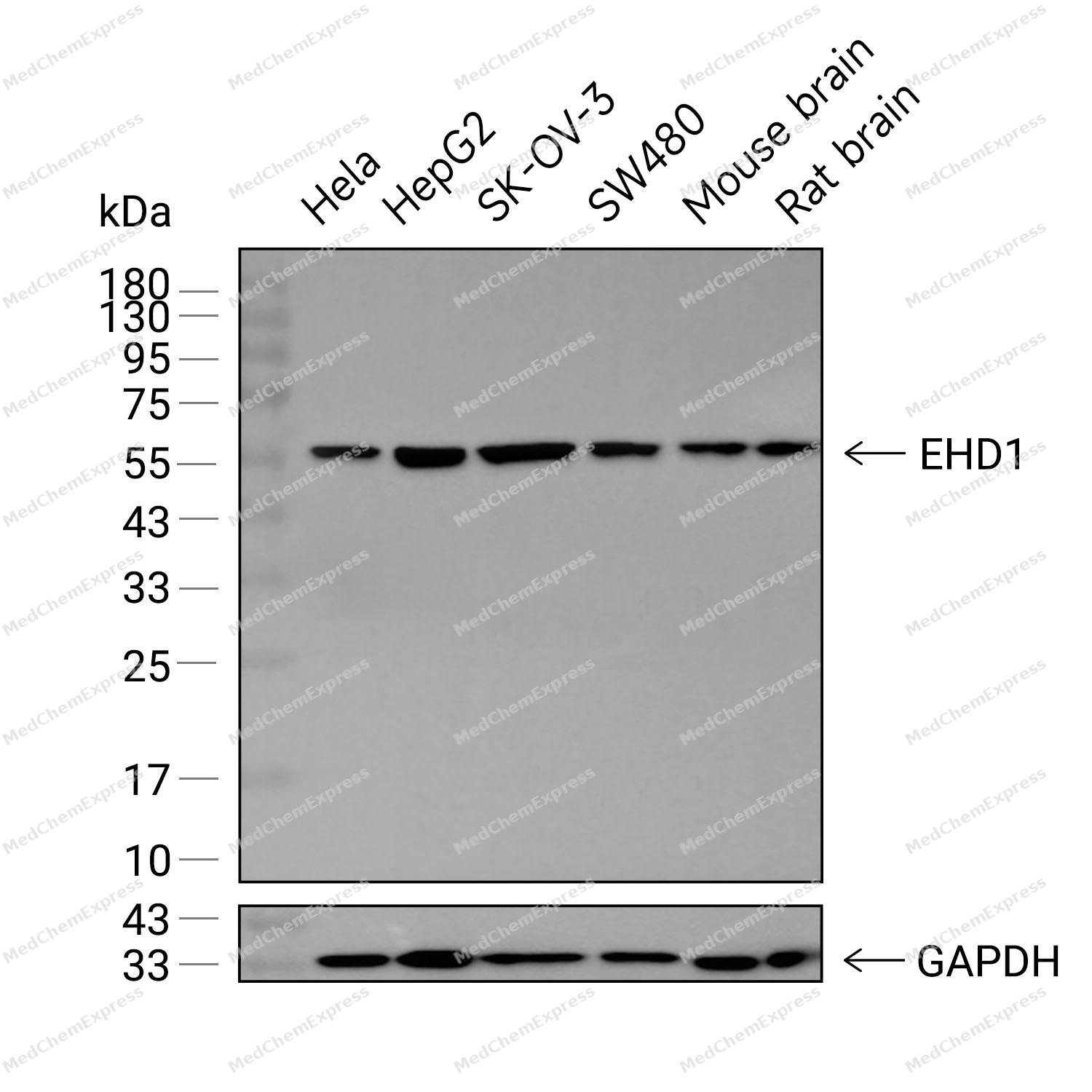 Western blot analysis was performed on extracts from Hela (lane 1, 15 μg), HepG2 (lane 2, 15 μg), SK-OV-3 (lane 3, 15 μg), SW480 (lane 4, 15 μg), Mouse brain (lane 5, 15 μg), and Rat brain (lane 6, 15 μg) using EHD1 Rabbit mAb. Proteins were transferred to a PVDF membrane and blocked with 5% non - fat milk in TBST at 4°C overnight. The primary antibody (1:1000 dilution) and the loading control antibody (GAPDH, HY-P80137, 1:60000 dilution) were incubated in 5% non-fat milk in TBST for 1 hour at 37°C. Goat Anti - Rabbit IgG - HRP Secondary Antibody (1:20000 dilution) was then applied for 40 minutes at 37°C.
 -
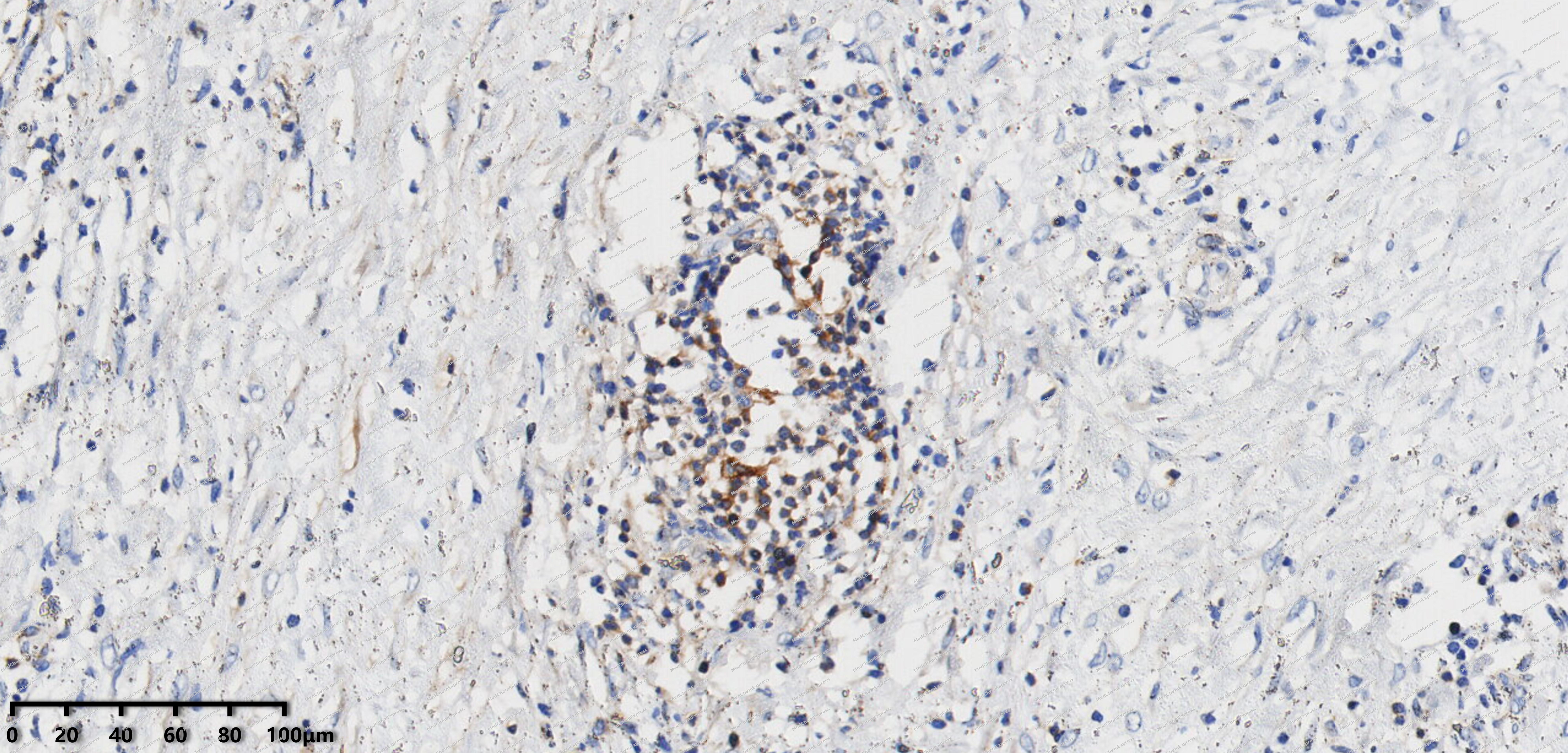 Immunohistochemical analysis of paraffin-embedded human lymphoma tissue using EHD1 antibody was performed. The section was pretreated using high-temperature mediated EDTA antigen retrieval buffer (pH 9.0), for 20 minutes. The tissues were incubated with primary antibody (HY-P82225, 1:200 dilution) at room temperature for 20 minutes. Detection was performed using an HRP conjugated compact polymer system. DAB was used as the chromogen. The tissues were counterstained with hematoxylin and mounted with neutral balsam mounting medium.
 -
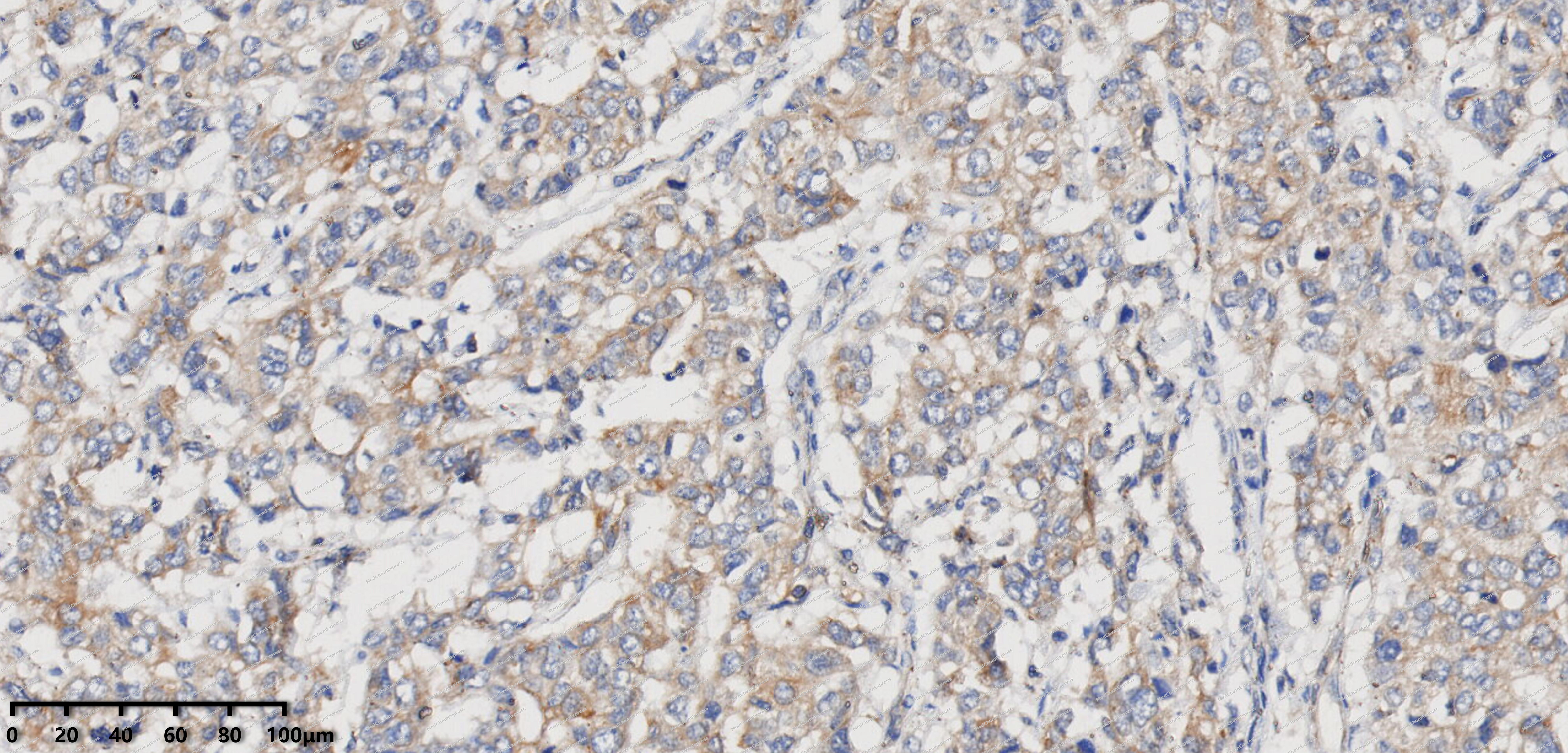 Immunohistochemical analysis of paraffin-embedded human breast cancer tissue using EHD1 antibody was performed. The section was pretreated using high-temperature mediated EDTA antigen retrieval buffer (pH 9.0), for 20 minutes. The tissues were incubated with primary antibody (HY-P82225, 1:200 dilution) at room temperature for 20 minutes. Detection was performed using an HRP conjugated compact polymer system. DAB was used as the chromogen. The tissues were counterstained with hematoxylin and mounted with neutral balsam mounting medium.
 -
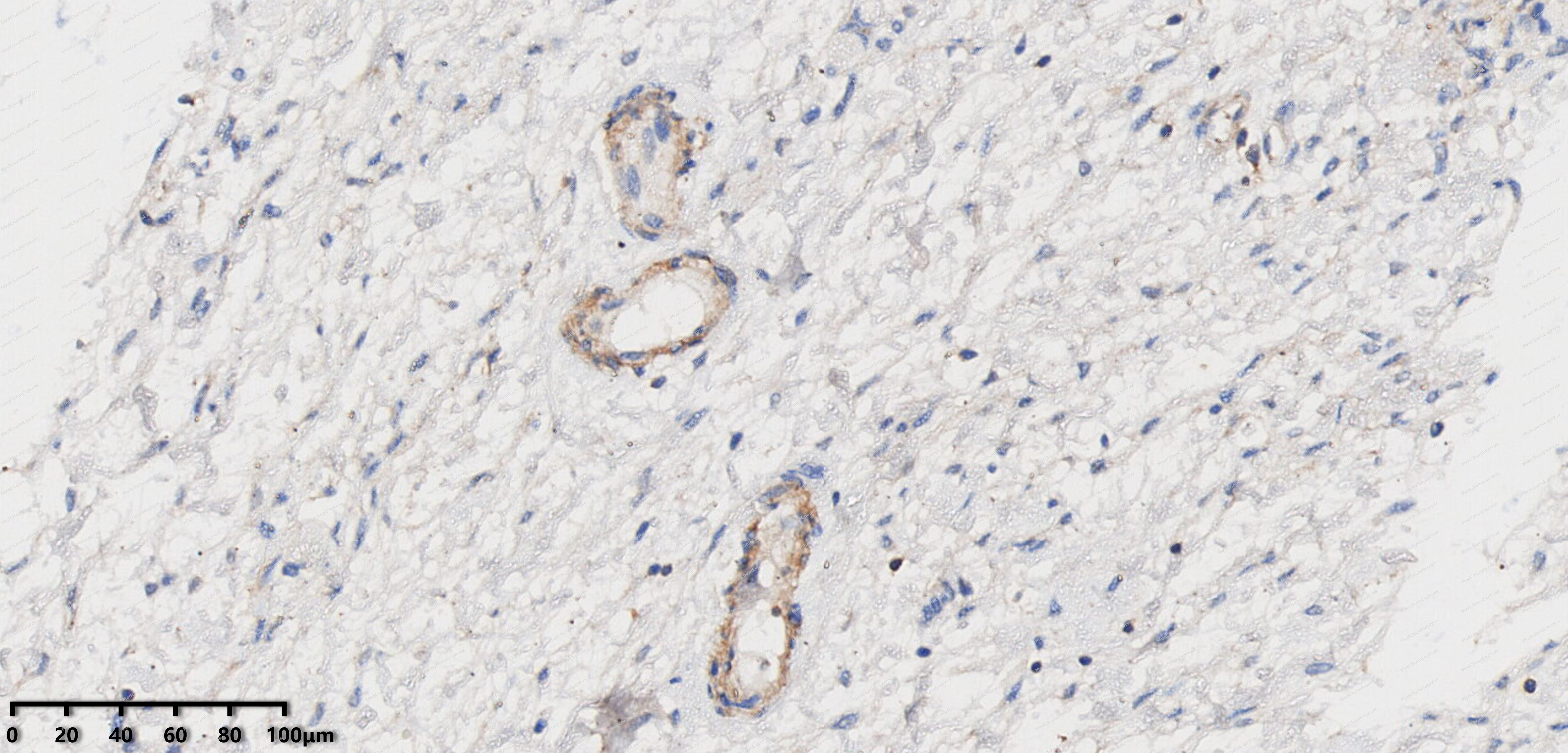 Immunohistochemical analysis of paraffin-embedded human uterine appendages tissue using EHD1 antibody was performed. The section was pretreated using high-temperature mediated EDTA antigen retrieval buffer (pH 9.0), for 20 minutes. The tissues were incubated with primary antibody (HY-P82225, 1:200 dilution) at room temperature for 20 minutes. Detection was performed using an HRP conjugated compact polymer system. DAB was used as the chromogen. The tissues were counterstained with hematoxylin and mounted with neutral balsam mounting medium.
 -
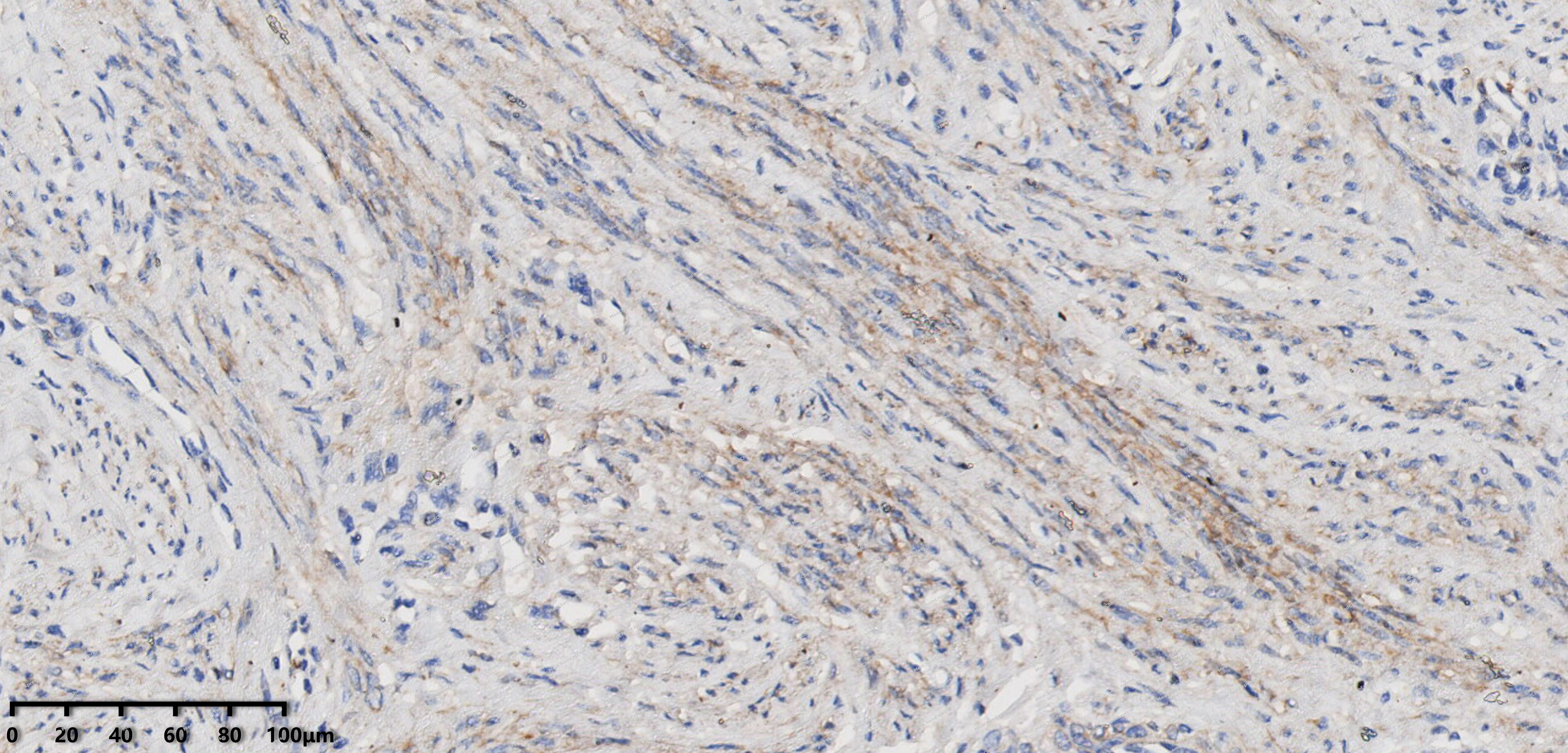 Immunohistochemical analysis of paraffin-embedded human cervix of uterus tissue using EHD1 antibody was performed. The section was pretreated using high-temperature mediated EDTA antigen retrieval buffer (pH 9.0), for 20 minutes. The tissues were incubated with primary antibody (HY-P82225, 1:200 dilution) at room temperature for 20 minutes. Detection was performed using an HRP conjugated compact polymer system. DAB was used as the chromogen. The tissues were counterstained with hematoxylin and mounted with neutral balsam mounting medium.
 -
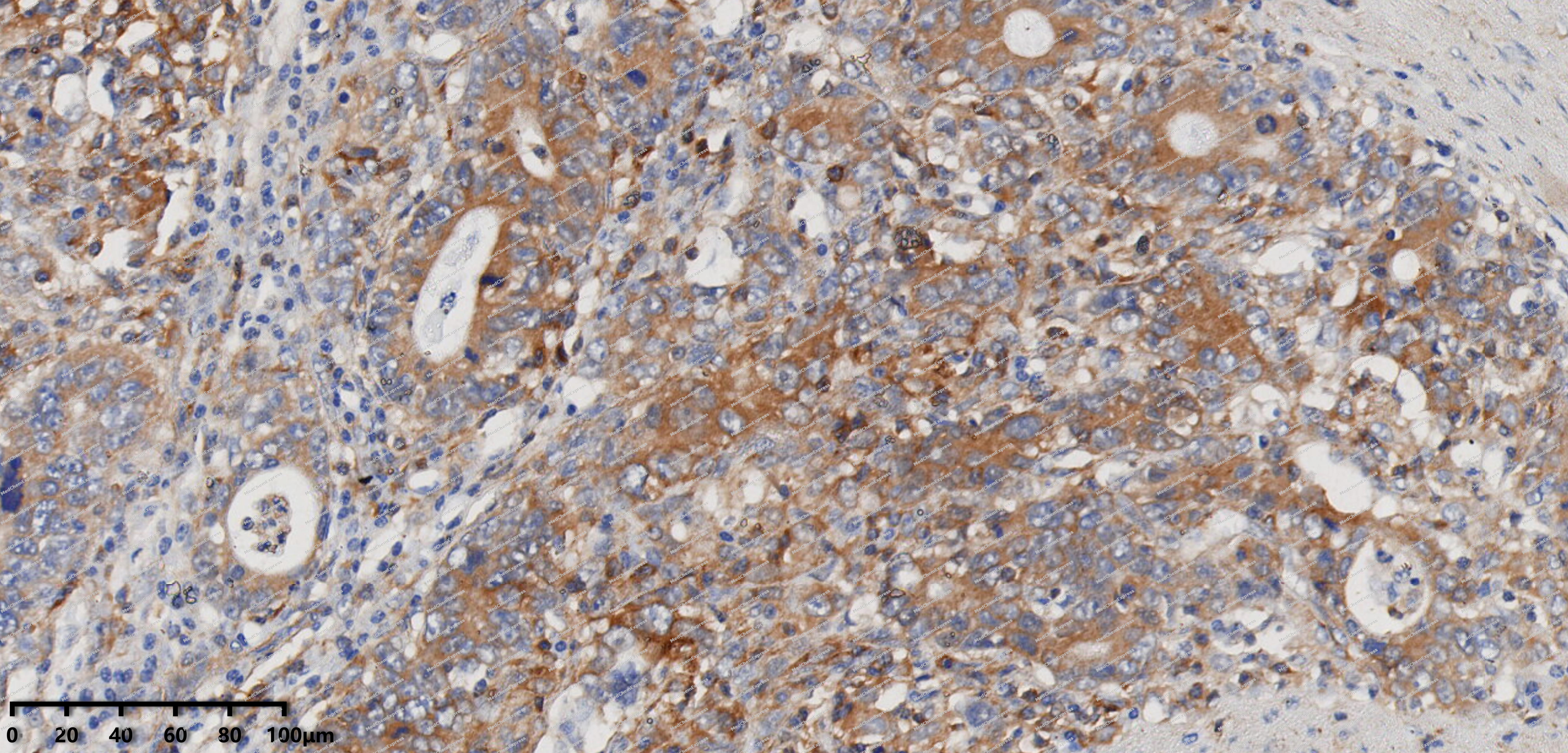 Immunohistochemical analysis of paraffin-embedded human gastric cancer tissue using EHD1 antibody was performed. The section was pretreated using high-temperature mediated EDTA antigen retrieval buffer (pH 9.0), for 20 minutes. The tissues were incubated with primary antibody (HY-P82225, 1:200 dilution) at room temperature for 20 minutes. Detection was performed using an HRP conjugated compact polymer system. DAB was used as the chromogen. The tissues were counterstained with hematoxylin and mounted with neutral balsam mounting medium.
 -
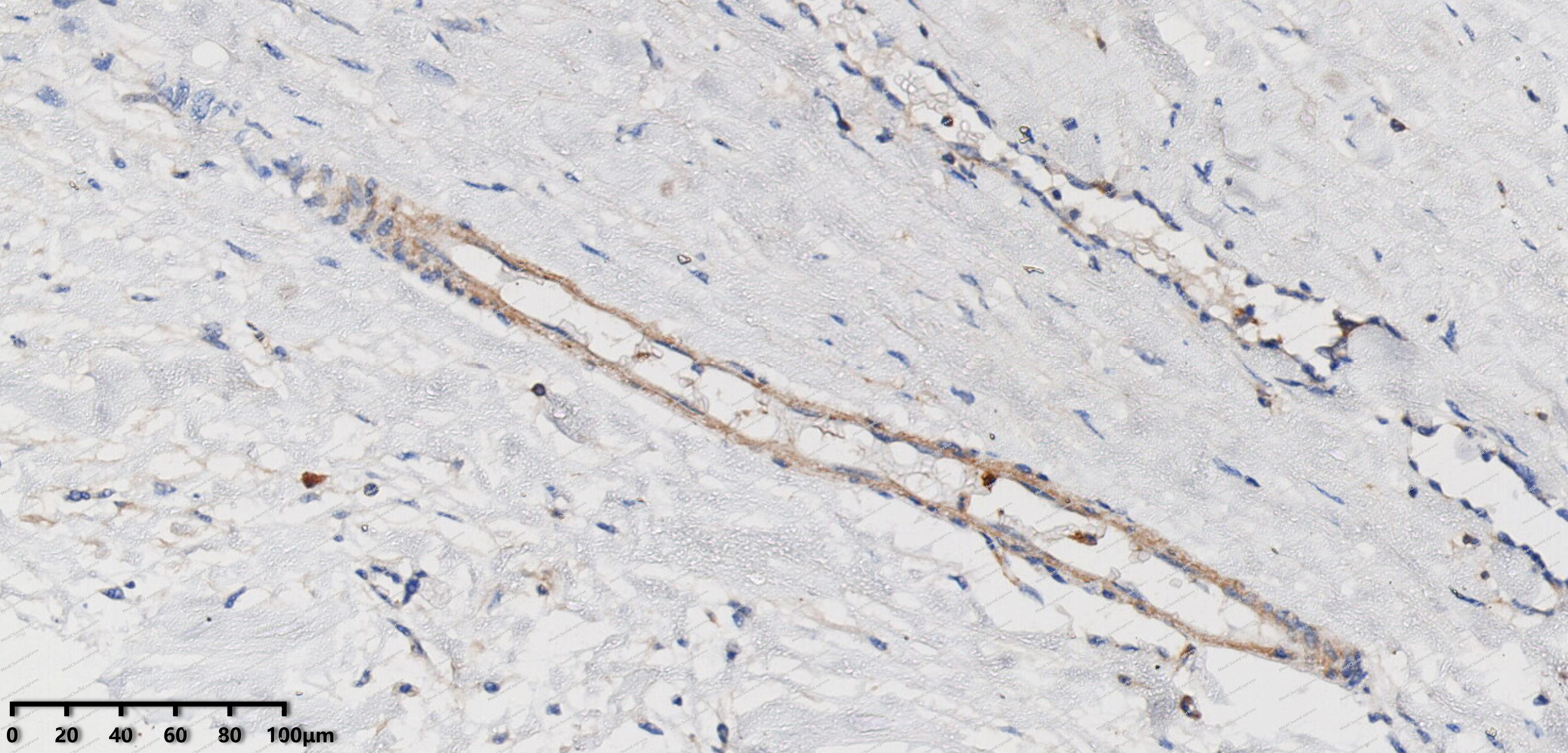 Immunohistochemical analysis of paraffin-embedded human mesothelioma tissue using EHD1 antibody was performed. The section was pretreated using high-temperature mediated EDTA antigen retrieval buffer (pH 9.0), for 20 minutes. The tissues were incubated with primary antibody (HY-P82225, 1:200 dilution) at room temperature for 20 minutes. Detection was performed using an HRP conjugated compact polymer system. DAB was used as the chromogen. The tissues were counterstained with hematoxylin and mounted with neutral balsam mounting medium.
 -
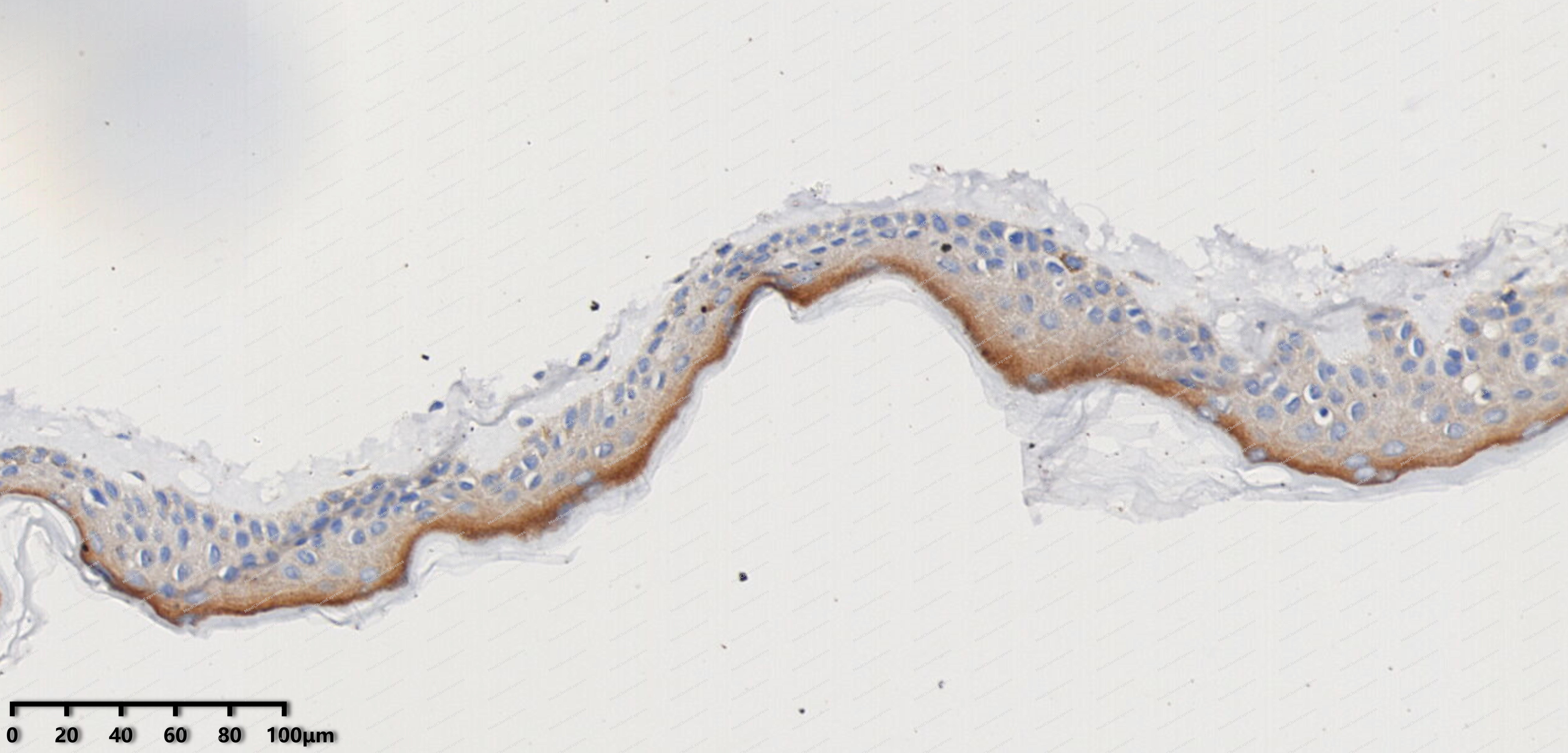 Immunohistochemical analysis of paraffin-embedded human melanoma tissue using EHD1 antibody was performed. The section was pretreated using high-temperature mediated EDTA antigen retrieval buffer (pH 9.0), for 20 minutes. The tissues were incubated with primary antibody (HY-P82225, 1:200 dilution) at room temperature for 20 minutes. Detection was performed using an HRP conjugated compact polymer system. DAB was used as the chromogen. The tissues were counterstained with hematoxylin and mounted with neutral balsam mounting medium.
 -
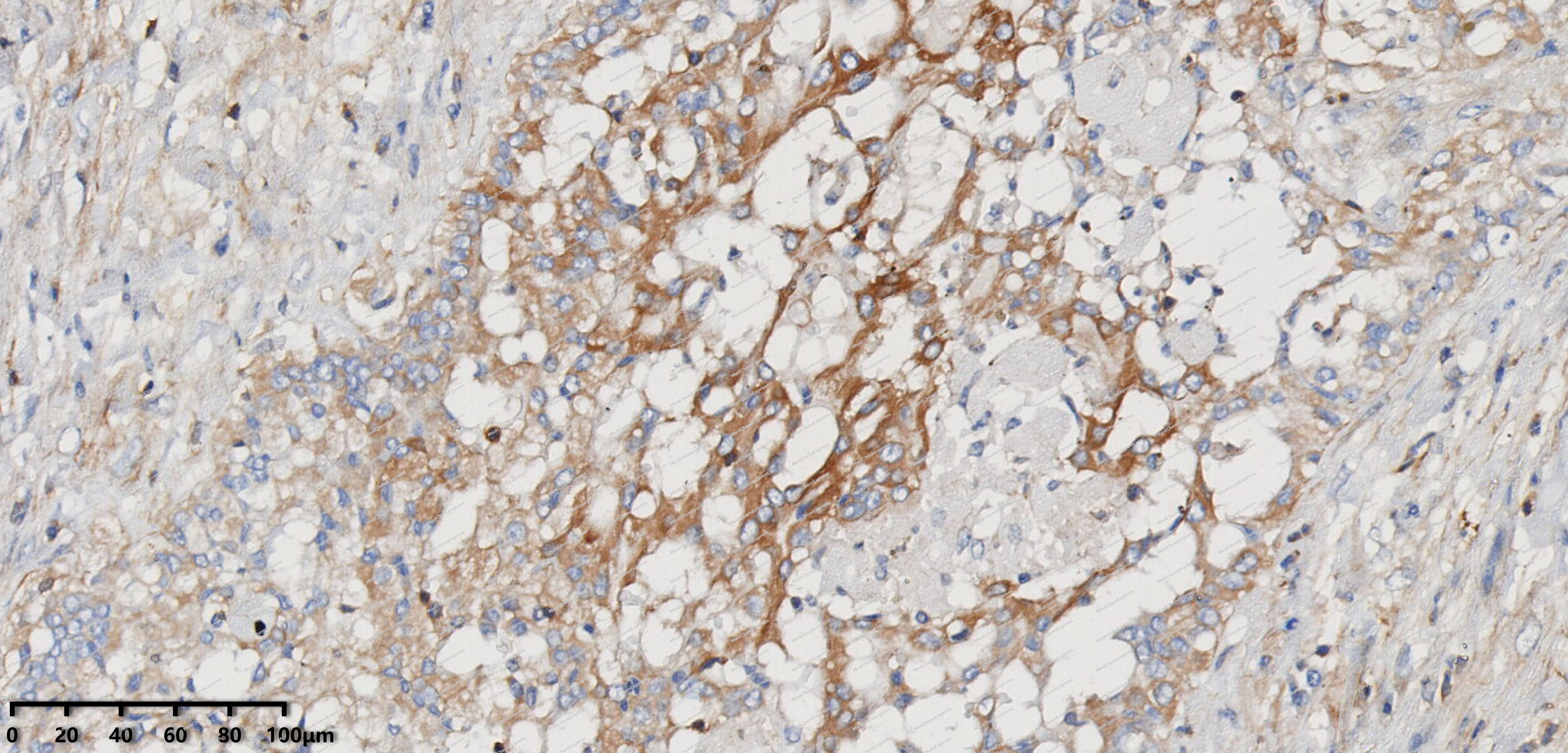 Immunohistochemical analysis of paraffin-embedded human embryonal carcinoma tissue using EHD1 antibody was performed. The section was pretreated using high-temperature mediated EDTA antigen retrieval buffer (pH 9.0), for 20 minutes. The tissues were incubated with primary antibody (HY-P82225, 1:200 dilution) at room temperature for 20 minutes. Detection was performed using an HRP conjugated compact polymer system. DAB was used as the chromogen. The tissues were counterstained with hematoxylin and mounted with neutral balsam mounting medium.
 -
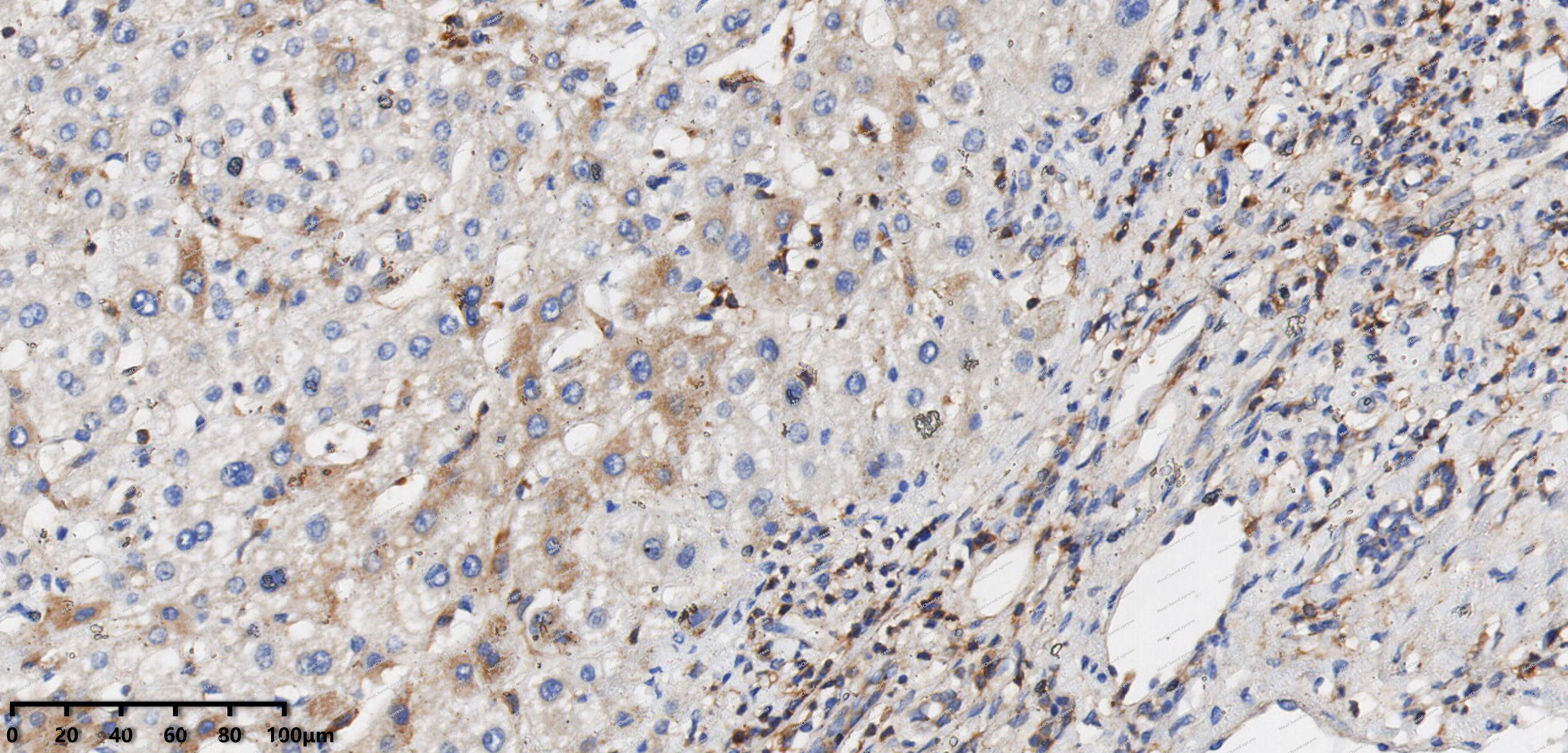 Immunohistochemical analysis of paraffin-embedded human Hepatocellular Carcinoma tissue using EHD1 antibody was performed. The section was pretreated using high-temperature mediated EDTA antigen retrieval buffer (pH 9.0), for 20 minutes. The tissues were incubated with primary antibody (HY-P82225, 1:200 dilution) at room temperature for 20 minutes. Detection was performed using an HRP conjugated compact polymer system. DAB was used as the chromogen. The tissues were counterstained with hematoxylin and mounted with neutral balsam mounting medium.
 -
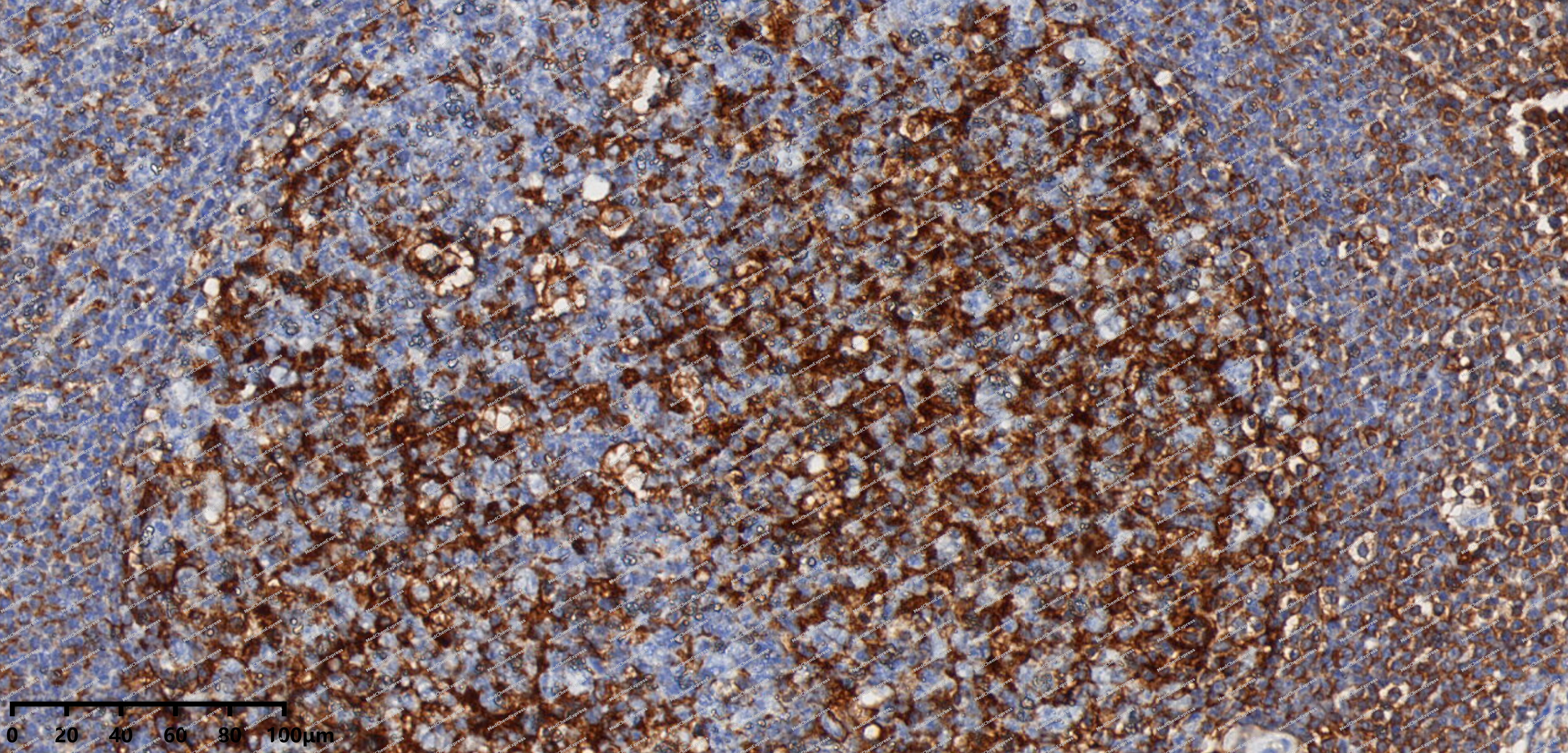 Immunohistochemical analysis of paraffin-embedded human Kimura's disease tissue using EHD1 antibody was performed. The section was pretreated using high-temperature mediated EDTA antigen retrieval buffer (pH 9.0), for 20 minutes. The tissues were incubated with primary antibody (HY-P82225, 1:200 dilution) at room temperature for 20 minutes. Detection was performed using an HRP conjugated compact polymer system. DAB was used as the chromogen. The tissues were counterstained with hematoxylin and mounted with neutral balsam mounting medium.
 -
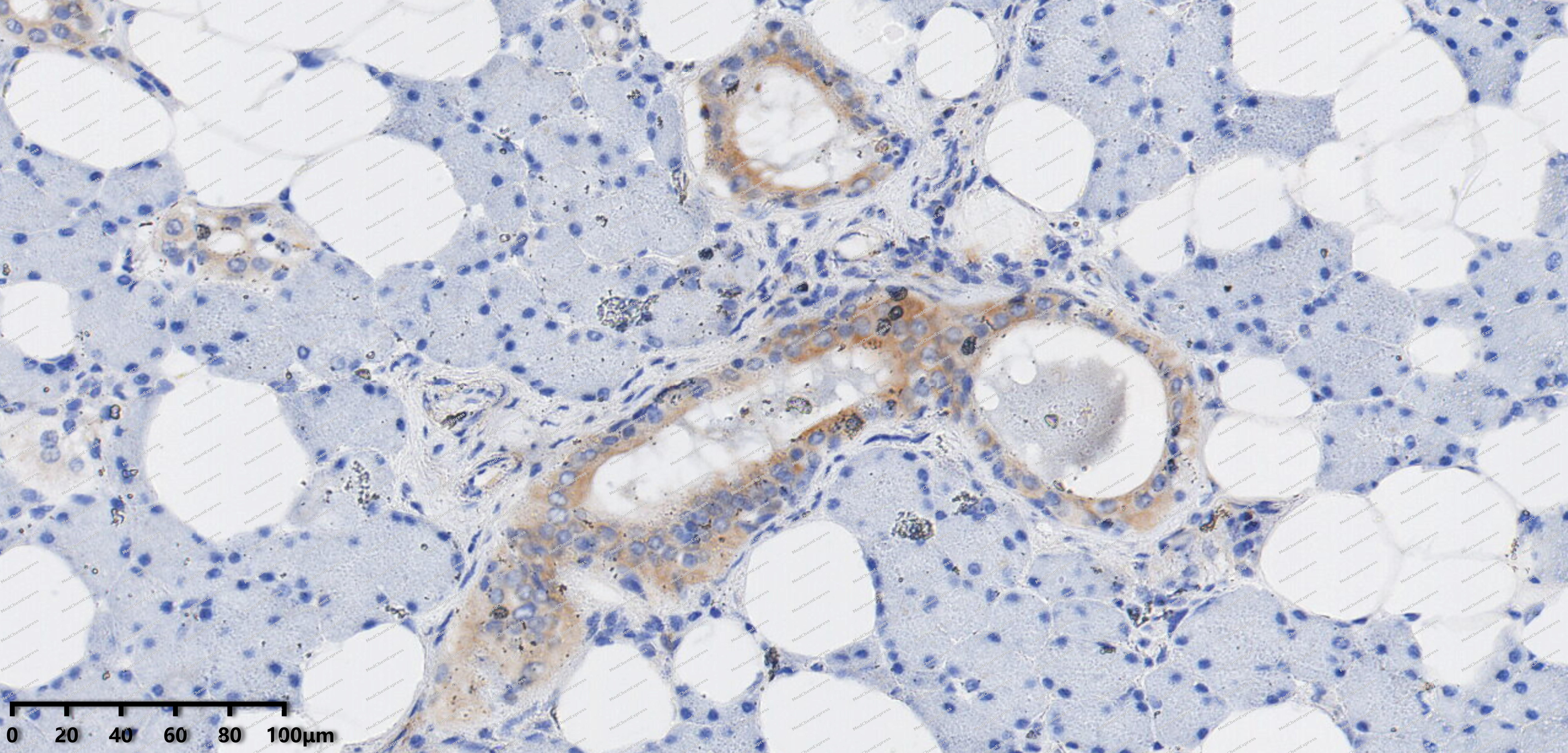 Immunohistochemical analysis of paraffin-embedded human adenolymphoma tissue using EHD1 antibody was performed. The section was pretreated using high-temperature mediated EDTA antigen retrieval buffer (pH 9.0), for 20 minutes. The tissues were incubated with primary antibody (HY-P82225, 1:200 dilution) at room temperature for 20 minutes. Detection was performed using an HRP conjugated compact polymer system. DAB was used as the chromogen. The tissues were counterstained with hematoxylin and mounted with neutral balsam mounting medium.
 -
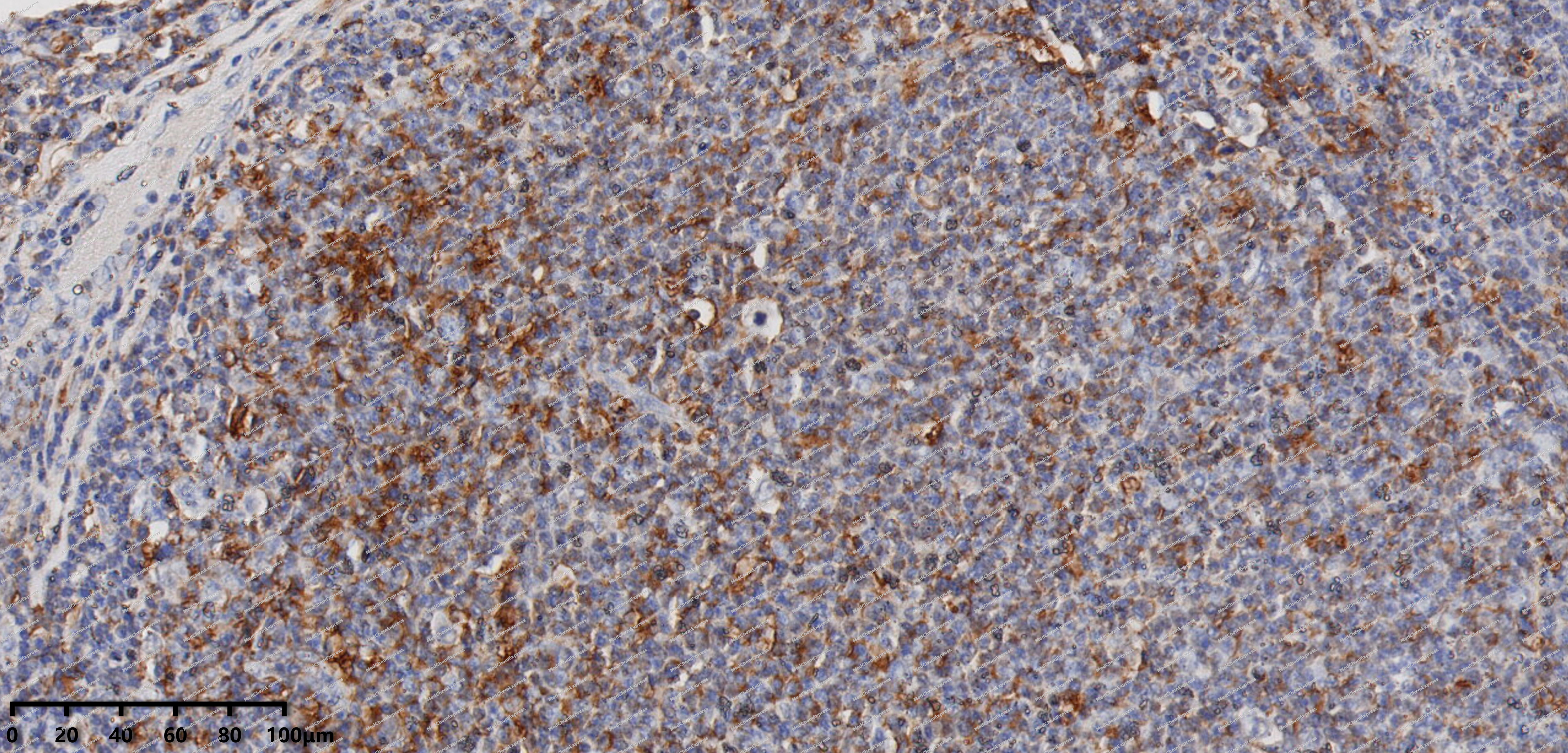 Immunohistochemical analysis of paraffin-embedded human Follicular Lymphoma tissue using EHD1 antibody was performed. The section was pretreated using high-temperature mediated EDTA antigen retrieval buffer (pH 9.0), for 20 minutes. The tissues were incubated with primary antibody (HY-P82225, 1:200 dilution) at room temperature for 20 minutes. Detection was performed using an HRP conjugated compact polymer system. DAB was used as the chromogen. The tissues were counterstained with hematoxylin and mounted with neutral balsam mounting medium.


Background
-
Function
EHD1 (Eps15 homology domain-containing 1) is a dynamin-like ATPase involved in endocytic trafficking and membrane protein recycling[1][2]. Mechanistically, EHD1 regulates endosome-mediated internalization and transcytosis, linking vesicular transport to the Wnt4/β-catenin signaling pathway in human endometrial stromal cells[3]. In disease models, EHD1 overexpression impairs decidualization and alters expression of prolactin and insulin-like growth factor binding protein-1, demonstrating relevance to recurrent implantation failure[3]. Compared with related isoforms, such as EHD3 and EHD4, EHD1 exhibits distinct cellular localization and specific regulatory effects on Wnt4/β-catenin signaling during embryo implantation[1][3]. EHD1’s ATPase activity and endocytic function provide opportunities for experimental modulation using adenoviral overexpression, while pathway-specific agonists like Wnt4 can restore impaired cellular responses[3]. These properties make EHD1 a practical target for investigating membrane trafficking dynamics, isoform-specific signaling, and the functional consequences of endocytic regulation in reproductive biology[1][3].
-
Subcellular Localization
Recycling endosome membrane; Peripheral membrane protein; Cytoplasmic side; Early endosome membrane; Peripheral membrane protein; Cytoplasmic side; Cell membrane; Peripheral membrane protein; Cytoplasmic side; Cell projection, cilium membrane; Peripheral membrane protein; Cytoplasmic side
-
Expression
Tissue_specificity:Highly expressed in testis -
Subunit
Homooligomer, and heterooligomer with EHD2, EHD3 and EHD4, ATP-binding is required for heterooligomerization (PubMed:16251358, PubMed:17233914, PubMed:18331452, PubMed:31615969). Interacts (via EH domain) with MICALL1 (via NPF1 motif); the interaction is direct and recruits EHD1 to membranes (PubMed:19864458, PubMed:20106972). Interacts with RAB35; the interaction is indirect through MICALL1 and recruits EHD1 to membranes (By similarity). Interacts (via EH domain) with PACSIN2 (via NPF motifs); regulates localization to tubular recycling endosome membranes (PubMed:23596323). Interacts with PACSIN1 (By similarity). Interacts with RAB8A (PubMed:19864458). Interacts with FER1L5 (via second C2 domain) (By similarity). Interacts with MYOF (By similarity). Interacts with ZFYVE20 (PubMed:15020713). Interacts (via EH domain) with RAB11FIP2 (PubMed:16251358)
-
SwissProt ID
-
Synonyms
EHD1; H PAST; hPAST1; PAST; PAST homolog 1; PAST1; Tilin
-
Research Field
Cardiovascular


Documentation
-
Data Sheet (262 KB)
-
SDS (251 KB)
- English - EN (251 KB)
- Français - FR (251 KB)
- Deutsch - DE (251 KB)
- Norwegian - NO (251 KB)
- Español - ES (251 KB)
- Swedish - SV (251 KB)
- Italian - IT (251 KB)
- Korean - KR (251 KB)
- Portuguese - PT (251 KB)
-
User Guide for Antibodies (1077 KB)
References
[1]. Gudmundsson H, et al. EH domain proteins regulate cardiac membrane protein targeting. Circ Res. 2010 Jul 9;107(1):84-95. [Content Brief]
[2]. Hermans A, et al. A 3D-Printed and Freely Available Device to Measure the Zebrafish Optokinetic Response Before and After Injury. Zebrafish. 2024 Apr;21(2):144-148. [Content Brief]
[3]. Zhou Q, et al. EHD1 impairs decidualization by regulating the Wnt4/β-catenin signaling pathway in recurrent implantation failure. EBioMedicine. 2019 Dec;50:343-354. [Content Brief]

 Powered by Bioz
