



HDAC5 Antibody (YA6590)
(Synonyms: Antigen NY CO 9 antibody; Antigen NY-CO-9 antibody; HD5 antibody; HDAC 5 antibody; HDAC5 antibody; HDAC5_HUMAN antibody; Histone deacetylase 5 antibody; NY CO 9 antibody; )HDAC5 Antibody (YA6590) is a Rabbit-derived and non-conjugated IgG monoclonal antibody, targeting to HDAC5.
-
Host:
Rabbit
-
Isotype:
IgG
-
Application:
IHC-P, ICC/IF, IP, ChIP
-
Reactivity :
Human, Mouse, Rat
-
Formulation:
Supplied in PBS (pH7.4), 0.1% BSA, 40% Glycerol. Preservative: 0.05% Sodium Azide.
-
Conjugation:
Non-conjugated


To place orders, for customer services and technical support, please contact: MedChemExpress USA
Tel: 609-228-6898 E-mail: [email protected] [email protected]





Applications
| Application |
IHC-P

IHC-P: Immunohistochemistry-Paraffin
|
ICC/IF
ICC/IF: Immunocytochemistry/
Immunofluorescence |
IP
IP: Immunoprecipitation
|
ChIP
ChIP: Chromatin Immunoprecipitation
|
|---|---|---|---|---|
| Dilution Ratio | 1:1000 | 1:100 | 1-2μg/sample | Use 0.5~2 μg for 25 μg of chromatin. |


Product Details
HDAC5 Antibody (YA6590) is a Rabbit-derived and non-conjugated IgG monoclonal antibody, targeting to HDAC5.
-
Host Rabbit

-
Clonality Recombinant,Monoclonal

-
Species ReactivityHuman, Mouse, Rat
-
Calculated Molecular Weight Predicted band size: 122 kDa

Synthetic peptide within human HDAC5 aa 171-220.
affinity purified.
Non-conjugated
Unmodified
IgG


Product Properties
-
Appearance
Solution
-
Formulation
Supplied in PBS (pH7.4), 0.1% BSA, 40% Glycerol. Preservative: 0.05% Sodium Azide.
-
Storage & Stability
Stored at -20°C for 1 year. Avoid repeated freeze / thaw cycles.
-
Shipping
Shipping with blue ice.


Verification Images
-
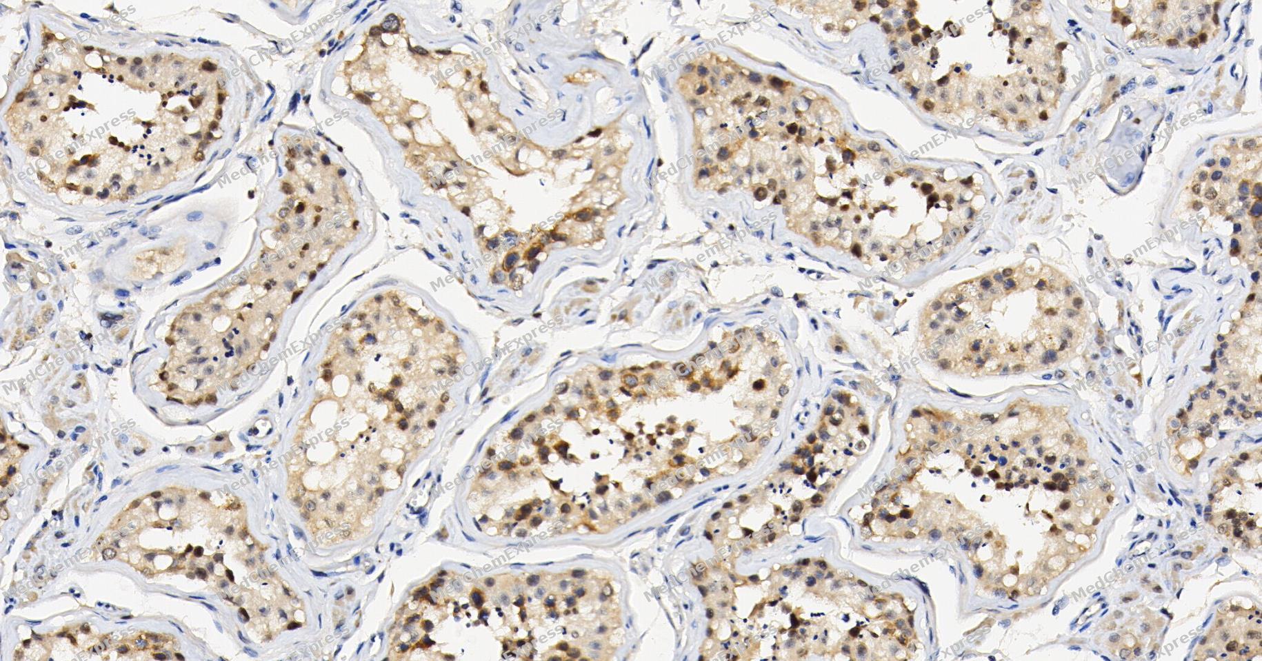 Immunohistochemical analysis of paraffin-embedded human testis tissue using HDAC5 Antibody (YA6590). The section was pre-treated using heat mediated antigen retrieval with Tris/EDTA buffer (pH 9.0) for 20 minutes. The tissues were probed with the primary antibody (HY-P86897, 1/200) overnight at 4℃. The detection was performed using an HRP conjugated compact polymer system. DAB was used as the chromogen. Tissues were counterstained with hematoxylin and mounted with neutral balsam.
 -
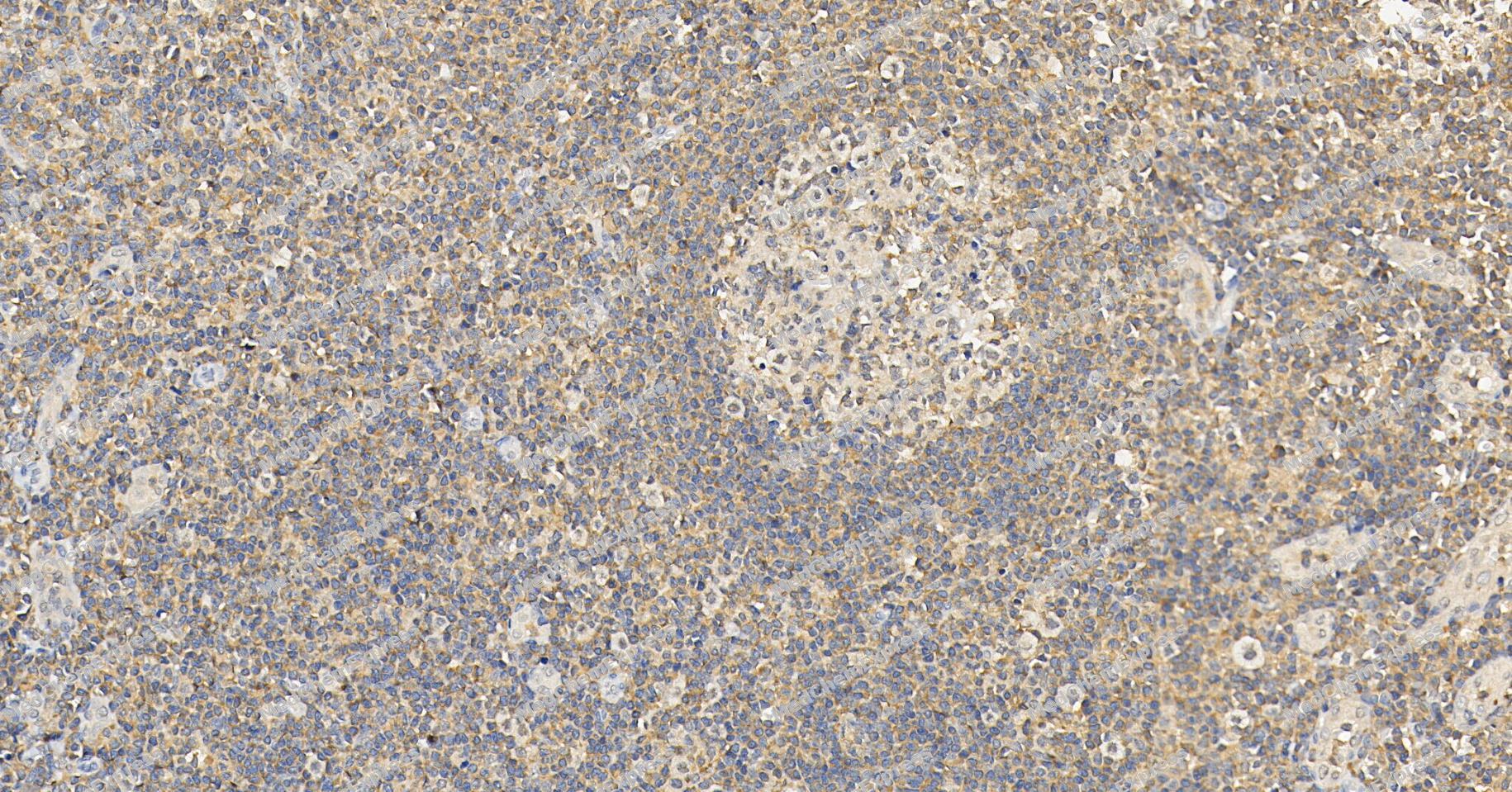 Immunohistochemical analysis of paraffin-embedded human tonsil tissue using HDAC5 Antibody (YA6590). The section was pre-treated using heat mediated antigen retrieval with Tris/EDTA buffer (pH 9.0) for 20 minutes. The tissues were probed with the primary antibody (HY-P86897, 1/200) overnight at 4℃. The detection was performed using an HRP conjugated compact polymer system. DAB was used as the chromogen. Tissues were counterstained with hematoxylin and mounted with neutral balsam.
 -
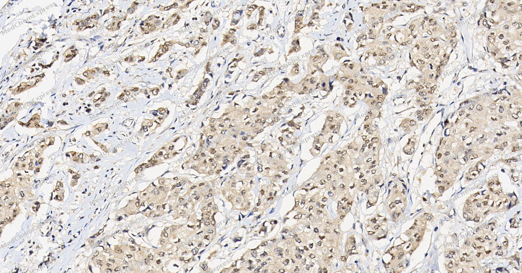 Immunohistochemical analysis of paraffin-embedded human breast cancer tissue using HDAC5 Antibody (YA6590). The section was pre-treated using heat mediated antigen retrieval with Tris/EDTA buffer (pH 9.0) for 20 minutes. The tissues were probed with the primary antibody (HY-P86897, 1/200) overnight at 4℃. The detection was performed using an HRP conjugated compact polymer system. DAB was used as the chromogen. Tissues were counterstained with hematoxylin and mounted with neutral balsam.
 -
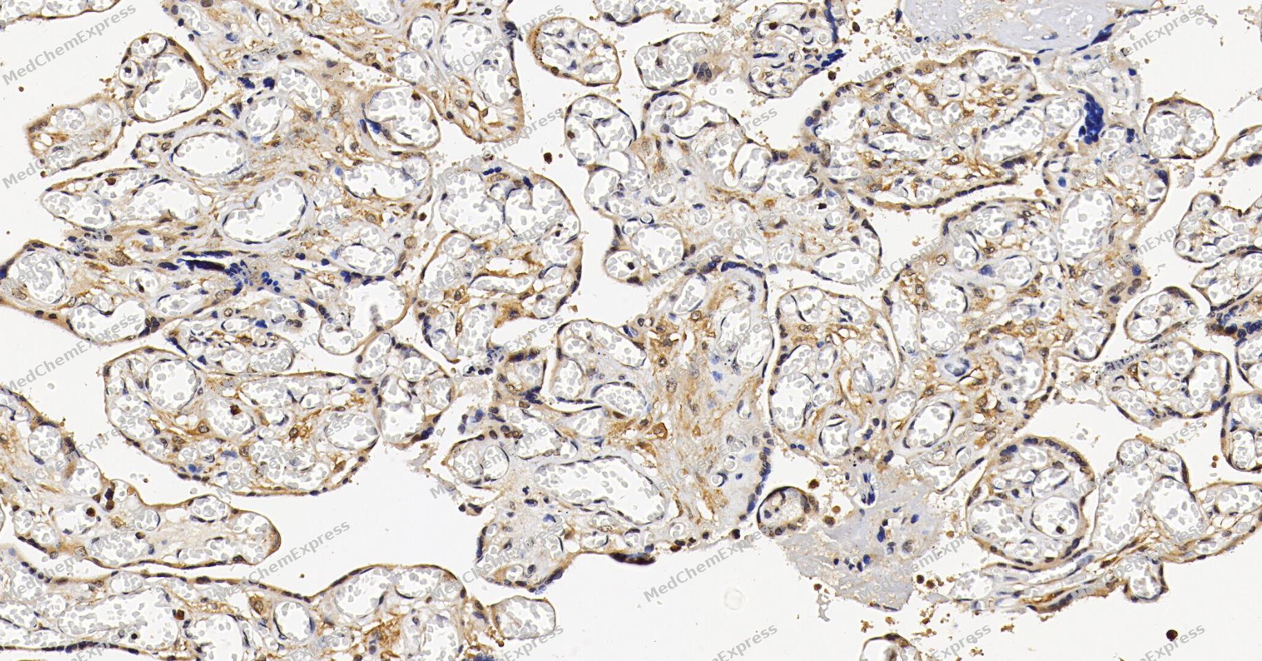 Immunohistochemical analysis of paraffin-embedded human placenta tissue using HDAC5 Antibody (YA6590). The section was pre-treated using heat mediated antigen retrieval with Tris/EDTA buffer (pH 9.0) for 20 minutes. The tissues were probed with the primary antibody (HY-P86897, 1/200) overnight at 4℃. The detection was performed using an HRP conjugated compact polymer system. DAB was used as the chromogen. Tissues were counterstained with hematoxylin and mounted with neutral balsam.
 -
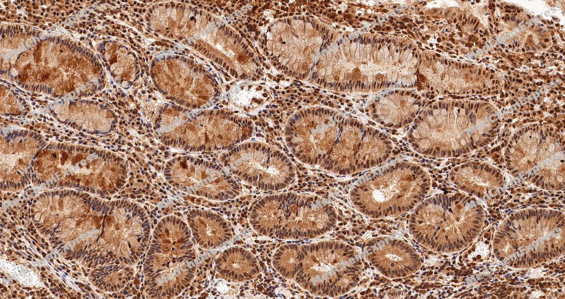 Immunohistochemical analysis of paraffin-embedded human colon tissue using HDAC5 Antibody (YA6590). The section was pre-treated using heat mediated antigen retrieval with Tris/EDTA buffer (pH 9.0) for 20 minutes. The tissues were probed with the primary antibody (HY-P86897, 1/200) overnight at 4℃. The detection was performed using an HRP conjugated compact polymer system. DAB was used as the chromogen. Tissues were counterstained with hematoxylin and mounted with neutral balsam.
 -
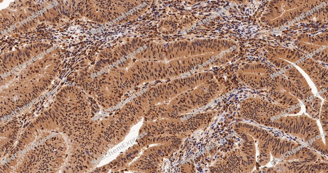 Immunohistochemical analysis of paraffin-embedded human colon cancer tissue using HDAC5 Antibody (YA6590). The section was pre-treated using heat mediated antigen retrieval with Tris/EDTA buffer (pH 9.0) for 20 minutes. The tissues were probed with the primary antibody (HY-P86897, 1/200) overnight at 4℃. The detection was performed using an HRP conjugated compact polymer system. DAB was used as the chromogen. Tissues were counterstained with hematoxylin and mounted with neutral balsam.


Background
-
Function
HDAC5 (histone deacetylase 5) is a member of the class IIa histone deacetylase family and functions as a signal-responsive transcriptional regulator through its conserved N-terminal transcription factor-binding domain and C-terminal catalytic domain[1][2]. HDAC5 interacts with transcription factors such as myocyte enhancer factor 2 (MEF2), and phosphorylation-dependent binding to 14-3-3 proteins promotes its nuclear export, thereby relieving transcriptional repression of target genes[1][2]. Mechanistically, HDAC5 participates in chromatin-dependent gene regulation and integrates extracellular signaling pathways that control cellular differentiation, survival, and tissue-specific transcriptional programs[2][3]. These properties place HDAC5 within a broader class IIa HDAC regulatory network that functions primarily through transcriptional repression complexes rather than strong intrinsic deacetylase activity[2]. In disease-related settings, aberrant HDAC5 expression has been reported across multiple cancer types, where it is associated with regulation of cell proliferation, invasion, stemness maintenance, immune-related processes, and cell-cycle progression[1]. Experimental studies summarized in cancer models further indicate that HDAC5 can influence oncogenic pathways including DLL4, Notch1, c-Met, and Six1 signaling, supporting its relevance as a molecular regulator of tumor biology[1]. Compared with related HDAC isoforms, HDAC5 shares the characteristic class IIa adaptor architecture and signal-dependent nucleocytoplasmic shuttling mechanism, yet retains distinct tissue distribution patterns and target-gene regulatory functions that contribute to biological specificity[1][2]. For experimental applications, HDAC5 is widely investigated as a candidate therapeutic target, and ongoing efforts to develop class IIa-selective HDAC modulators aim to improve isoform selectivity while minimizing off-target effects associated with pan-HDAC inhibition[2][3].
-
Subcellular Localization
Nucleus,Cytoplasm
-
Expression
Tissue_Specificity: Ubiquitous -
Isoforms & Post-Translational Modification
Q9UQL6 has three isomers: Q9UQL6-1: 121978 Da (predicted); Q9UQL6-2: 112191 Da (predicted); Q9UQL6-3: 122049 Da (predicted).
Phosphorylated by AMPK, CaMK1, SIK1 and PRKD1 at Ser-259 and Ser-498丨Ubiquitinated -
Subunit
Interacts with AHRR, BAHD1, BCOR, HDAC7, HDAC9, CTBP1, MEF2C, NCOR2, NRIP1, PHB2 and a 14-3-3 chaperone protein
-
SwissProt ID
-
Synonyms
Antigen NY CO 9 antibody; Antigen NY-CO-9 antibody; HD5 antibody; HDAC 5 antibody; HDAC5 antibody; HDAC5_HUMAN antibody; Histone deacetylase 5 antibody; NY CO 9 antibody;


Documentation
References
[1]. Yang J, et al. Insights Into the Function and Clinical Application of HDAC5 in Cancer Management. Front Oncol. 2021 Jun 10;11:661620. [Content Brief]
[2]. Park SY, et al. A short guide to histone deacetylases including recent progress on class II enzymes. Exp Mol Med. 2020 Feb;52(2):204-212. [Content Brief]
[3]. Ujlaky-Nagy L, et al. Disrupting EGFR-HER2 Transactivation by Pertuzumab in HER2-Positive Cancer: Quantitative Analysis Reveals EGFR Signal Input as Potential Predictor of Therapeutic Outcome. Int J Mol Sci. 2024 May 29;25(11):5978. [Content Brief]

 Powered by Bioz
