



HLA G Antibody (YA3225)
(Synonyms: HLA G antigen; MHC class I antigen G; HLA-G; HLA60; HLAG)
Based on 1 Customer Validation
HLA G Antibody (YA3225) is a Rabbit-derived and non-conjugated IgG monoclonal antibody, targeting to HLA G.
-
Host:
Rabbit
-
Isotype:
IgG
-
Application:
WB
-
Reactivity :
Human
-
Formulation:
Supplied in 10mM PBS, pH 7.4, 150mM sodium chloride, 0.05% BSA, 0.02% sodium azide and 50% glycerol.
-
Conjugation:
Non-conjugated


To place orders, for customer services and technical support, please contact: MedChemExpress USA
Tel: 609-228-6898 E-mail: [email protected] [email protected]





Applications
| Application |
WB

WB: Western Blot
|
|---|---|
| Dilution Ratio | 1:500-1:1000 |


Product Details
HLA G Antibody (YA3225) is a Rabbit-derived and non-conjugated IgG monoclonal antibody, targeting to HLA G.
-
Host Rabbit

-
Clonality Recombinant,Monoclonal

-
Species ReactivityHuman
-
Observed Molecular WeightObserved band size:45-60 kD
Note: Due to possible protein modifications or aggregation, the molecular weight should be confirmed by actual measurement, and the predicted value is for reference only.

A synthesized peptide derived from human HLA G aa200-265/338.
Endogenous
Affinity Chromatography
Non-conjugated
Unmodified
IgG


Product Properties
-
Appearance
Solution
-
Formulation
Supplied in 10mM PBS, pH 7.4, 150mM sodium chloride, 0.05% BSA, 0.02% sodium azide and 50% glycerol.
-
Concentration
Batch-dependent, Please check the COA for the concentration of each lot. Check Lot Concentration
-
Storage & Stability
Stored at -20°C for 1 year. Avoid repeated freeze / thaw cycles.
-
Shipping
Shipping with blue ice.


Verification Images
-
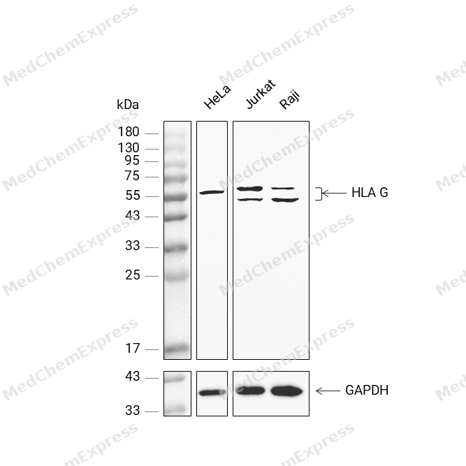 Western blot analysis was performed on protein extracts (40 μg) from HeLa (lane 2), Jurkat (lane 3), and Raji (lane 4) using HLA G antibody. Proteins were transferred onto a 0.45 μm PVDF membrane using the Trans-Blot® Turbo™ system for 13 min. The membrane was then blocked with 5% nonfat milk in TBST (HY-K1025) for 1 h at room temperature. The primary antibody (1:1000) and loading control antibody GAPDH Antibody (HRP) (HY-P80954A) (1:5000) were diluted in 5% nonfat milk in TBST and incubated with the membrane overnight at 4°C. After washing, the membrane of primary antibody was incubated with HRP-conjugated goat anti-rabbit/mouse IgG secondary antibody (HY-P8001/HY-P8004) (1:5000) diluted in 5% nonfat milk in TBST for 1 h at room temperature. Protein bands were visualized using an Ultra High Sensitivity ECL detection kit (HY-K1005).


Background
-
Function
HLA G is a Non-classical major histocompatibility class Ib molecule involved in immune regulatory processes at the maternal-fetal interface. In complex with B2M/beta-2 microglobulin binds a limited repertoire of nonamer self-peptides derived from intracellular proteins including histones and ribosomal proteins. Peptide-bound HLA-G-B2M complex acts as a ligand for inhibitory/activating KIR2DL4, LILRB1 and LILRB2 receptors on uterine immune cells to promote fetal development while maintaining maternal-fetal tolerance. Upon interaction with KIR2DL4 and LILRB1 receptors on decidual NK cells, it triggers NK cell senescence-associated secretory phenotype as a molecular switch to promote vascular remodeling and fetal growth in early pregnancy. Through interaction with KIR2DL4 receptor on decidual macrophages induces pro-inflammatory cytokine production mainly associated with tissue remodeling. Through interaction with LILRB2 receptor triggers differentiation of type 1 regulatory T cells and myeloid-derived suppressor cells, both of which actively maintain maternal-fetal tolerance. May play a role in balancing tolerance and antiviral-immunity at maternal-fetal interface by keeping in check the effector functions of NK, CD8+ T cells and B cells. Reprograms B cells toward an immune suppressive phenotype via LILRB1. May induce immune activation/suppression via intercellular membrane transfer (trogocytosis), likely enabling interaction with KIR2DL4, which resides mostly in endosomes. Through interaction with the inhibitory receptor CD160 on endothelial cells may control angiogenesis in immune privileged sites; Likely does not bind B2M and presents peptides. Negatively regulates NK cell- and CD8+ T cell-mediated cytotoxicity; Likely does not bind B2M and presents peptides. Negatively regulates NK cell- and CD8+ T cell-mediated cytotoxicity; Likely does not bind B2M and presents peptides. Negatively regulates NK cell- and CD8+ T cell-mediated cytotoxicity; Non-classical major histocompatibility class Ib molecule involved in immune regulatory processes at the maternal-fetal interface. In complex with B2M/beta-2 microglobulin binds a limited repertoire of nonamer self-peptides derived from intracellular proteins including histones and ribosomal proteins. Peptide-bound HLA-G-B2M complex acts as a ligand for inhibitory/activating KIR2DL4, LILRB1 and LILRB2 receptors on uterine immune cells to promote fetal development while maintaining maternal-fetal tolerance. Upon interaction with KIR2DL4 and LILRB1 receptors on decidual NK cells, it triggers NK cell senescence-associated secretory phenotype as a molecular switch to promote vascular remodeling and fetal growth in early pregnancy. Through interaction with KIR2DL4 receptor on decidual macrophages induces pro-inflammatory cytokine production mainly associated with tissue remodeling. Through interaction with LILRB2 receptor triggers differentiation of type 1 regulatory T cells and myeloid-derived suppressor cells, both of which actively maintain maternal-fetal tolerance. Reprograms B cells toward an immune suppressive phenotype via LILRB1; Likely does not bind B2M and presents peptides; Likely does not bind B2M and presents peptides[1][2][3][4][5][6][7][8][9][10][11][12][13][14].
-
Subcellular Localization
Cell membrane; Single-pass type I membrane protein; Endoplasmic reticulum membrane; Early endosome membrane; Secreted; Cell membrane; Single-pass type I membrane protein; Cell membrane; Single-pass type I membrane protein; Cell membrane; Single-pass type I membrane protein; Secreted; Early endosome; Secreted; Secreted; Cell projection, filopodium membrane
-
Expression
Tissue_specificity:Expressed in adult eye (PubMed:1570318) . Expressed in immune cell subsets including monocytes, myeloid and plasmacytoid dendritic cells and regulatory T cells (Tr1) (at protein level) (PubMed:20448110) . Secreted by follicular dendritic cell and follicular helper T cells (PubMed:24453251) ; Detected in physiological fluids including amniotic fluid and serum; Expressed in placenta, amniotic membrane, skin, cord blood and peripheral blood mononuclear cells
Induction:Up-regulated by immunosuppressive cytokine IL10 on dendritic cells and CD4+ T cells (PubMed:20448110) . Up-regulated by progesterone in cytotrophoblasts (PubMed:16210391) -
Isoforms & Post-Translational Modification
P17693 has 7 isomers: P17693-1: 38224 Da (predicted); P17693-2: 27680 Da (predicted); P17693-3: 17070 Da (predicted); P17693-4: 27613 Da (predicted); P17693-5: 36458 Da (predicted); P17693-6: 25928 Da (predicted); P17693-7: 13286 Da (predicted).
N-glycosylated;Produced by proteolytic cleavage at the cell surface (shedding) by matrix metalloproteinase MMP2 -
Subunit
Forms a heterotrimer with B2M and a self-peptide (peptide-bound HLA-G-B2M) (PubMed:7584149, PubMed:8805247). HLA-G-B2M complex interacts with components of the antigen processing machinery TAPBP and TAP1-TAP2 complex; this interaction is required for loading of high affinity peptides and heterotrimer translocation to the cell surface (PubMed:7584149). Interacts with CALCR; this interaction is required for appropriate folding (PubMed:9640257). Interacts with COPB1; this interaction mediates the endoplasmic reticulum (ER) retrieval of HLA-G-B2M complexes that bind low affinity peptides (PubMed:11520457, PubMed:12582157). On the cell surface, peptide-bound HLA-G-B2M molecules (referred to as monomers) can form disulfide-linked homomultimers, homodimers and homotrimers (PubMed:12454284, PubMed:12874224, PubMed:16455647). Interacts with KIR2DL4; this interaction is direct (PubMed:10190900, PubMed:16366734). Interacts with LILRB1 and LILRB2 receptors; this interaction is direct (PubMed:12853576, PubMed:16366734, PubMed:16455647, PubMed:17056715). Interacts with CD160; this interactions is direct (PubMed:16809620). Interacts with CD8A homodimer; this interaction is direct and might down-regulate T cell receptor signaling (PubMed:12853576). Isoform 2: Forms a non-disulfide-linked homodimer and interacts with LILRB2 (PubMed:28348268)
-
SwissProt ID
-
Synonyms
HLA G antigen; MHC class I antigen G; HLA-G; HLA60; HLAG
-
Research Field
Immunology


Documentation
References
[1]. Li C, et al. HLA-G homodimer-induced cytokine secretion through HLA-G receptors on human decidual macrophages and natural killer cells. Proc Natl Acad Sci U S A. 2009 Apr 7;106(14):5767-72. [Content Brief]
[2]. Rajagopalan S, et al. Cellular senescence induced by CD158d reprograms natural killer cells to promote vascular remodeling. Proc Natl Acad Sci U S A. 2012 Dec 11;109(50):20596-601. [Content Brief]
[3]. Fu B, et al. Natural Killer Cells Promote Fetal Development through the Secretion of Growth-Promoting Factors. Immunity. 2017 Dec 19;47(6):1100-1113.e6. [Content Brief]
[4]. Lee N, et al. The membrane-bound and soluble forms of HLA-G bind identical sets of endogenous peptides but differ with respect to TAP association. Immunity. 1995 Nov;3(5):591-600. [Content Brief]
[5]. Diehl M, et al. Nonclassical HLA-G molecules are classical peptide presenters. Curr Biol. 1996 Mar 1;6(3):305-14. [Content Brief]
[6]. Rajagopalan S, et al. Activation of NK cells by an endocytosed receptor for soluble HLA-G. PLoS Biol. 2006 Jan;4(1):e9. [Content Brief]
[7]. Gregori S, et al. Differentiation of type 1 T regulatory cells (Tr1) by tolerogenic DC-10 requires the IL-10-dependent ILT4/HLA-G pathway. Blood. 2010 Aug 12;116(6):935-44. [Content Brief]
[8]. Köstlin N, et al. HLA-G promotes myeloid-derived suppressor cell accumulation and suppressive activity during human pregnancy through engagement of the receptor ILT4. Eur J Immunol. 2017 Feb;47(2):374-384. [Content Brief]
[9]. Rajagopalan S, et al. A human histocompatibility leukocyte antigen (HLA)-G-specific receptor expressed on all natural killer cells. J Exp Med. 1999 Apr 5;189(7):1093-100. [Content Brief]
[10]. Riteau B, et al. HLA-G2, -G3, and -G4 isoforms expressed as nonmature cell surface glycoproteins inhibit NK and antigen-specific CTL cytolysis. J Immunol. 2001 Apr 15;166(8):5018-26. [Content Brief]
[11]. Naji A, et al. Binding of HLA-G to ITIM-bearing Ig-like transcript 2 receptor suppresses B cell responses. J Immunol. 2014 Feb 15;192(4):1536-46. [Content Brief]
[12]. Rajagopalan S, et al. DNA-PKcs controls an endosomal signaling pathway for a proinflammatory response by natural killer cells. Sci Signal. 2010 Feb 23;3(110):ra14. [Content Brief]
[13]. Tilburgs T, et al. The HLA-G cycle provides for both NK tolerance and immunity at the maternal-fetal interface. Proc Natl Acad Sci U S A. 2015 Oct 27;112(43):13312-7. [Content Brief]
[14]. Fons P, et al. Soluble HLA-G1 inhibits angiogenesis through an apoptotic pathway and by direct binding to CD160 receptor expressed by endothelial cells. Blood. 2006 Oct 15;108(8):2608-15. [Content Brief]

 Powered by Bioz
