



Melanoma gp100 Antibody (YA5625)
(Synonyms: 95 kDa melanocyte specific secreted glycoprotein; 95 kDa melanocyte-specific secreted glycoprotein; D12S53E; gp100; M-beta; ME20; ME20 M/ME20 S; ME20-M; ME20-S; ME20M; ME20M/ME20S; ME20S; Melanocyte lineage specific antigen GP100; Melanocyte protein mel 17; Melanocyte protein Pmel 17; Melanocyte protein Pmel 17 precursor; Melanocytes lineage-specific antigen GP100; Melanoma associated ME20 antigen; Melanoma gp100; Melanoma-associated ME20 antigen; Melanosomal matrix protein 17; Melanosomal matrix protein17; P1; p100; p26; PMEL 17; PMEL; PMEL_HUMAN; PMEL17; Premelanosome protein; Secreted melanoma-associated ME20 antigen; SI; SIL; SILV; Silver; Mouse homolog; like; Silver homolog; Silver locus protein homolog; Silver, Mouse, homolog of)
Based on 1 Customer Validation
Melanoma gp100 Antibody (YA5625) is a Mouse-derived and non-conjugated IgG1 monoclonal antibody, targeting to Melanoma gp100.
-
Host:
Mouse
-
Isotype:
IgG
-
Application:
IHC-P, WB, ICC/IF, ELISA
-
Reactivity :
Human, Mouse
-
Formulation:
Supplied in PBS, 50% glycerol, 0.05% Proclin 300, 0.05%BSA
-
Conjugation:
Non-conjugated


To place orders, for customer services and technical support, please contact: MedChemExpress USA
Tel: 609-228-6898 E-mail: [email protected] [email protected]





Applications
| Application |
IHC-P

IHC-P: Immunohistochemistry-Paraffin
|
WB
WB: Western Blot
|
ICC/IF
ICC/IF: Immunocytochemistry/
Immunofluorescence |
ELISA
ELISA: Enzyme Linked Immunosorbent Assay
|
|---|---|---|---|---|
| Dilution Ratio | 1:200-1000 | 1:500-2000 | 1:100-500 | 1:1000-5000 |


Product Details
Melanoma gp100 Antibody (YA5625) is a Mouse-derived and non-conjugated IgG1 monoclonal antibody, targeting to Melanoma gp100.
-
Host Mouse

-
Clonality Monoclonal

-
Species ReactivityHuman, Mouse
-
Observed Molecular WeightObserved band size: 80 kDa
Note: Due to possible protein modifications or aggregation, the molecular weight should be confirmed by actual measurement, and the predicted value is for reference only.

-
Calculated Molecular Weight Predicted band size: 70 kDa

Synthesized peptide derived from human PMEL AA range: 400-500
Protein G
Non-conjugated
Unmodified
IgG


Product Properties
-
Appearance
Solution
-
Formulation
Supplied in PBS, 50% glycerol, 0.05% Proclin 300, 0.05%BSA
-
Concentration
Batch-dependent, Please check the COA for the concentration of each lot. Check Lot Concentration
-
Storage & Stability
Stored at -20°C for 1 year. Avoid repeated freeze / thaw cycles.
-
Shipping
Shipping with blue ice.


Verification Images
-
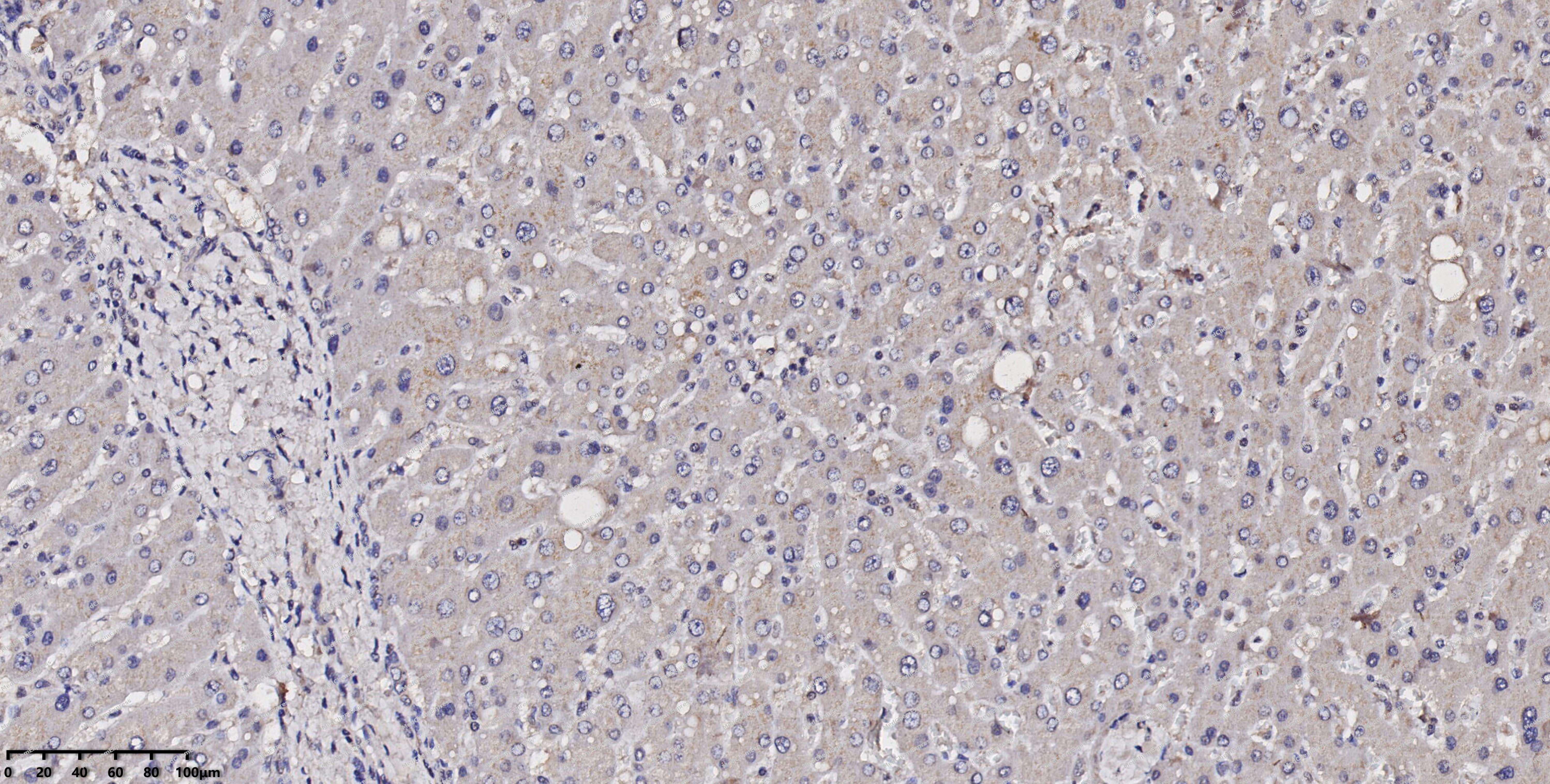 Immunohistochemical analysis of paraffin-embedded human liver using Melanoma gp100 antibody. The section was pre-treated using heat mediated antigen retrieval with EDTA (pH 9.0) for 14 minutes. The tissues were blocked in 5% BSA for 30 minutes at room temperature, washed with TBST, and then probed with the primary antibody (HY-P85933, 1/1000) for 30 minutes at room temperature. The detection was performed using Polymer HRP-conjugated Goat Anti-Mouse/Rabbit lgG(H&L) secondary antibody (HY-P83652). DAB was used as the chromogen. Tissues were counterstained with hematoxylin and mounted with DPX.
 -
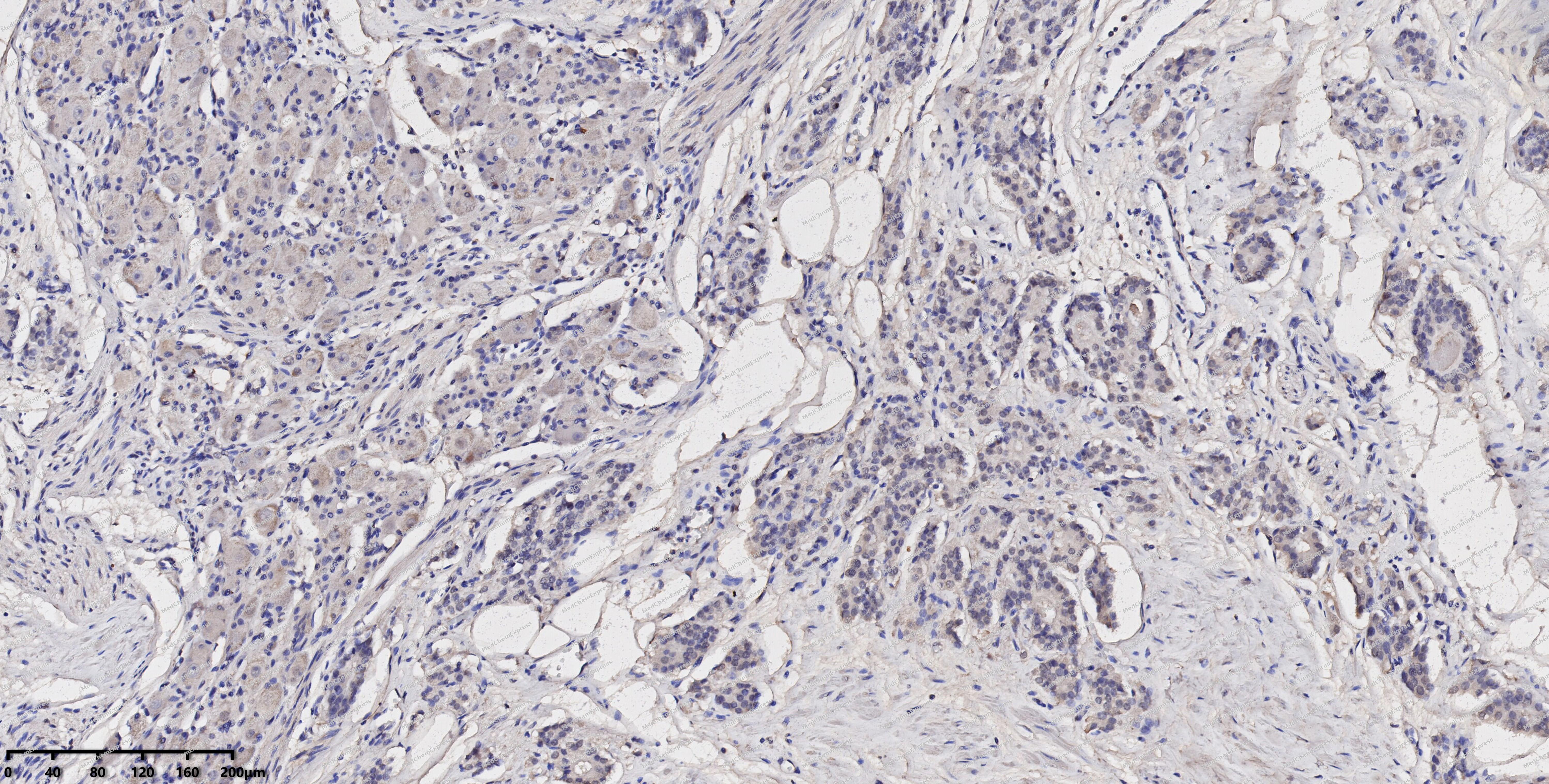 Immunohistochemical analysis of paraffin-embedded human prostate cancer using Melanoma gp100 antibody. The section was pre-treated using heat mediated antigen retrieval with EDTA (pH 9.0) for 14 minutes. The tissues were blocked in 5% BSA for 30 minutes at room temperature, washed with TBST, and then probed with the primary antibody (HY-P85933, 1/1000) for 30 minutes at room temperature. The detection was performed using Polymer HRP-conjugated Goat Anti-Mouse/Rabbit lgG(H&L) secondary antibody (HY-P83652). DAB was used as the chromogen. Tissues were counterstained with hematoxylin and mounted with DPX.
 -
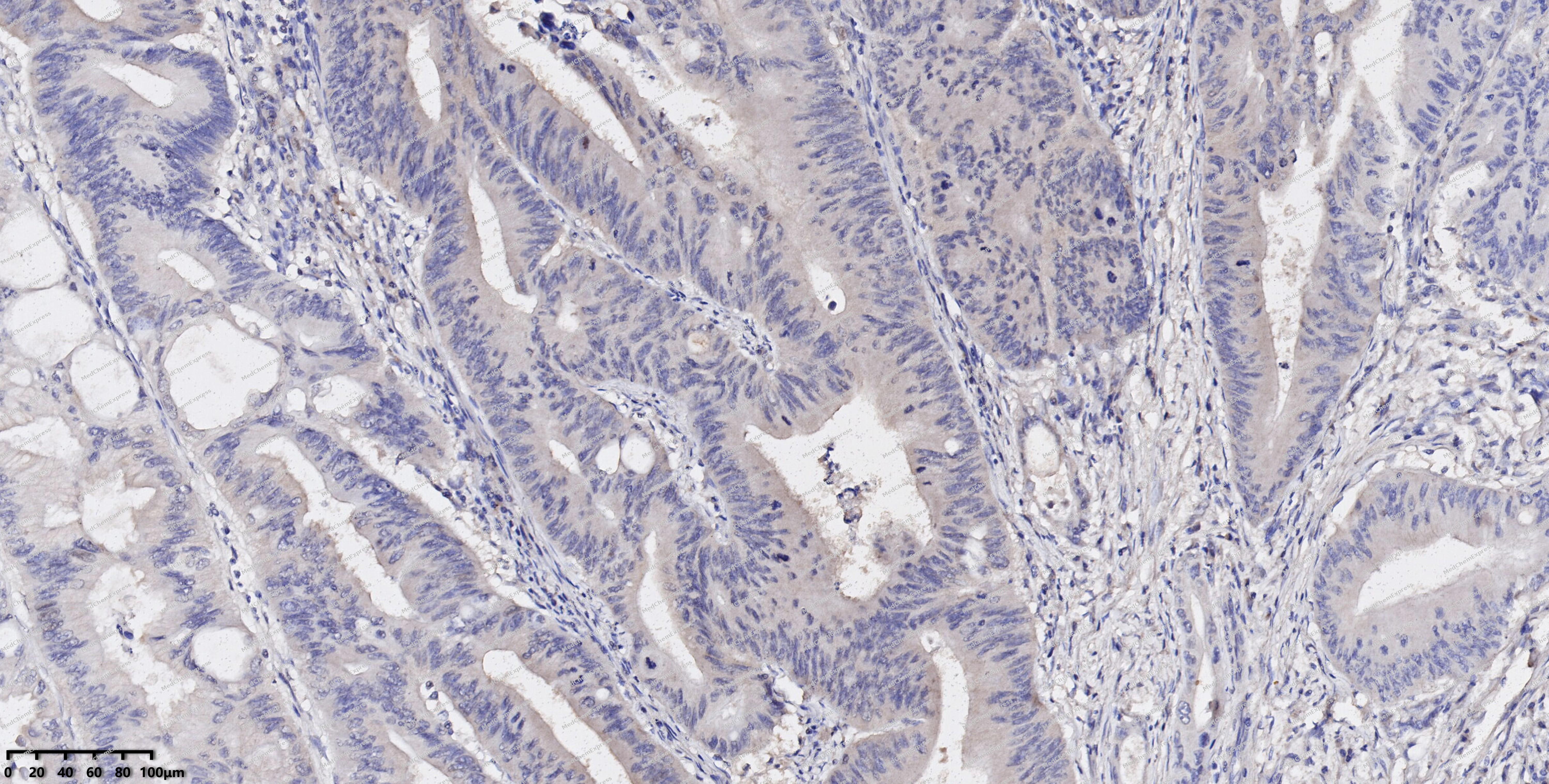 Immunohistochemical analysis of paraffin-embedded human colon cancer using Melanoma gp100 antibody. The section was pre-treated using heat mediated antigen retrieval with EDTA (pH 9.0) for 14 minutes. The tissues were blocked in 5% BSA for 30 minutes at room temperature, washed with TBST, and then probed with the primary antibody (HY-P85933, 1/1000) for 30 minutes at room temperature. The detection was performed using Polymer HRP-conjugated Goat Anti-Mouse/Rabbit lgG(H&L) secondary antibody (HY-P83652). DAB was used as the chromogen. Tissues were counterstained with hematoxylin and mounted with DPX.
 -
 Immunohistochemical analysis of paraffin-embedded human melanoma using Melanoma gp100 antibody. The section was pre-treated using heat mediated antigen retrieval with EDTA (pH 9.0) for 14 minutes. The tissues were blocked in 5% BSA for 30 minutes at room temperature, washed with TBST, and then probed with the primary antibody (HY-P85933, 1/1000) for 30 minutes at room temperature. The detection was performed using Polymer HRP-conjugated Goat Anti-Mouse/Rabbit lgG(H&L) secondary antibody (HY-P83652). DAB was used as the chromogen. Tissues were counterstained with hematoxylin and mounted with DPX.


Background
-
Function
Melanoma gp100 forms physiological amyloids that play a central role in melanosome morphogenesis and pigmentation. The maturation of unpigmented premelanosomes from stage I to II is marked by assembly of processed amyloidogenic fragments into parallel fibrillar sheets, which elongate the vesicle into a striated ellipsoidal shape. In pigmented stage III and IV melanosomes, the amyloid matrix serves as a platform where eumelanin precursors accumulate at high local concentrations for pigment formation. May prevent pigmentation-associated toxicity by sequestering toxic reaction intermediates of eumelanin biosynthesis pathway; Represents a potent melanoma-specific antigen. Among melanoma non-mutated self-peptides, G9-154 (KTWGQYWQV), G9-209 (ITDQVPFSV) and G9-280 (YLEPGPVTA), appear to act as immunodominant common epitopes that stimulate anti-tumor immune response mediated by HLA-A-restricted cytotoxic T cells
-
Subcellular Localization
Endoplasmic reticulum membrane; Single-pass type I membrane protein; Golgi apparatus, cis-Golgi network membrane; Single-pass type I membrane protein; Endosome, multivesicular body; Melanosome; Extracellular vesicle; Secreted
-
Expression
Tissue_specificity:It is typically expressed at low levels in quiescent adult melanocytes, but is overexpressed in proliferating neonatal melanocytes and during tumor growth. It is also overexpressed in melanoma. Some expression has also been found in dysplastic nevi. -
Isoforms & Post-Translational Modification
P40967 has 5 isomers: P40967-1: 70255 Da (predicted); P40967-2: 70949 Da (predicted); P40967-3: 60581 Da (predicted); P40967-4: 66035 Da (predicted); P40967-5: 66729 Da (predicted).
N- and O-glycosylated. A small amount of P1/P100 (major form) undergoes glycosylation in ER and Golgi compartments to yield P2/P120 (minor form). The mature P2 form leaves the trans-Golgi network and is mainly targeted to stage I melanosomes via the plasma membrane and clathrin-mediated endocytosis. Stage II melanosomes harbor only Golgi-modified fragments that are derived from M-alpha and that bear sialylated O-linked oligosaccharides. O-glycosylation of the RPT region is a conserved feature likely involved in amyloid sheet separation via electrostatic repulsion;Undergoes multiple proteolytic processing. In a post-Golgi prelysosomal compartment, P2 is cleaved by a furin-like proprotein convertase (PC) into two disulfide-linked subunits: a large lumenal subunit, M-alpha/ME20-S, and an integral membrane subunit, M-beta. Despite cleavage, only a small fraction of M-alpha is secreted, as most M-alpha and M-beta remain associated with each other intracellularly via a disulfide bond (PubMed:11694580, PubMed:12732614, PubMed:15096515, PubMed:15695812, PubMed:17991747). Once targeted to stage I melanosomes, beta-secretase BACE2 cleaves the M-beta fragment to release the amyloidogenic luminal fragment containing M-alpha and a small portion of M-beta N-terminus (PubMed:23754390). M-alpha is further cleaved by metalloproteinases, likely ADAM10 or ADAM17, and still unknown proteases to yield subfragments that ultimately assemble into amyloid fibrils (PubMed:19047044). The C-terminal fragment of M-beta is processed by the gamma-secretase complex to release a short intracytoplasmic domain (PubMed:19047044) -
Subunit
Homodimer; disulfide-linked. Dimerization in the endoplasmic reticulum and early Golgi prevents premature fibril formation. The dimers are resolved to monomers in late- or post-Golgi compartments (PubMed:26694611). Heterooligomer; amyloid-type. Processed amyloidogenic fragments assemble into fibrils that further organize into beta-sheet quaternary amyloid structures (PubMed:28272432, PubMed:30988362). Interacts (via luminal C-terminal fragment) with CD63; this is important for sorting of the luminal fragment in tetraspanin rich microdomains in stage I melanosomes to prevent premature lysosomal degradation (PubMed:21962903). Interacts with APOE; this allows the loading of the luminal fragment on ILVs to induce fibril nucleation (PubMed:26387950). Interacts with MLANA (PubMed:15695812)
-
SwissProt ID
-
Synonyms
95 kDa melanocyte specific secreted glycoprotein; 95 kDa melanocyte-specific secreted glycoprotein; D12S53E; gp100; M-beta; ME20; ME20 M/ME20 S; ME20-M; ME20-S; ME20M; ME20M/ME20S; ME20S; Melanocyte lineage specific antigen GP100; Melanocyte protein mel 17; Melanocyte protein Pmel 17; Melanocyte protein Pmel 17 precursor; Melanocytes lineage-specific antigen GP100; Melanoma associated ME20 antigen; Melanoma gp100; Melanoma-associated ME20 antigen; Melanosomal matrix protein 17; Melanosomal matrix protein17; P1; p100; p26; PMEL 17; PMEL; PMEL_HUMAN; PMEL17; Premelanosome protein; Secreted melanoma-associated ME20 antigen; SI; SIL; SILV; Silver; Mouse homolog; like; Silver homolog; Silver locus protein homolog; Silver, Mouse, homolog of


Documentation
-
Data Sheet (235 KB)
-
SDS (254 KB)
- English - EN (254 KB)
- Français - FR (254 KB)
- Deutsch - DE (254 KB)
- Norwegian - NO (254 KB)
- Español - ES (254 KB)
- Swedish - SV (254 KB)
- Italian - IT (254 KB)
- Korean - KR (254 KB)
- Portuguese - PT (254 KB)
-
User Guide for Antibodies (1077 KB)

 Powered by Bioz
