



Mast Cell Tryptase Antibody (YA1533)
(Synonyms: TPS1; TPS2; TPSAB1; TPSB1; MCP 7; Mast cell protease 7; Tryptase alpha 1)
Based on 1 Customer Validation
Mast Cell Tryptase Antibody (YA1533) is a Rabbit-derived and non-conjugated IgG monoclonal antibody, targeting to Mast Cell Tryptase.
-
Host:
Rabbit
-
Isotype:
IgG
-
Application:
WB, IHC-P, IP
-
Reactivity :
Human, Mouse, Rat
-
Formulation:
Supplied in 50mM Tris-Glycine(pH 7.4), 0.15M NaCl, 40%Glycerol, 0.01% sodium azide and 0.05% BSA.
-
Conjugation:
Non-conjugated


To place orders, for customer services and technical support, please contact: MedChemExpress USA
Tel: 609-228-6898 E-mail: [email protected] [email protected]





Applications
| Application |
WB

WB: Western Blot
|
IHC-P
IHC-P: Immunohistochemistry-Paraffin
|
IP
IP: Immunoprecipitation
|
|---|---|---|---|
| Dilution Ratio | 1:500-1:1000 | 1:50-1:100 | 1:50 |


Product Details
Mast Cell Tryptase Antibody (YA1533) is a Rabbit-derived and non-conjugated IgG monoclonal antibody, targeting to Mast Cell Tryptase.
-
Host Rabbit

-
Clonality Recombinant,Monoclonal

-
Species ReactivityHuman, Mouse, Rat
-
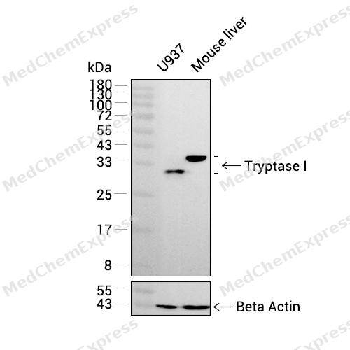
Observed Molecular WeightObserved band size: 31 kDa
Note: Due to possible protein modifications or aggregation, the molecular weight should be confirmed by actual measurement, and the predicted value is for reference only.

-
Calculated Molecular Weight Predicted band size: 31 kDa

Entrez Gene: 7177 Human ; 100503895 Mouse ; 54271 Rat
SwissProt: Q15661 Human ; Q02844 Mouse ; P27435 Rat
OMIM: 191080 Human
A synthesized peptide derived from human Mast Cell Tryptase aa10-275.
Endogenous
Affinity Chromatography
Non-conjugated
Unmodified
IgG


Product Properties
-
Appearance
Liquid
-
Formulation
Supplied in 50mM Tris-Glycine(pH 7.4), 0.15M NaCl, 40%Glycerol, 0.01% sodium azide and 0.05% BSA.
-
Concentration
Batch-dependent, Please check the COA for the concentration of each lot. Check Lot Concentration
-
Storage & Stability
Stored at -20°C for 1 year. Avoid repeated freeze / thaw cycles.
-
Shipping
Shipping with blue ice.


Verification Images
-
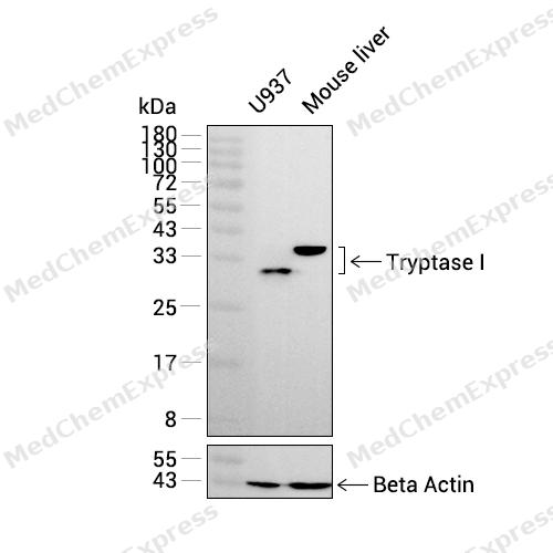 Western blot analysis was performed on extracts from U937 (lane 1, 15 μg), and Mouse liver (lane 2, 15 μg) using Tryptase I Rabbit mAb.Proteins were transferred to a PVDF membrane and blocked with 5% non-fat milk in TBST at 4°C overnight.The primary antibody (1:1000 dilution) and the loading control antibody (beta-Actin, HY-P83730, 1:20000 dilution) were incubated in 5% non-fat milk in TBST for 1 hour at 37°C.Goat Anti-Rabbit IgG-HRP Secondary Antibody (1:20000 dilution) was then applied for 40 minutes at 37°C.
 -
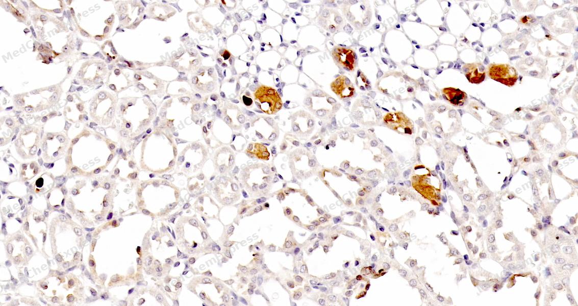 Immunohistochemical analysis of paraffin-embedded rat kidney tissue using Mast Cell Tryptase Antibody. The section was pre-treated using heat mediated antigen retrieval with sodium citrate buffer (pH 6.0) for 8 minutes. The tissues were blocked in QuickBlock for 20 minutes at room temperature, washed with ddH2O and PBS, and then probed with the primary antibody at 1/100 dilution in 4℃ overnight. The detection was performed using an HRP conjugated compact polymer system. DAB was used as the chromogen. Tissues were counterstained with hematoxylin and mounted with DPX.
 -
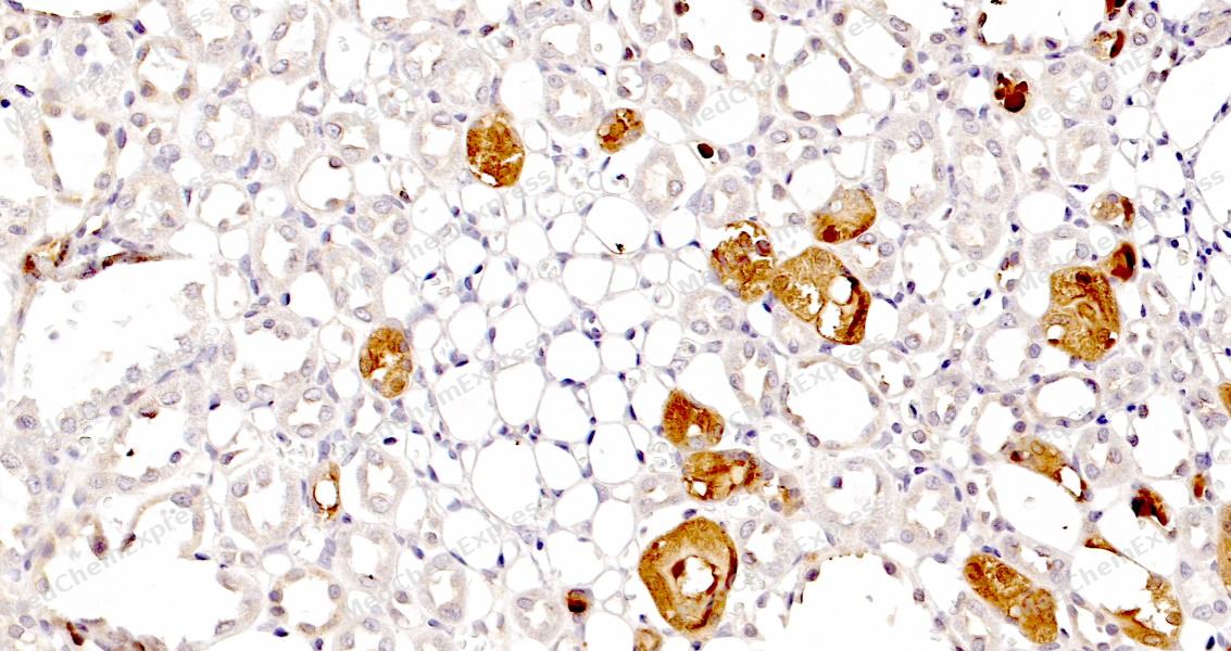 Immunohistochemical analysis of paraffin-embedded rat kidney tissue using Mast Cell Tryptase Antibody. The section was pre-treated using heat mediated antigen retrieval with sodium citrate buffer (pH 6.0) for 8 minutes. The tissues were blocked in QuickBlock for 20 minutes at room temperature, washed with ddH2O and PBS, and then probed with the primary antibody at 1/100 dilution in 4℃ overnight. The detection was performed using an HRP conjugated compact polymer system. DAB was used as the chromogen. Tissues were counterstained with hematoxylin and mounted with DPX.


Background
-
Function
Mast Cell Tryptase is a Tryptase is the major neutral protease present in mast cells and is secreted upon the coupled activation-degranulation response of this cell type. May play a role in innate immunity. Isoform 2 cleaves large substrates, such as fibronectin, more efficiently than isoform 1, but seems less efficient toward small substrates[1].
-
Subcellular Localization
Secreted
-
Expression
Tissue_specificity:Isomer 1 and isomer 2 are expressed in the lungs, stomach, spleen, heart, and skin; in these tissues, isomer 1 is dominant. Isomer 2 is expressed in aortic, splenic, and breast tumors, with the highest expression levels in endothelial cells of certain vessels around the aorta and in endothelial cells around tumors, while expression levels are very low (at the protein level) in mast cells. -
Isoforms & Post-Translational Modification
Q15661 has 2 isomers: Q15661-1: 30515 Da (predicted); Q15661-2: 29533 Da (predicted).
-
Subunit
Homotetramer. The active tetramer is converted to inactive monomers at neutral and acidic pH in the absence of heparin. Low concentrations of inactive monomers become active monomers at pH 6.0 in the presence of heparin. When the concentration of active monomers is higher, they convert to active monomers and then to active tetramers. These monomers are active and functionally distinct from the tetrameric enzyme. In contrast to the hidden active sites in the tetrameric form, the active site of the monomeric form is accessible for macromolecular proteins and inhibitors, e.g. fibrinogen which is a substrate for the monomeric but not for the tetrameric form. The monomeric form forms a complex with SERPINB6
-
SwissProt ID
-
Synonyms
TPS1; TPS2; TPSAB1; TPSB1; MCP 7; Mast cell protease 7; Tryptase alpha 1
-
Research Field
Immunology


Documentation
-
Data Sheet (235 KB)
-
SDS (251 KB)
- English - EN (251 KB)
- Français - FR (251 KB)
- Deutsch - DE (251 KB)
- Norwegian - NO (251 KB)
- Español - ES (251 KB)
- Swedish - SV (251 KB)
- Italian - IT (251 KB)
- Korean - KR (251 KB)
- Portuguese - PT (251 KB)
-
User Guide for Antibodies (1077 KB)

 Powered by Bioz
