



Met (C-Met) Antibody (YA5930)
(Synonyms: MET; Hepatocyte growth factor receptor; HGF receptor; HGF/SF receptor; Proto-oncogene c-Met; Scatter factor receptor; SF receptor; Tyrosine-protein kinase Met)
Based on 1 Customer Validation
Met (C-Met) Antibody (YA5930) is a Rabbit-derived and non-conjugated IgG monoclonal antibody, targeting to Met (C-Met).
-
Host:
Rabbit
-
Isotype:
IgG
-
Application:
WB, IHC-P, ICC/IF, IP, ELISA
-
Reactivity :
Human, Mouse, Rat
-
Formulation:
Supplied in PBS, 50% glycerol, 0.05% Proclin 300, 0.05%BSA
-
Conjugation:
Non-conjugated


To place orders, for customer services and technical support, please contact: MedChemExpress USA
Tel: 609-228-6898 E-mail: [email protected] [email protected]





Applications
| Application |
IHC-P

IHC-P: Immunohistochemistry-Paraffin
|
WB
WB: Western Blot
|
ICC/IF
ICC/IF: Immunocytochemistry/
Immunofluorescence |
ELISA
ELISA: Enzyme Linked Immunosorbent Assay
|
IP
IP: Immunoprecipitation
|
|---|---|---|---|---|---|
| Dilution Ratio | 1:200-1:1000 | 1:2000-1:10000 | 1:200-1:1000 | 1:5000-1:20000 | 1:50-1:200 |


Product Details
Met (C-Met) Antibody (YA5930) is a Rabbit-derived and non-conjugated IgG monoclonal antibody, targeting to Met (C-Met).
-
Host Rabbit

-
Clonality Monoclonal

-
Species ReactivityHuman, Mouse, Rat
-
Observed Molecular WeightObserved band size: 180 kDa
Note: Due to possible protein modifications or aggregation, the molecular weight should be confirmed by actual measurement, and the predicted value is for reference only.

-
Calculated Molecular Weight Predicted band size: 156 kDa

Protein A
Non-conjugated
Unmodified
IgG


Product Properties
-
Appearance
Solution
-
Formulation
Supplied in PBS, 50% glycerol, 0.05% Proclin 300, 0.05%BSA
-
Concentration
Batch-dependent, Please check the COA for the concentration of each lot. Check Lot Concentration
-
Storage & Stability
Stored at -20°C for 1 year. Avoid repeated freeze / thaw cycles.
-
Shipping
Shipping with blue ice.


Verification Images
-
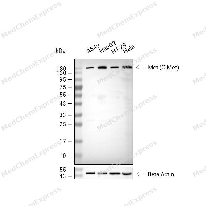 Western blot analysis of extracts from A549 (lane2, 20μg), HepG2 (lane3, 20μg), HT-29 (lane4, 20μg) and Hela (lane5, 20μg) using Met (C-Met) Antibody (HY-P86238). Proteins were transferred to a PVDF membrane and blocked with 5% non-fat milk in TBST for 2 hour at room temperature. The primary antibody (1/5000) and Loading control antibody (Beta Actin, HY-P80993, 1/10,000) was used in 5% non-fat milk in TBST at 4°C overnight. Goat Anti-Rabbit IgG-HRP Secondary Antibody (HY-P8001 ,1/10,000) was used for 1 hour at room temperature.
 -
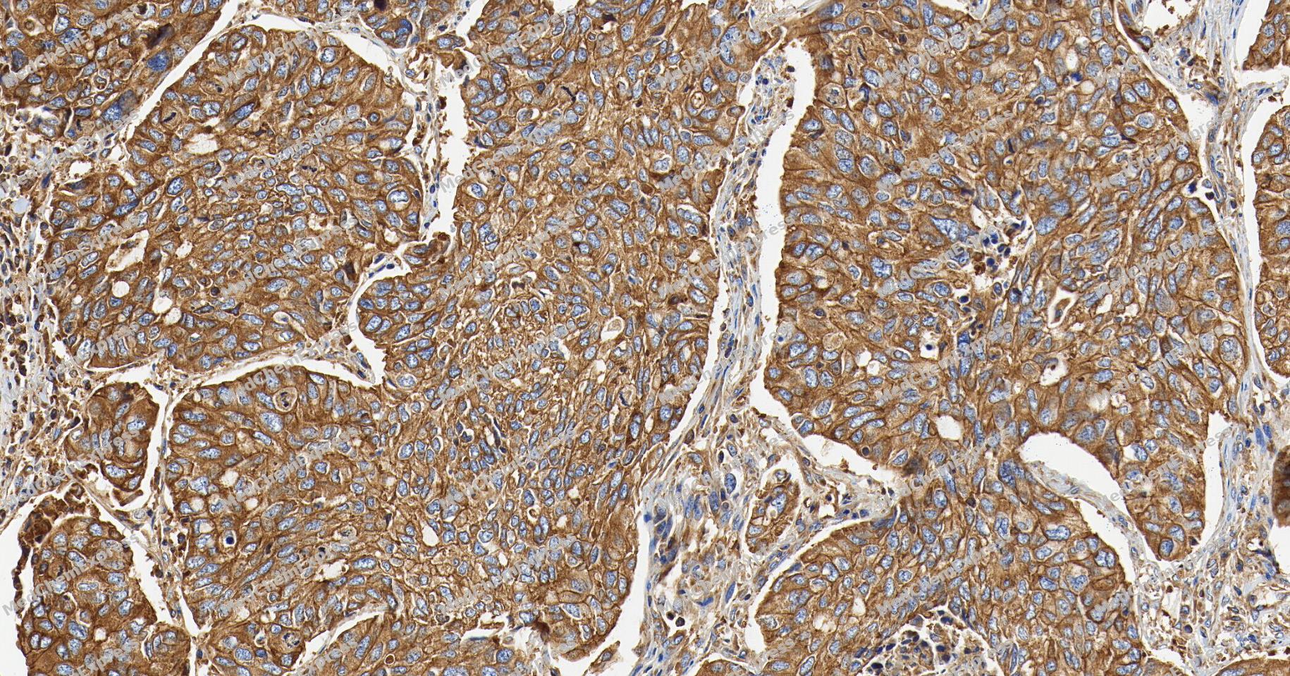 Immunohistochemical analysis of paraffin-embedded human lung cancer tissue using Met (C-Met) Antibody (YA5930). The section was pre-treated using heat mediated antigen retrieval with Tris/EDTA buffer (pH 9.0) for 20 minutes. The tissues were probed with the primary antibody (HY-P86238,1/200) overnight at 4℃. The detection was performed using an HRP conjugated compact polymer system. DAB was used as the chromogen. Tissues were counterstained with hematoxylin and mounted with neutral balsam.
 -
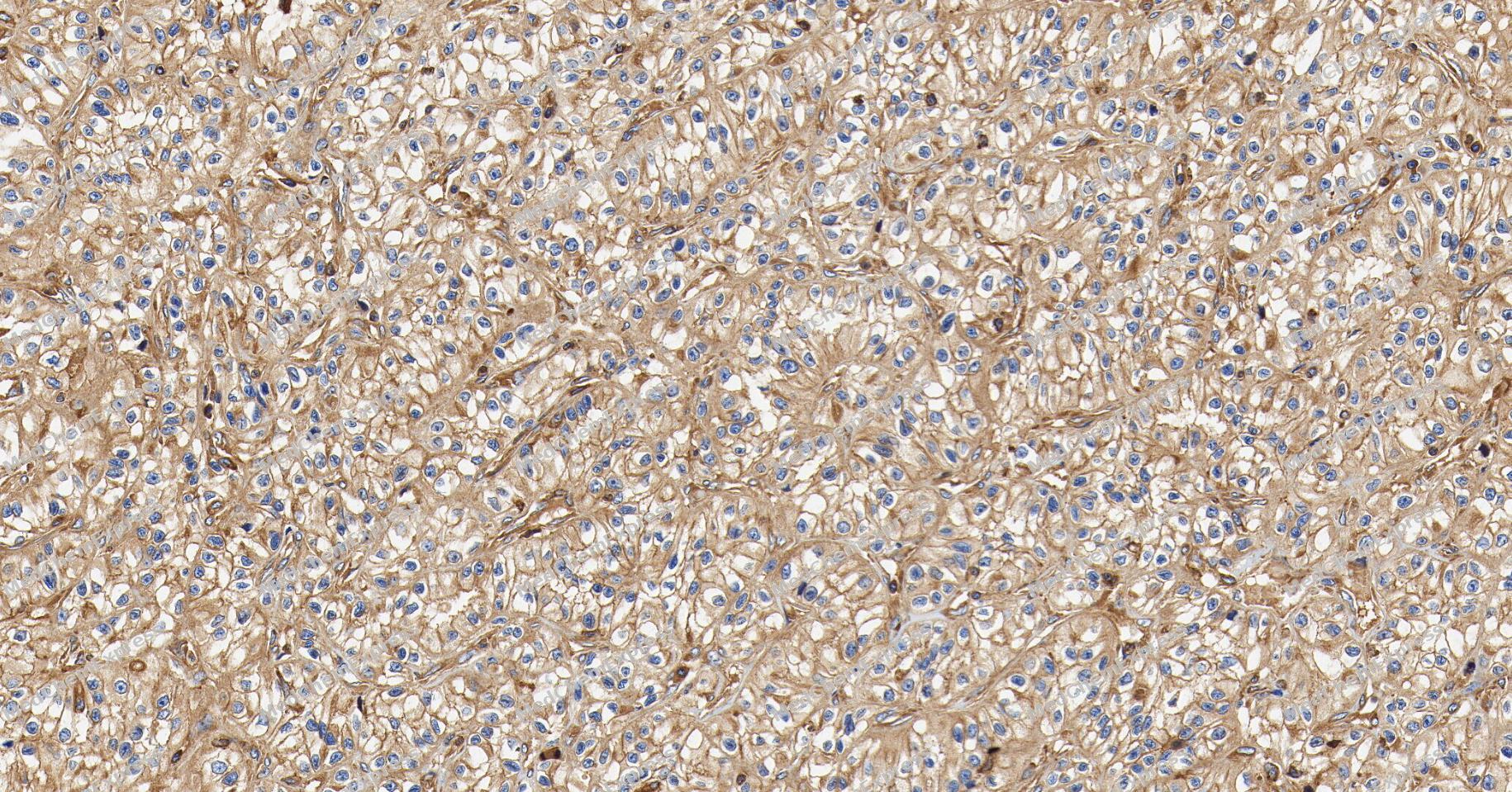 Immunohistochemical analysis of paraffin-embedded human renal cell cancer tissue using Met (C-Met) Antibody (YA5930). The section was pre-treated using heat mediated antigen retrieval with Tris/EDTA buffer (pH 9.0) for 20 minutes. The tissues were probed with the primary antibody (HY-P86238,1/200) overnight at 4℃. The detection was performed using an HRP conjugated compact polymer system. DAB was used as the chromogen. Tissues were counterstained with hematoxylin and mounted with neutral balsam.
 -
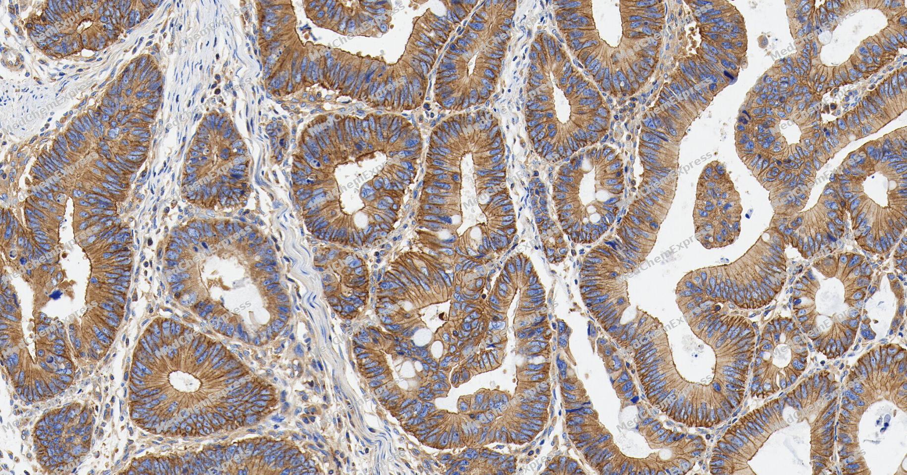 Immunohistochemical analysis of paraffin-embedded human colon cancer tissue using Met (C-Met) Antibody (YA5930). The section was pre-treated using heat mediated antigen retrieval with Tris/EDTA buffer (pH 9.0) for 20 minutes. The tissues were probed with the primary antibody (HY-P86238,1/200) overnight at 4℃. The detection was performed using an HRP conjugated compact polymer system. DAB was used as the chromogen. Tissues were counterstained with hematoxylin and mounted with neutral balsam.
 -
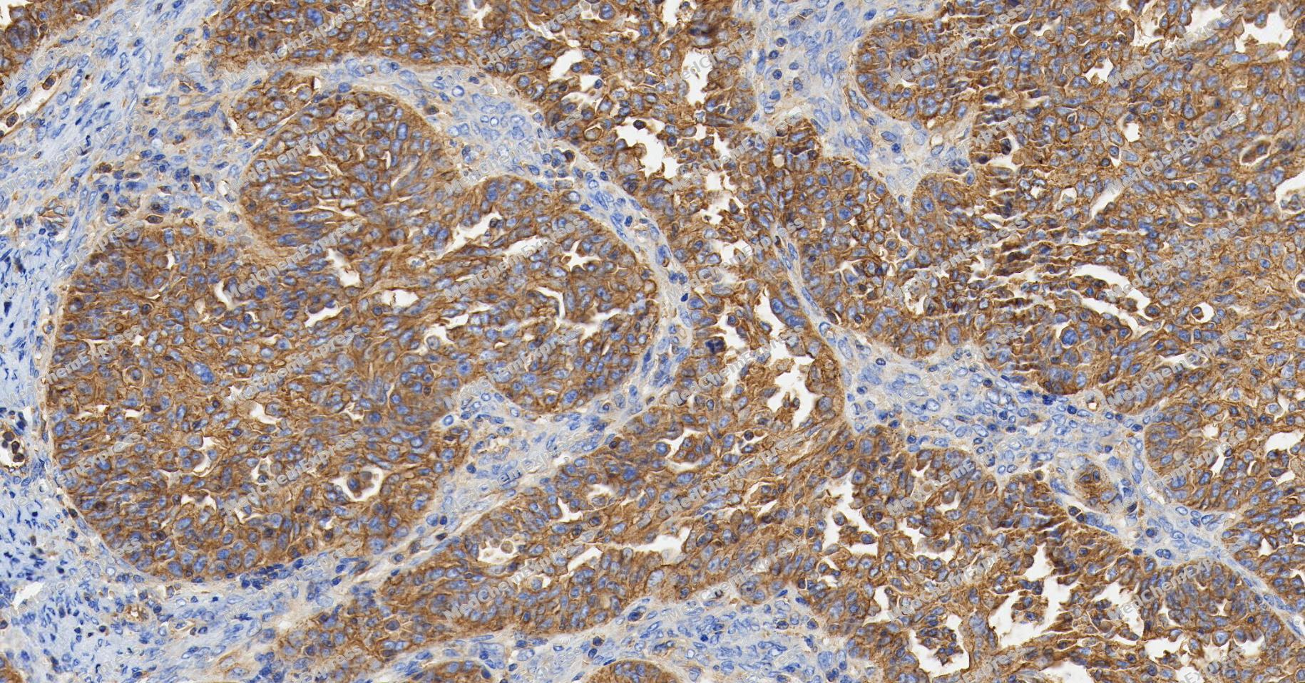 Immunohistochemical analysis of paraffin-embedded human ovarian cancer tissue using Met (C-Met) Antibody (YA5930). The section was pre-treated using heat mediated antigen retrieval with Tris/EDTA buffer (pH 9.0) for 20 minutes. The tissues were probed with the primary antibody (HY-P86238,1/200) overnight at 4℃. The detection was performed using an HRP conjugated compact polymer system. DAB was used as the chromogen. Tissues were counterstained with hematoxylin and mounted with neutral balsam.
 -
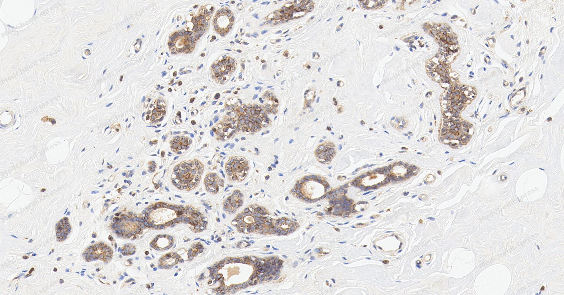 Immunohistochemical analysis of paraffin-embedded human breast tissue using Met (C-Met) Antibody (YA5930). The section was pre-treated using heat mediated antigen retrieval with Tris/EDTA buffer (pH 9.0) for 20 minutes. The tissues were probed with the primary antibody (HY-P86238,1/200) overnight at 4℃. The detection was performed using an HRP conjugated compact polymer system. DAB was used as the chromogen. Tissues were counterstained with hematoxylin and mounted with neutral balsam.
 -
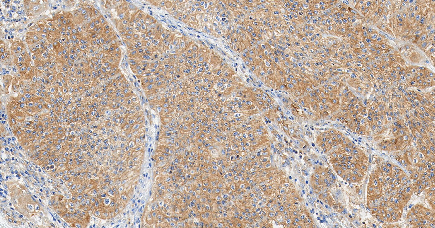 Immunohistochemical analysis of paraffin-embedded human bladder cancer tissue using Met (C-Met) Antibody (YA5930). The section was pre-treated using heat mediated antigen retrieval with Tris/EDTA buffer (pH 9.0) for 20 minutes. The tissues were probed with the primary antibody (HY-P86238,1/200) overnight at 4℃. The detection was performed using an HRP conjugated compact polymer system. DAB was used as the chromogen. Tissues were counterstained with hematoxylin and mounted with neutral balsam.
 -
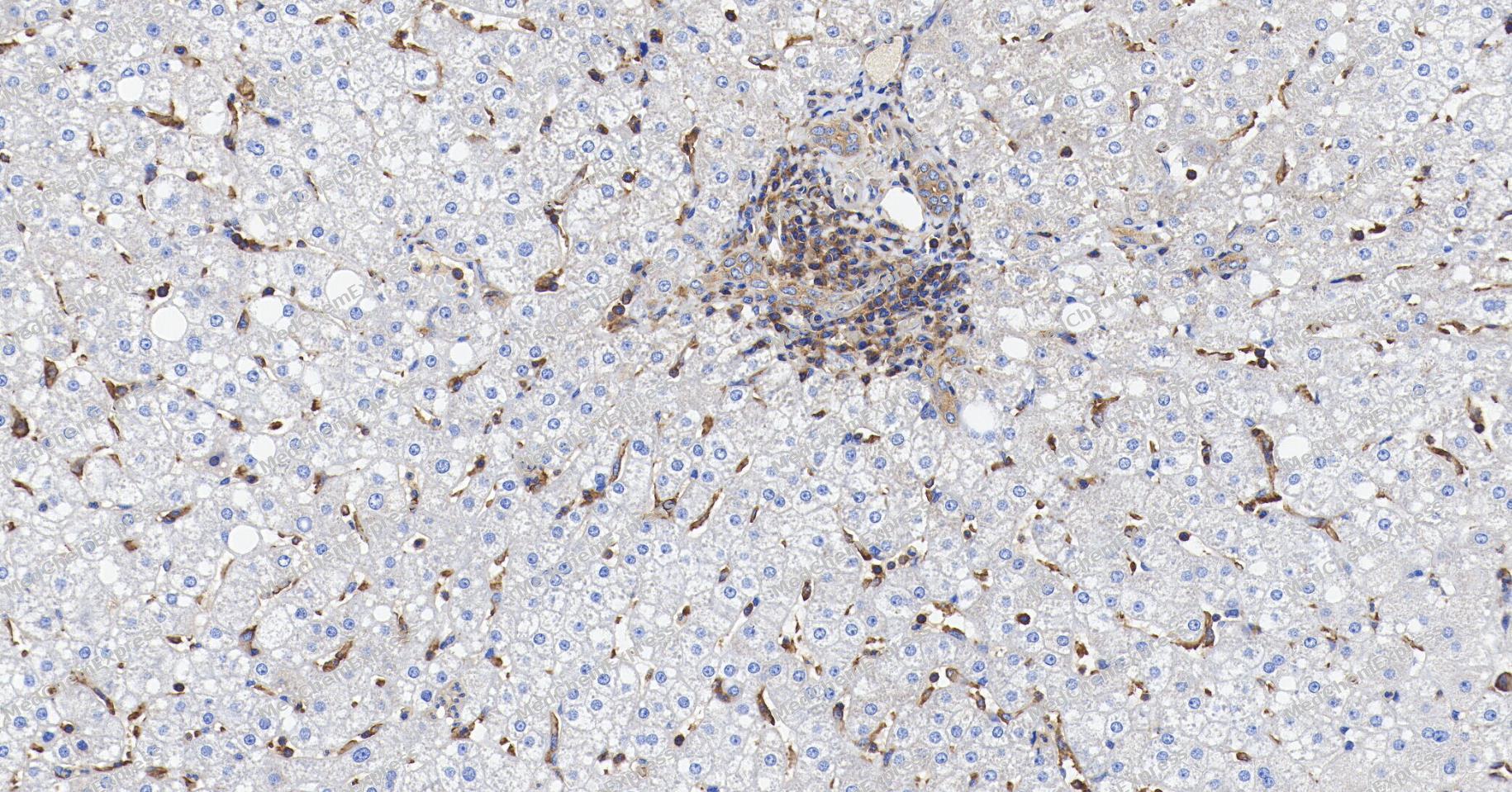 Immunohistochemical analysis of paraffin-embedded human liver cancer tissue using Met (C-Met) Antibody (YA5930). The section was pre-treated using heat mediated antigen retrieval with Tris/EDTA buffer (pH 9.0) for 20 minutes. The tissues were probed with the primary antibody (HY-P86238,1/200) overnight at 4℃. The detection was performed using an HRP conjugated compact polymer system. DAB was used as the chromogen. Tissues were counterstained with hematoxylin and mounted with neutral balsam.
 -
 Immunohistochemical analysis of paraffin-embedded mouse duodenum tissue using Met (C-Met) Antibody (HY-P86238, 1/800). The section was pretreated using heat mediated antigen retrieval with sodium citrate buffer (pH 6.0) for 8 minutes. The tissues were blocked with quick block buffer for 0.5 hours at room temperature, washed with PBS and PBST, and then incubated with the primary antibody overnight at 4℃. The detection was performed using an HRP conjugated compact polymer system. DAB was used as the chromogen. Tissues were counterstained with hematoxylin and mounted with DPX.
 -
 Immunohistochemical analysis of paraffin-embedded mouse rectum tissue using Met (C-Met) Antibody (HY-P86238, 1/800). The section was pretreated using heat mediated antigen retrieval with sodium citrate buffer (pH 6.0) for 8 minutes. The tissues were blocked with quick block buffer for 0.5 hours at room temperature, washed with PBS and PBST, and then incubated with the primary antibody overnight at 4℃. The detection was performed using an HRP conjugated compact polymer system. DAB was used as the chromogen. Tissues were counterstained with hematoxylin and mounted with DPX.
 -
 Immunohistochemical analysis of paraffin-embedded mouse small intestine tissue using Met (C-Met) Antibody (HY-P86238, 1/800). The section was pretreated using heat mediated antigen retrieval with sodium citrate buffer (pH 6.0) for 8 minutes. The tissues were blocked with quick block buffer for 0.5 hours at room temperature, washed with PBS and PBST, and then incubated with the primary antibody overnight at 4℃. The detection was performed using an HRP conjugated compact polymer system. DAB was used as the chromogen. Tissues were counterstained with hematoxylin and mounted with DPX.
 -
 Immunohistochemical analysis of paraffin-embedded mouse stomach tissue using Met (C-Met) Antibody (HY-P86238, 1/800). The section was pretreated using heat mediated antigen retrieval with sodium citrate buffer (pH 6.0) for 8 minutes. The tissues were blocked with quick block buffer for 0.5 hours at room temperature, washed with PBS and PBST, and then incubated with the primary antibody overnight at 4℃. The detection was performed using an HRP conjugated compact polymer system. DAB was used as the chromogen. Tissues were counterstained with hematoxylin and mounted with DPX.
 -
 Immunohistochemical analysis of paraffin-embedded rat small intestine tissue using Met (C-Met) Antibody (HY-P86238, 1/800). The section was pretreated using heat mediated antigen retrieval with sodium citrate buffer (pH 6.0) for 8 minutes. The tissues were blocked with quick block buffer for 0.5 hours at room temperature, washed with PBS and PBST, and then incubated with the primary antibody overnight at 4℃. The detection was performed using an HRP conjugated compact polymer system. DAB was used as the chromogen. Tissues were counterstained with hematoxylin and mounted with DPX.
 -
 Immunohistochemical analysis of paraffin-embedded mouse lung tissue using Met (C-Met) Antibody (HY-P86238, 1/800). The section was pretreated using heat mediated antigen retrieval with sodium citrate buffer (pH 6.0) for 8 minutes. The tissues were blocked with quick block buffer for 0.5 hours at room temperature, washed with PBS and PBST, and then incubated with the primary antibody overnight at 4℃. The detection was performed using an HRP conjugated compact polymer system. DAB was used as the chromogen. Tissues were counterstained with hematoxylin and mounted with DPX.


Background
-
Function
c-Met (hepatocyte growth factor receptor, HGFR) is a protein possesses tyrosine kinase activity. The primary single chain precursor protein is post-translationally cleaved to produce the alpha and beta subunits, which are disulfide linked to form the mature receptor. c-Met is a membrane receptor that is essential for embryonic development and wound healing. Hepatocyte growth factor (HGF) is the only known ligand of the c-Met receptor. c-Met is normally expressed by cells of epithelial origin, while expression of HGF is restricted to cells of mesenchymalorigin. Upon HGF stimulation, c-Met induces several biological responses that collectively give rise to a program known as invasive growth.
-
Subcellular Localization
Membrane; Single-pass type I membrane protein; Secreted
-
Expression
Tissue_specificity:It is expressed in normal hepatocytes, as well as in the epithelial cells of the stomach, small intestine, and large intestine. It is also present in the basal keratinocytes of the esophagus and skin. Higher concentrations are found in the liver, gastrointestinal tract, thyroid gland, and kidneys. It is also expressed in brain tissue. It is also expressed in the metaphysis (protein level) (PubMed:26637977) . -
Isoforms & Post-Translational Modification
c-Met has 3 isomers: P08581-1: 155541 Da (predicted); P08581-2: 157712 Da (predicted); P08581-3: 85745 Da (predicted).
Autophosphorylated in response to ligand binding on Tyr-1234 and Tyr-1235 in the kinase domain leading to further phosphorylation of Tyr-1349 and Tyr-1356 in the C-terminal multifunctional docking site. Dephosphorylated by PTPRJ at Tyr-1349 and Tyr-1365. Dephosphorylated by PTPN1 and PTPN2;Ubiquitinated. Ubiquitination by CBL regulates MET endocytosis, resulting in decreasing plasma membrane receptor abundance, and in endosomal degradation and/or recycling of internalized receptors;O-mannosylation of IPT/TIG domains by TMEM260 is required for protein maturation (PubMed:37186866). O-mannosylated residues are composed of single mannose glycans that are not elongated or modified (PubMed:37186866);(Microbial infection) Tyrosine phosphorylation is stimulated by L.monocytogenes InlB. Tyrosine phosphorylation is maximal 10-20 minutes after treatment with InlB and disappears by 60 minutes. The phosphorylated residues were not identified -
Subunit
Heterodimer made of an alpha chain (50 kDa) and a beta chain (145 kDa) which are disulfide linked. Binds PLXNB1. Interacts when phosphorylated with downstream effectors including STAT3, PIK3R1, SRC, PCLG1, GRB2 and GAB1.
-
SwissProt ID
-
Synonyms
MET; Hepatocyte growth factor receptor; HGF receptor; HGF/SF receptor; Proto-oncogene c-Met; Scatter factor receptor; SF receptor; Tyrosine-protein kinase Met


Documentation
-
Data Sheet (235 KB)
-
SDS (252 KB)
- English - EN (252 KB)
- Français - FR (252 KB)
- Deutsch - DE (252 KB)
- Norwegian - NO (252 KB)
- Español - ES (252 KB)
- Swedish - SV (252 KB)
- Italian - IT (252 KB)
- Korean - KR (252 KB)
- Portuguese - PT (252 KB)
-
User Guide for Antibodies (1077 KB)

 Powered by Bioz
