



Osteopontin Antibody (YA6362)
(Synonyms: BNSP; Bone sialoprotein 1; BSP I; BSPI; Early T lymphocyte activation 1; ETA 1; ETA1; MGC110940; Nephropontin; OPN; osteopontin/immunoglobulin alpha 1 heavy chain constant region fusion protein; OSTP_HUMAN; secreted phosphoprotein 1(osteopontin, bone sialoprotein I, early T-lymphocyte activation 1); Secreted phosphoprotein 1; SPP 1; SPP-1; SPP1; SPP1/CALPHA1 fusion; Urinary stone protein; Uropontin.)Based on 1 Customer Validation
Osteopontin Antibody (YA6362) is a Rabbit-derived and non-conjugated IgG monoclonal antibody, targeting to Osteopontin.
-
Host:
Rabbit
-
Isotype:
IgG
-
Application:
IHC-P, WB, IHC-F, IF-Tissue
-
Reactivity :
Human
-
Formulation:
Supplied in 0.01M TBS (pH7.4) with 1% BSA, 0.02% Proclin300 and 50% Glycerol.
-
Conjugation:
Non-conjugated


To place orders, for customer services and technical support, please contact: MedChemExpress USA
Tel: 609-228-6898 E-mail: [email protected] [email protected]





Applications
| Application |
IHC-P

IHC-P: Immunohistochemistry-Paraffin
|
WB
WB: Western Blot
|
IHC-F
IHC-F: Immunohistochemistry-Frozen
|
ICC/IF
ICC/IF: Immunocytochemistry/
Immunofluorescence |
|---|---|---|---|---|
| Dilution Ratio | 1:100-500 | 1:500-2000 | 1:100-500 | 1:100-500 |


Product Details
Osteopontin Antibody (YA6362) is a Rabbit-derived and non-conjugated IgG monoclonal antibody, targeting to Osteopontin.
-
Host Rabbit

-
Clonality Monoclonal

-
Species ReactivityHuman
-
Observed Molecular WeightObserved band size: 63 kDa
Note: Due to possible protein modifications or aggregation, the molecular weight should be confirmed by actual measurement, and the predicted value is for reference only.

-
Calculated Molecular Weight Predicted band size: 35 kDa

Recombinant human Osteopontin protein aa 17-314/314
affinity purified.
Non-conjugated
Unmodified
IgG


Product Properties
-
Appearance
Solution
-
Formulation
Supplied in 0.01M TBS (pH7.4) with 1% BSA, 0.02% Proclin300 and 50% Glycerol.
-
Concentration
Batch-dependent, Please check the COA for the concentration of each lot. Check Lot Concentration
-
Storage & Stability
Stored at -20°C for 1 year. Avoid repeated freeze / thaw cycles.
-
Shipping
Shipping with blue ice.


Verification Images
-
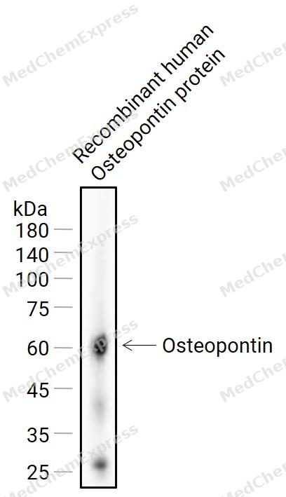 Western blot analysis of extracts from Recombinant human Osteopontin protein(50ng)using Osteopontin Antibody (HY-P86670). Proteins were transferred to a NC membrane and blocked with 5% Non-fat milk powder in TBST for 1 hour at room temperature. The primary antibody (1/1000) was used in 5% Non-fat milk powder in TBST at 4°C overnight. HRP-conjugated Goat Anti-Rabbit Secondary Antibody (1/20,000) was used for 1 hour at room temperature.
 -
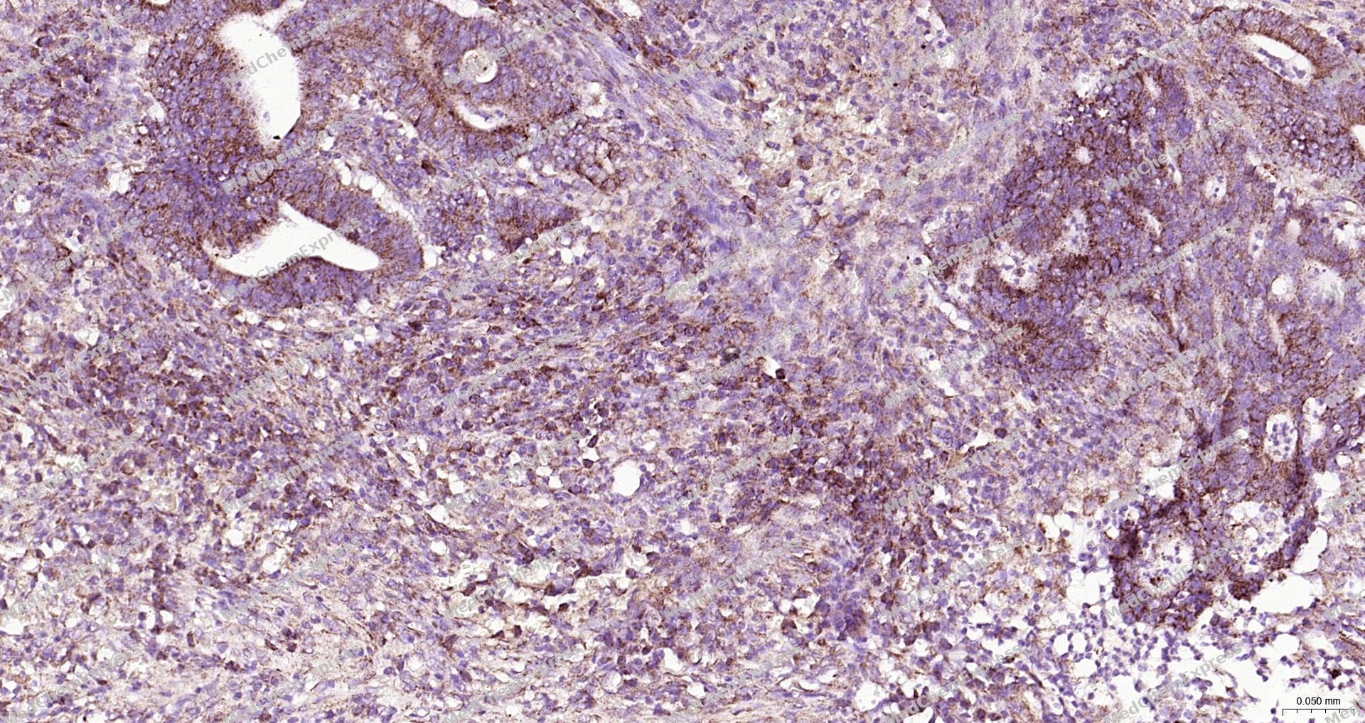 Immunohistochemical analysis of paraffin-embedded human colon cancer tissue using Osteopontin antibody. The section was pre-treated using heat mediated antigen retrieval with sodium citrate buffer (pH 6.0) for 15 minutes. The tissues were blocked in 10% normal goat serum for 30 minutes at room temperature, washed with ddH2O and PBS, and then probed with the primary antibody (Osteopontin,HY-P86670, 1/200) at 4°C overnight. The detection was performed using an HRP conjugated compact polymer system. DAB was used as the chromogen. Tissues were counterstained with hematoxylin and mounted with DPX.
 -
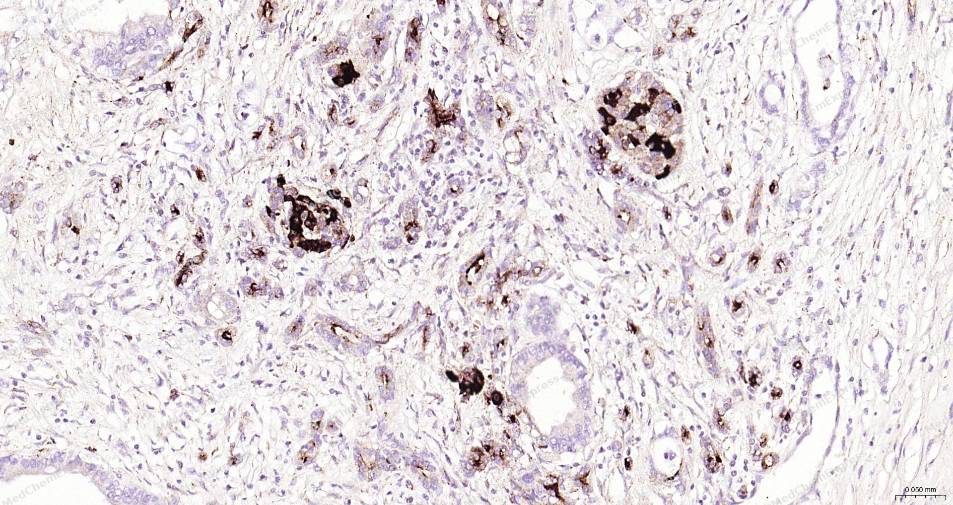 Immunohistochemical analysis of paraffin-embedded human pancreatic cancer tissue using Osteopontin antibody. The section was pre-treated using heat mediated antigen retrieval with sodium citrate buffer (pH 6.0) for 15 minutes. The tissues were blocked in 10% normal goat serum for 30 minutes at room temperature, washed with ddH2O and PBS, and then probed with the primary antibody (Osteopontin,HY-P86670, 1/200) at 4°C overnight. The detection was performed using an HRP conjugated compact polymer system. DAB was used as the chromogen. Tissues were counterstained with hematoxylin and mounted with DPX.
 -
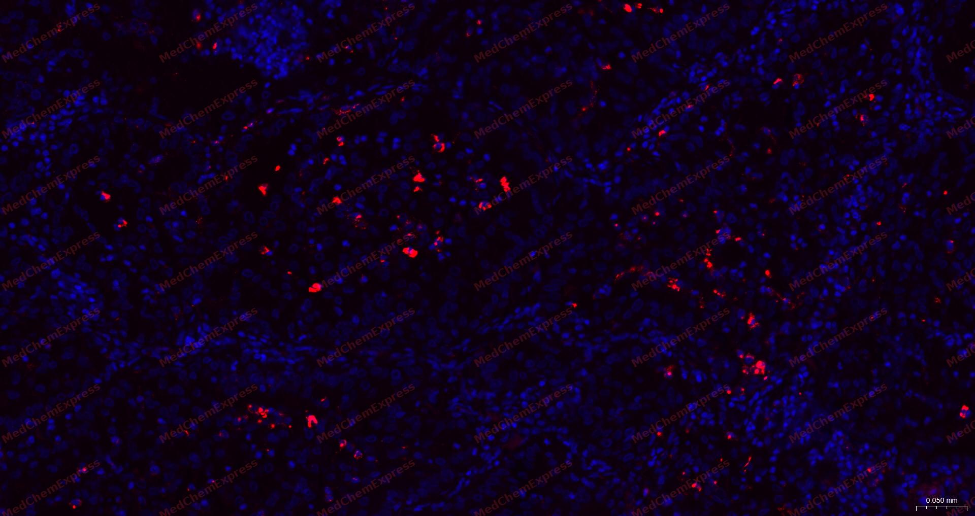 Immunofluorescence analysis of paraffin-embedded human lung cancer using Osteopontin antibody. The section was pre-treated using heat mediated antigen retrieval with sodium citrate buffer (pH 6.0) for 15 minutes. The tissues were blocked in 10% normal goat serum for 30 minutes at room temperature, then incubated with Osteopontin antibody (HY-P86670) at 1/200 dilution overnight at 4 ℃. Alexa Fluor® 594-conjugated AffiniPure Goat Anti-Rabbit IgG H&L(HY-P8003) was used as the secondary antibody at 1/200 dilution. The Nuclear counterstain was DAPI (Blue).
 -
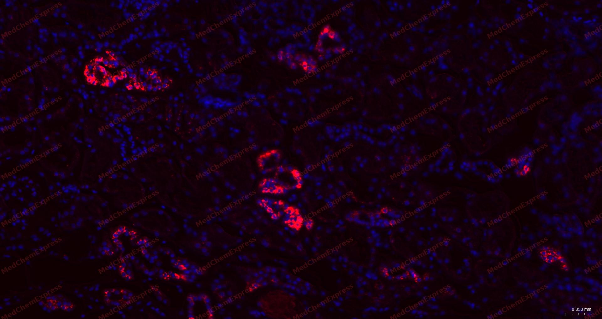 Immunofluorescence analysis of paraffin-embedded human kidney using Osteopontin antibody. The section was pre-treated using heat mediated antigen retrieval with sodium citrate buffer (pH 6.0) for 15 minutes. The tissues were blocked in 10% normal goat serum for 30 minutes at room temperature, then incubated with Osteopontin antibody (HY-P86670) at 1/200 dilution overnight at 4 ℃. Alexa Fluor® 594-conjugated AffiniPure Goat Anti-Rabbit IgG H&L(HY-P8003) was used as the secondary antibody at 1/200 dilution. The Nuclear counterstain was DAPI (Blue).
 -
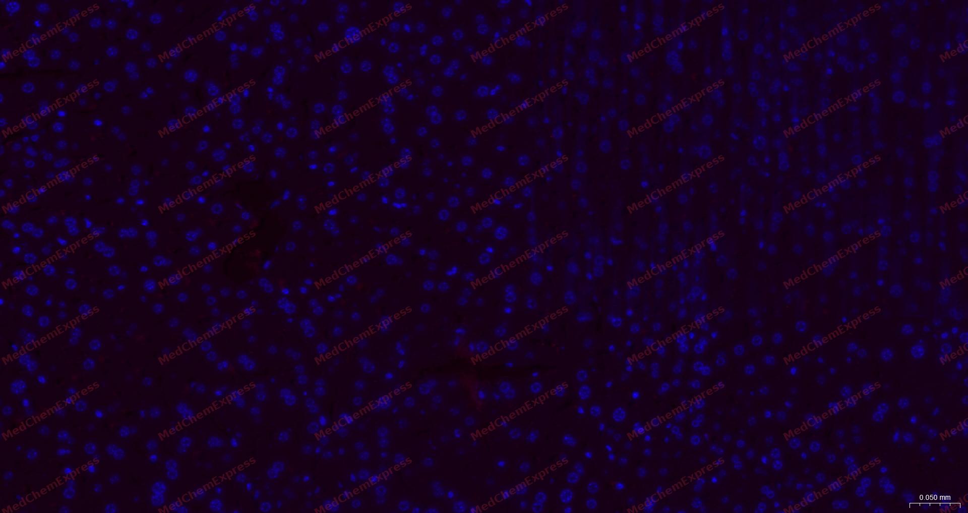 Immunofluorescence analysis of paraffin-embedded mouse liver using Osteopontin antibody. The section was pre-treated using heat mediated antigen retrieval with sodium citrate buffer (pH 6.0) for 15 minutes. The tissues were blocked in 10% normal goat serum for 30 minutes at room temperature, then incubated with Osteopontin antibody (HY-P86670) at 1/200 dilution overnight at 4 ℃. Alexa Fluor® 594-conjugated AffiniPure Goat Anti-Rabbit IgG H&L(HY-P8003) was used as the secondary antibody at 1/200 dilution. The Nuclear counterstain was DAPI (Blue).


Background
-
Function
Osteopontin (OPN) is a secreted, multifunctional, extracellular matrix (ECM), calcium-binding, glycosylated phosphoprotein that is associated with various normal biological functions and pathological processes. OPN is physiologically produced by a wide range of cells and has been identified in various body fluids, playing a crucial role in bone remodeling and vascularization. OPN is also expressed by multiple immune cells, acting as a chemotactic factor, a mediator of cell activation and cytokine production, and an anti-apoptotic factor, thereby regulating inflammation and immune responses. OPN can be involved in the tumorigenesis of various cancer types, including head and neck squamous cell carcinoma (HNSCC), by promoting cell proliferation, adhesion, invasion, and migration through multiple signaling pathways[1][2].
-
Subcellular Localization
Secreted
-
Expression
Tissue_specificity:Protein levels were detected in cerebrospinal fluid and urine (PubMed:25326458, PubMed:36213313, PubMed:37453717) . In bone. Found in plasma. -
Isoforms & Post-Translational Modification
P10451 has 4 isomers: P10451-1: 35423 Da (predicted); P10451-3: 32355 Da (predicted); P10451-4: 33017 Da (predicted); P10451-5: 33843 Da (predicted).
Forms covalent cross-links mediated by transglutaminase TGM2, between a glutamine and the epsilon-amino group of a lysine residue, forming homopolymers and heteropolymers, increasing its collagen binding properties;Extensively phosphorylated by FAM20C in the extracellular medium at multiple sites within the S-x-E/pS motif (PubMed:15869464, PubMed:22582013, PubMed:26091039). The phosphorylated form inhibits hydroxyapatite crystallization. Dephosphorylation via a mechanism involving ALPL/TNAP promotes hydroxyapatite crystallization (By similarity);O-glycosylated. Isoform 5 is GalNAc O-glycosylated at Thr-59 or Ser-62 -
Subunit
Interacts (via N-terminus) with integrin ITGA9:ITGB1
-
SwissProt ID
-
Synonyms
BNSP; Bone sialoprotein 1; BSP I; BSPI; Early T lymphocyte activation 1; ETA 1; ETA1; MGC110940; Nephropontin; OPN; osteopontin/immunoglobulin alpha 1 heavy chain constant region fusion protein; OSTP_HUMAN; secreted phosphoprotein 1(osteopontin, bone sialoprotein I, early T-lymphocyte activation 1); Secreted phosphoprotein 1; SPP 1; SPP-1; SPP1; SPP1/CALPHA1 fusion; Urinary stone protein; Uropontin.


Documentation
-
Data Sheet (260 KB)
-
SDS (254 KB)
- English - EN (254 KB)
- Français - FR (254 KB)
- Deutsch - DE (254 KB)
- Norwegian - NO (254 KB)
- Español - ES (254 KB)
- Swedish - SV (254 KB)
- Italian - IT (254 KB)
- Korean - KR (254 KB)
- Portuguese - PT (254 KB)
-
User Guide for Antibodies (1077 KB)
References

 Powered by Bioz
