



Phospho-MDM2 (Ser166) Antibody (YA1888)
(Synonyms: Double minute 2 protein; Hdm2; Oncoprotein Mdm2)
Based on 1 Customer Validation
Phospho-MDM2 (Ser166) Antibody (YA1888) is a Rabbit-derived and non-conjugated IgG monoclonal antibody, targeting to Phospho-MDM2 (Ser166).
-
Host:
Rabbit
-
Isotype:
IgG
-
Application:
WB, IHC-P
-
Reactivity :
Human, Rat
-
Formulation:
Supplied in 50mM Tris-Glycine(pH 7.4), 0.15M NaCl, 40% Glycerol, 0.01% Sodium azide and 0.05% BSA
-
Conjugation:
Non-conjugated


To place orders, for customer services and technical support, please contact: MedChemExpress USA
Tel: 609-228-6898 E-mail: [email protected] [email protected]





Applications
| Application |
WB

WB: Western Blot
|
IHC-P
IHC-P: Immunohistochemistry-Paraffin
|
|---|---|---|
| Dilution Ratio | 1:500-1:1000 | 1:50-1:100 |


Product Details
Phospho-MDM2 (Ser166) Antibody (YA1888) is a Rabbit-derived and non-conjugated IgG monoclonal antibody, targeting to Phospho-MDM2 (Ser166).
-
Host Rabbit

-
Clonality Recombinant,Monoclonal

-
Species ReactivityHuman, Rat
-
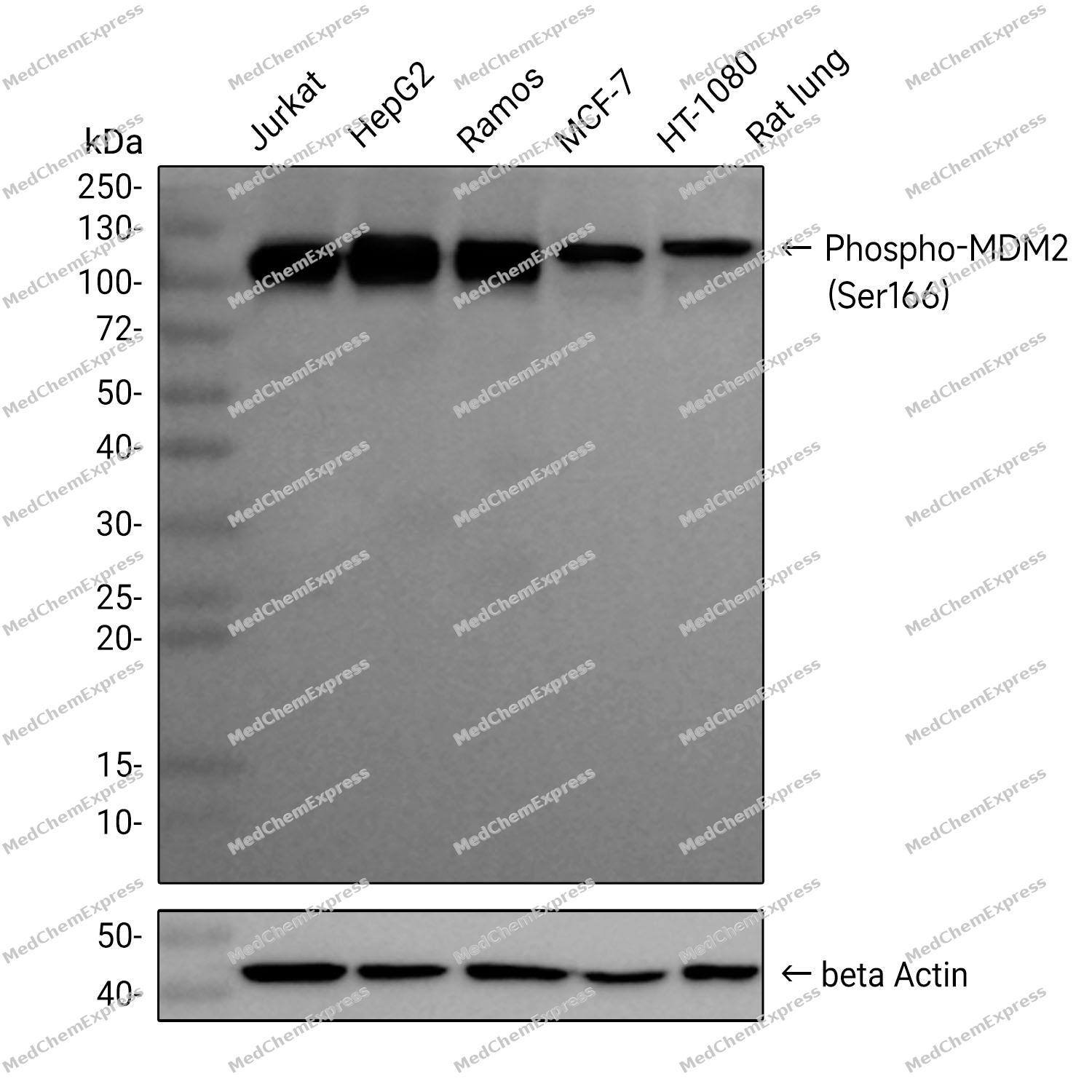
Observed Molecular WeightObserved band size: 90 kDa
Note: Due to possible protein modifications or aggregation, the molecular weight should be confirmed by actual measurement, and the predicted value is for reference only.

-
Calculated Molecular Weight Predicted band size: 55 kDa

A synthetic phosphopeptide corresponding to residues surrounding Ser166 of human MDM2
Endogenous
Affinity Purified
Non-conjugated
Phosphorylated
IgG


Product Properties
-
Appearance
Solution
-
Formulation
Supplied in 50mM Tris-Glycine(pH 7.4), 0.15M NaCl, 40% Glycerol, 0.01% Sodium azide and 0.05% BSA
-
Concentration
Batch-dependent, Please check the COA for the concentration of each lot. Check Lot Concentration
-
Storage & Stability
Stored at -20°C for 1 year. Avoid repeated freeze / thaw cycles.
-
Shipping
Shipping with blue ice.


Verification Images
-
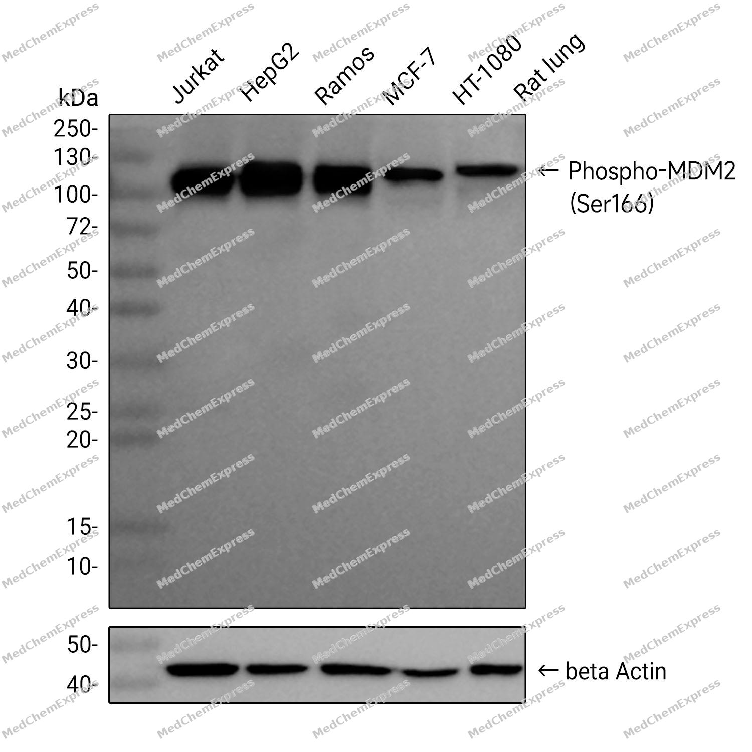 Western blot analysis was performed on extracts from Jurkat (lane 1, 15 μg), HepG2 (lane 2, 15 μg), Ramos (lane 3, 15 μg), MCF-7 (lane 4, 15 μg), HT-1080 (lane 5, 15 μg) using Phospho-MDM2 (Ser166) Rabbit mAb.Proteins were transferred to a PVDF membrane and blocked with 5% non-fat milk in TBST at 4°C overnight.The primary antibody (1:1000 dilution) and the loading control antibody (beta-Actin, HY-P80438, 1:5000 dilution) were incubated in 5% non-fat milk in TBST for 1 hour at 37°C.Goat Anti-Rabbit IgG-HRP Secondary Antibody (1:20000 dilution) was then applied for 40 minutes at 37°C.
 -
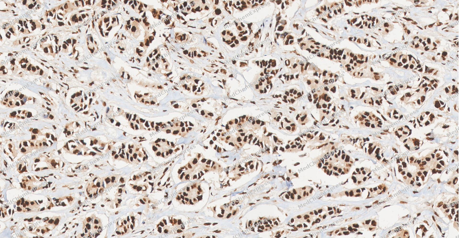 Immunohistochemical analysis of paraffin-embedded human Breast Cancer tissue using Phospho-MDM2 (Ser166) antibody was performed. The section was pretreated using high-temperature and high-pressure mediated EDTA antigen retrieval buffer (pH 9.0), for 5 minutes. The tissues were incubated with primary antibody (HY-P82143,1:100 dilution) at room temperature for 60 minutes. Detection was performed using an HRP conjugated compact polymer system. DAB was used as the chromogen. The tissues were counterstained with hematoxylin and mounted with neutral balsam mounting medium.
 -
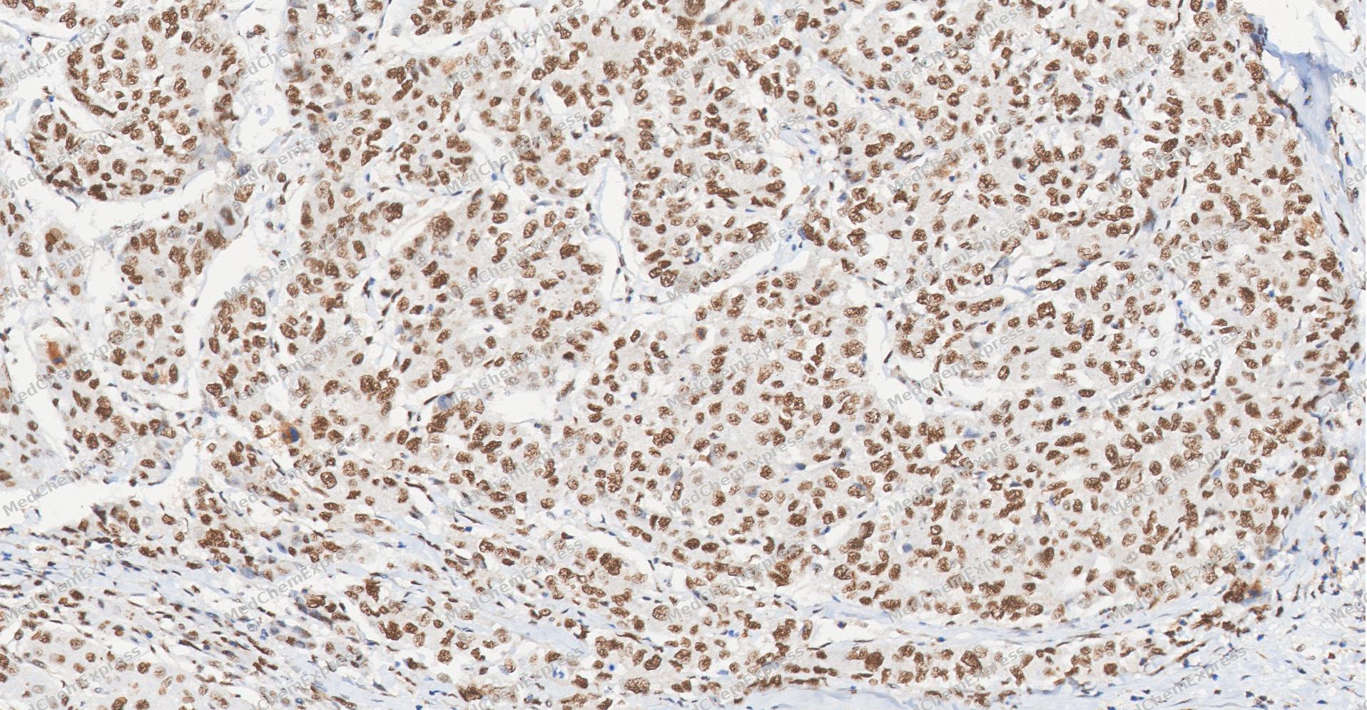 Immunohistochemical analysis of paraffin-embedded human Liver Cancer tissue using Phospho-MDM2 (Ser166) antibody was performed. The section was pretreated using high-temperature and high-pressure mediated EDTA antigen retrieval buffer (pH 9.0), for 5 minutes. The tissues were incubated with primary antibody (HY-P82143,1:100 dilution) at room temperature for 60 minutes. Detection was performed using an HRP conjugated compact polymer system. DAB was used as the chromogen. The tissues were counterstained with hematoxylin and mounted with neutral balsam mounting medium.
 -
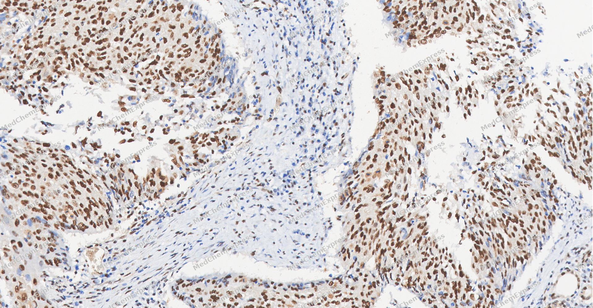 Immunohistochemical analysis of paraffin-embedded human Esophageal Carcinoma tissue using Phospho-MDM2 (Ser166) antibody was performed. The section was pretreated using high-temperature and high-pressure mediated EDTA antigen retrieval buffer (pH 9.0), for 5 minutes. The tissues were incubated with primary antibody (HY-P82143,1:100 dilution) at room temperature for 60 minutes. Detection was performed using an HRP conjugated compact polymer system. DAB was used as the chromogen. The tissues were counterstained with hematoxylin and mounted with neutral balsam mounting medium.
 -
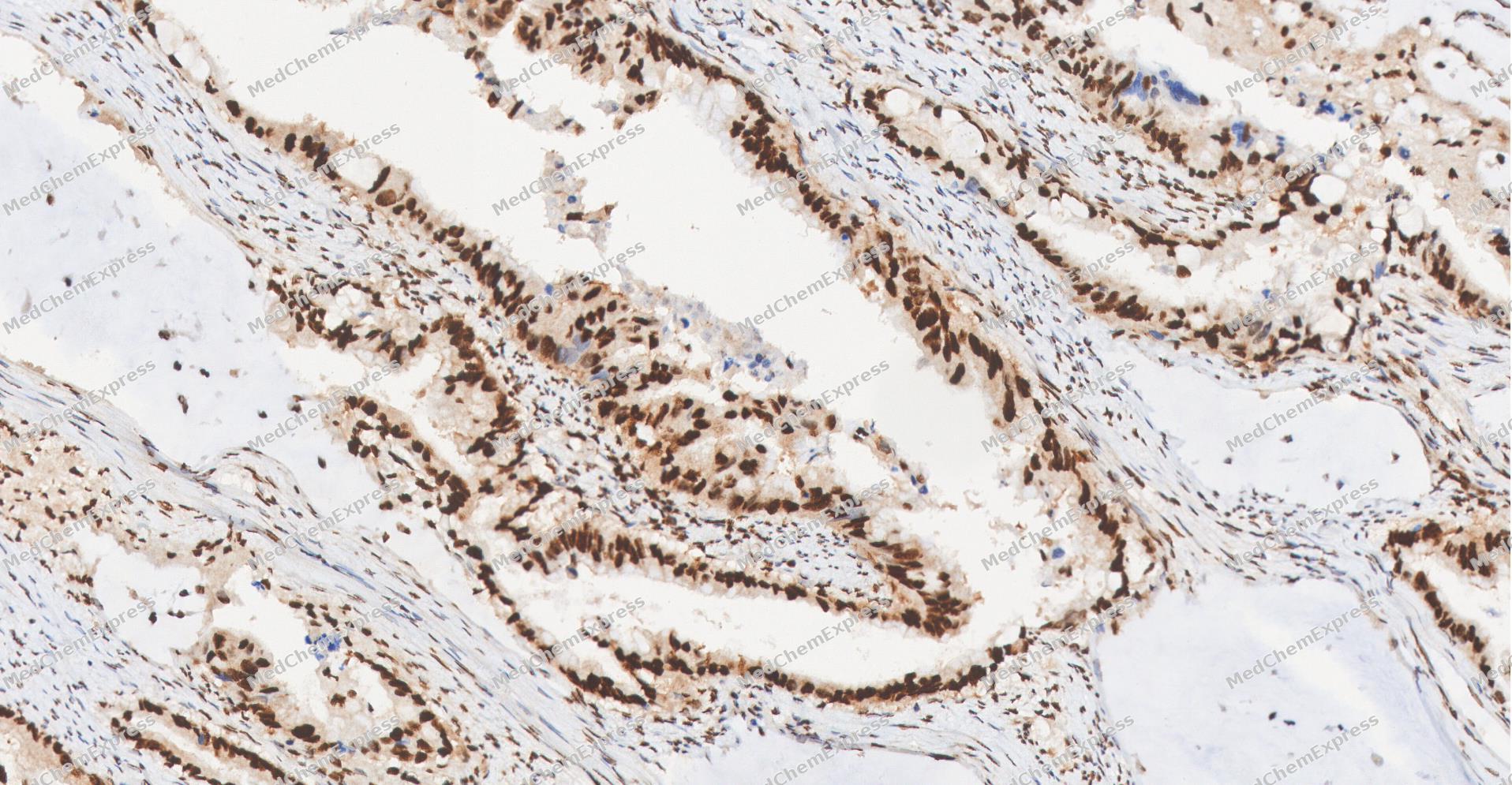 Immunohistochemical analysis of paraffin-embedded human Gastric Cancer tissue using Phospho-MDM2 (Ser166) antibody was performed. The section was pretreated using high-temperature and high-pressure mediated EDTA antigen retrieval buffer (pH 9.0), for 5 minutes. The tissues were incubated with primary antibody (HY-P82143,1:100 dilution) at room temperature for 60 minutes. Detection was performed using an HRP conjugated compact polymer system. DAB was used as the chromogen. The tissues were counterstained with hematoxylin and mounted with neutral balsam mounting medium.
 -
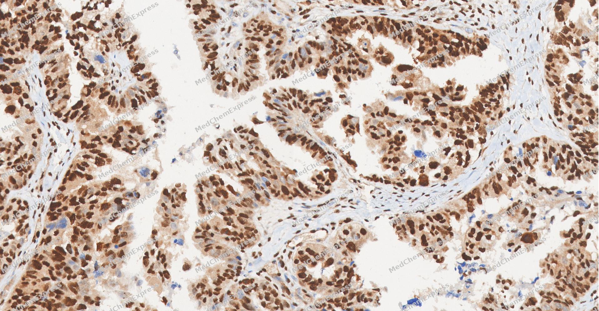 Immunohistochemical analysis of paraffin-embedded human Ovarian Cancer tissue using Phospho-MDM2 (Ser166) antibody was performed. The section was pretreated using high-temperature and high-pressure mediated EDTA antigen retrieval buffer (pH 9.0), for 5 minutes. The tissues were incubated with primary antibody (HY-P82143,1:100 dilution) at room temperature for 60 minutes. Detection was performed using an HRP conjugated compact polymer system. DAB was used as the chromogen. The tissues were counterstained with hematoxylin and mounted with neutral balsam mounting medium.
 -
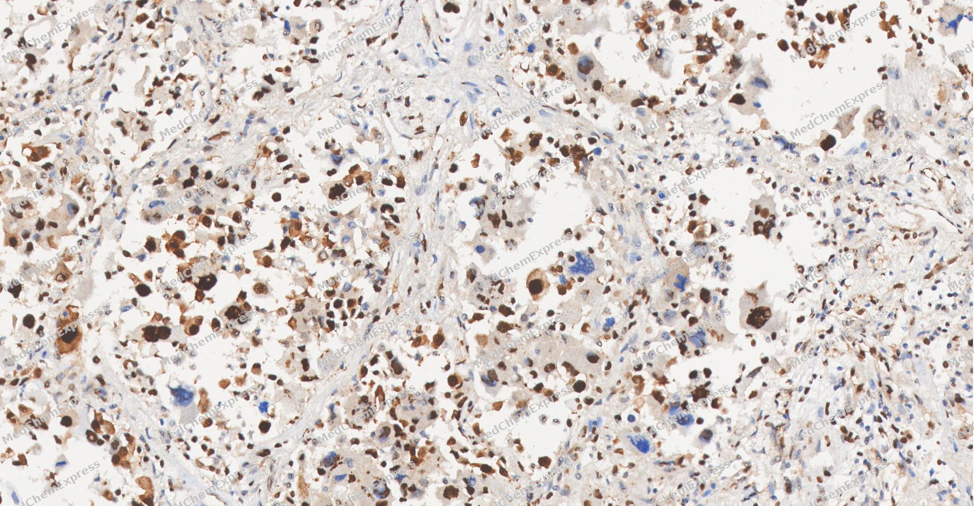 Immunohistochemical analysis of paraffin-embedded human Lung Adenocarcinoma tissue using Phospho-MDM2 (Ser166) antibody was performed. The section was pretreated using high-temperature and high-pressure mediated EDTA antigen retrieval buffer (pH 9.0), for 5 minutes. The tissues were incubated with primary antibody (HY-P82143,1:100 dilution) at room temperature for 60 minutes. Detection was performed using an HRP conjugated compact polymer system. DAB was used as the chromogen. The tissues were counterstained with hematoxylin and mounted with neutral balsam mounting medium.
 -
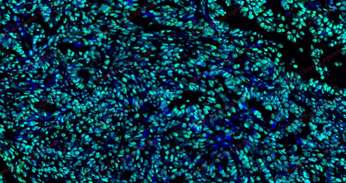 Tyramide signaling amplification based immunofluorescence was performed on paraffin-embedded human Ovarian Cancer tissue using Phospho-MDM2 (Ser166) antibody. Antigen retrieval was performed in EDTA buffer pH 9.0 (95 °C, 20 min) followed by cooling to RT. Then incubated with primary antibody (HY-P82143, 1:300 dilution) at room temperature for 60 minutes and HRP conjugated secondary antibody for 10 minutes. Fluorescence was then developed with Vari Fluor 532 TSA (200×)(HY-D1832). The tissues were counterstained with DAPI and mounted with Anti-fade mounting medium.
 -
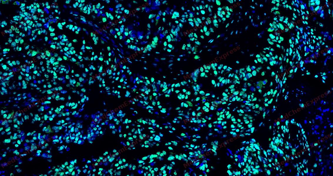 Tyramide signaling amplification based immunofluorescence was performed on paraffin-embedded human Ovarian Cancer tissue using Phospho-MDM2 (Ser166) antibody. Antigen retrieval was performed in EDTA buffer pH 9.0 (95 °C, 20 min) followed by cooling to RT. Then incubated with primary antibody (HY-P82143, 1:300 dilution) at room temperature for 60 minutes and HRP conjugated secondary antibody for 10 minutes. Fluorescence was then developed with Vari Fluor 532 TSA (200×)(HY-D1832). The tissues were counterstained with DAPI and mounted with Anti-fade mounting medium.
 -
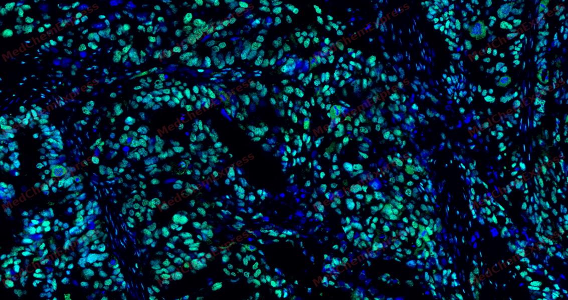 Tyramide signaling amplification based immunofluorescence was performed on paraffin-embedded human Ovarian Cancer tissue using Phospho-MDM2 (Ser166) antibody. Antigen retrieval was performed in EDTA buffer pH 9.0 (95 °C, 20 min) followed by cooling to RT. Then incubated with primary antibody (HY-P82143, 1:300 dilution) at room temperature for 60 minutes and HRP conjugated secondary antibody for 10 minutes. Fluorescence was then developed with Vari Fluor 532 TSA (200×)(HY-D1832). The tissues were counterstained with DAPI and mounted with Anti-fade mounting medium.
 -
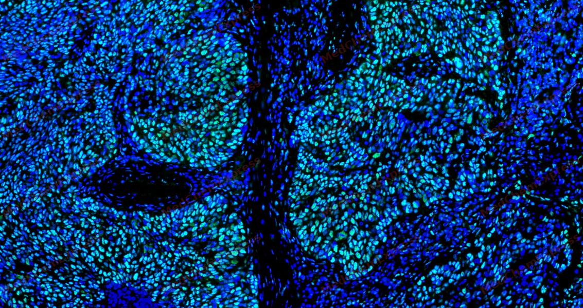 Tyramide signaling amplification based immunofluorescence was performed on paraffin-embedded human Esophageal Carcinoma tissue using Phospho-MDM2 (Ser166) antibody. Antigen retrieval was performed in EDTA buffer pH 9.0 (95 °C, 20 min) followed by cooling to RT. Then incubated with primary antibody (HY-P82143, 1:300 dilution) at room temperature for 60 minutes and HRP conjugated secondary antibody for 10 minutes. Fluorescence was then developed with Vari Fluor 532 TSA (200×)(HY-D1832). The tissues were counterstained with DAPI and mounted with Anti-fade mounting medium.
 -
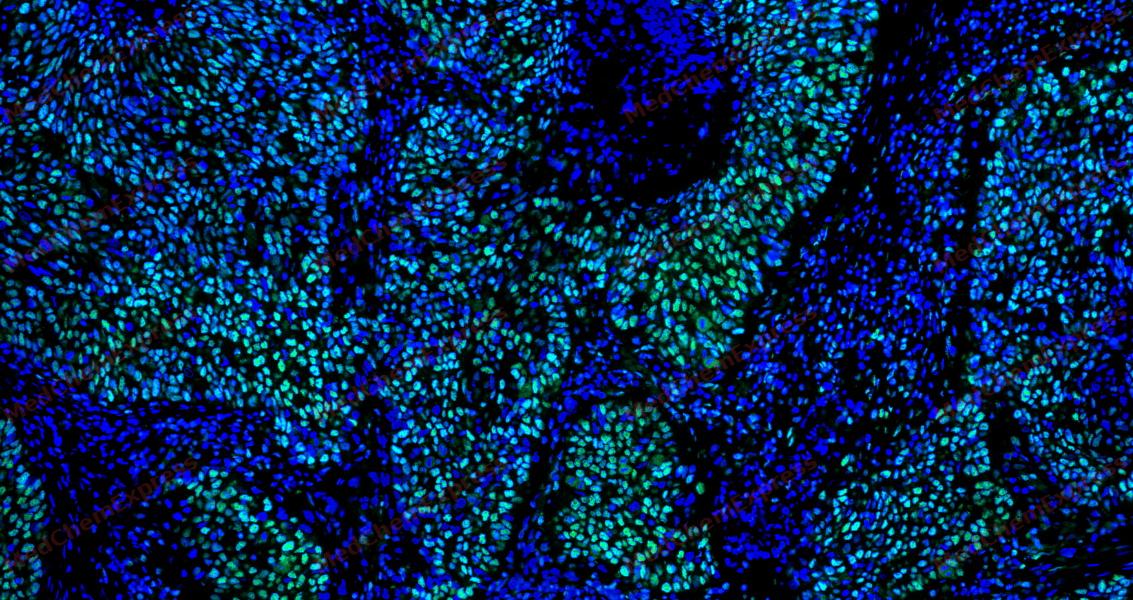 Tyramide signaling amplification based immunofluorescence was performed on paraffin-embedded human Esophageal Carcinoma tissue using Phospho-MDM2 (Ser166) antibody. Antigen retrieval was performed in EDTA buffer pH 9.0 (95 °C, 20 min) followed by cooling to RT. Then incubated with primary antibody (HY-P82143, 1:300 dilution) at room temperature for 60 minutes and HRP conjugated secondary antibody for 10 minutes. Fluorescence was then developed with Vari Fluor 532 TSA (200×)(HY-D1832). The tissues were counterstained with DAPI and mounted with Anti-fade mounting medium.
 -
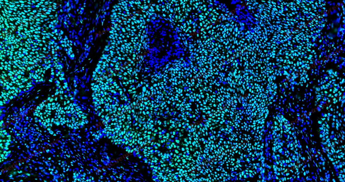 Tyramide signaling amplification based immunofluorescence was performed on paraffin-embedded human Esophageal Carcinoma tissue using Phospho-MDM2 (Ser166) antibody. Antigen retrieval was performed in EDTA buffer pH 9.0 (95 °C, 20 min) followed by cooling to RT. Then incubated with primary antibody (HY-P82143, 1:300 dilution) at room temperature for 60 minutes and HRP conjugated secondary antibody for 10 minutes. Fluorescence was then developed with Vari Fluor 532 TSA (200×)(HY-D1832). The tissues were counterstained with DAPI and mounted with Anti-fade mounting medium.


Background
-
Function
Murine double minute 2 (MDM2) is an E3 ubiquitin ligase that functions as a primary negative regulator of the tumor suppressor p53[1][2]. Mechanistically, MDM2 ubiquitinates p53, targeting it for proteasomal degradation, while also regulating additional transcription factors and post-transcriptional processes independently of p53[1][2][3]. MDM2 interacts with its homolog MDM4 (MDMX) through RING domain heterodimers, modulating protein stability and p53 activity[1][4]. Compared with MDM4, MDM2 possesses intrinsic E3 ligase activity and can degrade p53 even in the absence of MDM4, whereas MDM4 stabilizes the heterodimer complex without ubiquitin ligase function[1][4][5]. Dysregulation of MDM2 contributes to oncogenesis across multiple malignancies, including leukemia, neuroblastoma, breast cancer, and Theileria parva-induced lymphoproliferative disorders[4][6][7][8][9]. In experimental models, inhibition of MDM2 with small molecules such as RG7112, MI-63, CGM097, or XR-2 stabilizes p53, induces apoptosis, and can synergize with other pathway modulators to enhance anti-tumor activity[6][9][10][11][12]. Recent strategies exploit protein-protein interfaces, such as the MDM2-CK1α interaction, to induce targeted protein modifications and p53 activation independently of p53 status, providing tools for mechanistic studies and therapeutic design[13][14]. MDM2 inhibitors and dual MDM2/MDM4-targeting agents demonstrate isoform-specific effects, highlighting the importance of distinguishing functional contributions of MDM2 versus MDM4 in both experimental and therapeutic contexts[15][16][17].
-
Subcellular Localization
Nucleus, nucleoplasm; Cytoplasm; Nucleus, nucleolus; Nucleus
-
Expression
Tissue_specificity:They are ubiquitous. Mdm2-A, Mdm2-B, Mdm2-C, Mdm2-D, Mdm2-E, Mdm2-F, and Mdm2-G subtypes have been found in a variety of cancers, but are not present in normal tissues.
Induction:By DNA damage -
Isoforms & Post-Translational Modification
Q00987 has 11 isomers: Q00987-1: 55233 Da (predicted); Q00987-2: 33140 Da (predicted); Q00987-3: 30265 Da (predicted); Q00987-4: 24467 Da (predicted); Q00987-5: 35980 Da (predicted); Q00987-6: 14689 Da (predicted); Q00987-7: 11587 Da (predicted); Q00987-8: 48488 Da (predicted); Q00987-9: 49899 Da (predicted); Q00987-10: 49249 Da (predicted); Q00987-11: 55991 Da (predicted).
Phosphorylation on Ser-166 by SGK1 activates ubiquitination of p53/TP53 (PubMed:19756449). Phosphorylated at multiple sites near the RING domain by ATM upon DNA damage; this promotes its ubiquitination and degradation, preventing p53/TP53 degradation (PubMed:10611322, PubMed:12167711, PubMed:18382127, PubMed:19816404, PubMed:26124108);Autoubiquitination leads to proteasomal degradation; resulting in p53/TP53 activation it may be regulated by SFN (PubMed:18382127, PubMed:30879903). Also ubiquitinated by TRIM13 (PubMed:21333377). ATM-phosphorylated MDM2 is ubiquitinated by the SCF(FBXO31) complex in response to genotoxic stress, promoting its degradation and p53/TP53-mediated DNA damage response (PubMed:26124108). Deubiquitinated by USP2 leads to its accumulation and increases deubiquitination and degradation of p53/TP53 (PubMed:17290220). Deubiquitinated by USP7 leading to its stabilization (PubMed:15053880) -
Subunit
Interacts with p53/TP53, TP73/p73, RBL5 and RP11. Binds specifically to RNA. Can interact with RB1, E1A-associated protein EP300 and the E2F1 transcription factor. Forms a ternary complex with p53/TP53 and WWOX. Interacts with CDKN2AIP, RFWD3, USP7, PYHIN1, and RBBP6. Interacts with ARRB1 and ARRB2. Interacts with PSMA3. Found in a trimeric complex with MDM2, MDM4 and USP2. Interacts with USP2 (via N-terminus and C-terminus). Interacts with MDM4. Part of a complex with MDM2, DAXX, RASSF1 and USP7. Part of a complex with DAXX, MDM2 and USP7. Interacts directly with DAXX and USP7. Interacts (via C-terminus) with RASSF1 isoform A (via N-terminus); the interaction is independent of TP53. Interacts with APEX1; leading to its ubiquitination and degradation. Interacts with RYBP; this inhibits ubiquitination of TP53. Identified in a complex with RYBP and p53/TP53. Also a component of the TRIM28/KAP1-MDM2-p53/TP53 complex involved in regulating p53/TP53 stabilization and activity. Binds directly both p53/TP53 and TRIM28. Component of the TRIM28/KAP1-ERBB4-MDM2 complex involved in connecting growth factor responses with DNA damage. Interacts directly with both TRIM28 and ERBB4 in the complex. Interacts with DYRK2. Interacts with IGF1R. Interacts with TRIM13; the interaction ubiquitinates MDM2 leading to its proteasomal degradation. Interacts with SNAI1; this interaction promotes SNAI1 ubiquitination. Interacts with NOTCH1 (via intracellular domain). Interacts with FHIT. Interacts with RFFL and RNF34; the interaction stabilizes MDM2. Interacts with CDK5RAP3 and CDKN2A/ARF; form a ternary complex involved in regulation of p53/TP53 (PubMed:16173922). Interacts with MTA1. Interacts with AARB2. Interacts with MTBP. Interacts with PML. Interacts with TBRG1. Interacts (via its RanBP2-type zinc finger domain) with RPL11 in the 5S RNP complex composed of 5S RNA, RPL5 and RPL11; this interaction occurs in the nucleoplasm and negatively regulates MDM2-mediated TP53 ubiquitination and degradation (PubMed:15195100, PubMed:24120868, PubMed:37291423). Interacts with ADGRB1; the interaction results in inhibition of MDM2-mediated ubiquitination and degradation of DLG4/PSD95, promoting DLG4 stability and regulating synaptic plasticity (By similarity). Interacts with RPL23A; this interaction may promote p53/TP53 polyubiquitination (PubMed:26203195). Interacts with NDUFS1 (PubMed:30879903). Interacts with MORN3; the interaction enhances the ubiquitination of p53/TP53 (PubMed:29681526)
-
SwissProt ID
-
Synonyms
Double minute 2 protein; Hdm2; Oncoprotein Mdm2
-
Research Field
Neuroscience


Documentation
-
Data Sheet (264 KB)
-
SDS (251 KB)
- English - EN (251 KB)
- Français - FR (251 KB)
- Deutsch - DE (251 KB)
- Norwegian - NO (251 KB)
- Español - ES (251 KB)
- Swedish - SV (251 KB)
- Italian - IT (251 KB)
- Korean - KR (251 KB)
- Portuguese - PT (251 KB)
-
User Guide for Antibodies (1077 KB)
References
[1]. Zhu IY, et al. Structure and function of MDM2 and MDM4 in health and disease. Biochem J. 2025 Feb 17;482(4):BCJ20240757. [Content Brief]
[2]. Biderman L, et al. Mdm2 and MdmX as Regulators of Gene Expression. Genes Cancer. 2012 Mar;3(3-4):264-73. [Content Brief]
[3]. Thomasova D, et al. p53-independent roles of MDM2 in NF-κB signaling: implications for cancer therapy, wound healing, and autoimmune diseases. Neoplasia. 2012 Dec;14(12):1097-101. [Content Brief]
[4]. Lama R, et al. Small molecule MMRi62 targets MDM4 for degradation and induces leukemic cell apoptosis regardless of p53 status. Front Oncol. 2022 Aug 5;12:933446. [Content Brief]
[5]. Strachan GD, et al. A transcriptionally inactive E2F-1 targets the MDM family of proteins for proteolytic degradation. J Biol Chem. 2001 Dec 7;276(49):45677-85. [Content Brief]
[6]. Mitobe Y, et al. Antagonizing MDM2 Overexpression Induced by MDM4 Inhibitor CEP-1347 Effectively Reactivates Wild-Type p53 in Malignant Brain Tumor Cells. Cancers (Basel). 2023 Aug 30;15(17):4326. [Content Brief]
[7]. Hayashida K, et al. MDM2 regulates a novel form of incomplete neoplastic transformation of Theileria parva infected lymphocytes. Exp Mol Pathol. 2013 Feb;94(1):228-38. [Content Brief]
[8]. Qin JJ, et al. Experimental Therapy of Advanced Breast Cancer: Targeting NFAT1-MDM2-p53 Pathway. Prog Mol Biol Transl Sci. 2017;151:195-216. [Content Brief]
[9]. Maser T, et al. The MDM2 inhibitor CGM097 combined with the BET inhibitor OTX015 induces cell death and inhibits tumor growth in models of neuroblastoma. Cancer Med. 2020 Nov;9(21):8144-8158. [Content Brief]
[10]. Daniele S, et al. Long lasting inhibition of Mdm2-p53 interaction potentiates mesenchymal stem cell differentiation into osteoblasts. Biochim Biophys Acta Mol Cell Res. 2019 May;1866(5):737-749. [Content Brief]
[11]. Li Y, et al. Nonsense-mediated mRNA decay inhibition synergizes with MDM2 inhibition to suppress TP53 wild-type cancer cells in p53 isoform-dependent manner. Cell Death Discov. 2022;8(1):402. [Content Brief]
[12]. Gu D, et al. Inhibition of the MDM2 E3 Ligase induces apoptosis and autophagy in wild-type and mutant p53 models of multiple myeloma, and acts synergistically with ABT-737. PLoS One. 2014 Sep 2;9(9):e103015. [Content Brief]
[14]. Huart AS, et al. Exploiting the MDM2-CK1α protein-protein interface to develop novel biologics that induce UBL-kinase-modification and inhibit cell growth. PLoS One. 2012;7(8):e43391. [Content Brief]
[15]. Surmiak E, et al. A Unique Mdm2-Binding Mode of the 3-Pyrrolin-2-one- and 2-Furanone-Based Antagonists of the p53-Mdm2 Interaction. ACS Chem Biol. 2016 Dec 16;11(12):3310-3318. [Content Brief]
[17]. Jiang L, et al. Protoporphyrin IX is a dual inhibitor of p53/MDM2 and p53/MDM4 interactions and induces apoptosis in B-cell chronic lymphocytic leukemia cells. Cell Death Discov. 2019 Mar 11;5:77. [Content Brief]

 Powered by Bioz
