



Phospho-STAT5(Tyr694) Antibody (YA1513)
(Synonyms: STAT5A; STAT5; Signal transducer and activator of transcription 5A)Based on 1 Customer Validation
Phospho-STAT5(Tyr694) Antibody (YA1513) is a Rabbit-derived and non-conjugated IgG monoclonal antibody, targeting to Phospho-STAT5(Tyr694).
-
Host:
Rabbit
-
Isotype:
IgG
-
Application:
WB, IHC-P
-
Reactivity :
Human, Mouse
-
Formulation:
Supplied in 50mM Tris-Glycine(pH 7.4), 0.15M NaCl, 40%Glycerol, 0.01% sodium azide and 0.05% BSA.
-
Conjugation:
Non-conjugated


To place orders, for customer services and technical support, please contact: MedChemExpress USA
Tel: 609-228-6898 E-mail: [email protected] [email protected]





Applications
| Application |
WB

WB: Western Blot
|
IHC-P
IHC-P: Immunohistochemistry-Paraffin
|
|---|---|---|
| Dilution Ratio | 1:500-1:1000 | 1:50-1:100 |


Product Details
Phospho-STAT5(Tyr694) Antibody (YA1513) is a Rabbit-derived and non-conjugated IgG monoclonal antibody, targeting to Phospho-STAT5(Tyr694).
-
Host Rabbit

-
Clonality Recombinant,Monoclonal

-
Species ReactivityHuman, Mouse
-
Observed Molecular WeightObserved band size: 91 kDa
Note: Due to possible protein modifications or aggregation, the molecular weight should be confirmed by actual measurement, and the predicted value is for reference only.

-
Calculated Molecular Weight Predicted band size: 91 kDa

A synthesized peptide derived from human Phospho-Stat5 (Y694) DG-pY-VK.
Endogenous
Affinity Chromatography
Non-conjugated
Phosphorylated
IgG


Product Properties
-
Appearance
Solution
-
Formulation
Supplied in 50mM Tris-Glycine(pH 7.4), 0.15M NaCl, 40%Glycerol, 0.01% sodium azide and 0.05% BSA.
-
Concentration
Batch-dependent, Please check the COA for the concentration of each lot. Check Lot Concentration
-
Storage & Stability
Stored at -20°C for 1 year. Avoid repeated freeze / thaw cycles.
-
Shipping
Shipping with blue ice.


Verification Images
-
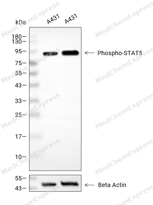 Western blot analysis of extracts from A431 (lane 2(20μg), A431 (lane 3(40μg), using Phospho-STAT5(Tyr694) Antibody. Proteins were transferred to a PVDF membrane and blocked with 5% BSA in TBST for 2 hour at room temperature. The primary antibody and Loading control antibody (Beta Actin, HY-P80438, 1/3000) was used in 5% BSA in TBST at 4°C overnight. Goat Anti-Mouse/Rabbit IgG-HRP Secondary Antibody (HY-P8004/HY-P8001, 1/10,000) was used for 1 hour at room temperature.
 -
 Immunohistochemical analysis of paraffin-embedded mouse ovary tissue using Phospho-STAT5(Tyr694) Antibody (HY-P81768, 1/100). The section was pretreated using heat mediated antigen retrieval with sodium citrate buffer (pH 6.0) for 8 minutes. The tissues were blocked with quick block buffer for 0.5 hours at room temperature, washed with PBS and PBST, and then incubated with the primary antibody overnight at 4℃. The detection was performed using an HRP conjugated compact polymer system. DAB was used as the chromogen. Tissues were counterstained with hematoxylin and mounted with DPX.
 -
 Immunohistochemical analysis of paraffin-embedded mouse spleen tissue using Phospho-STAT5(Tyr694) Antibody (HY-P81768, 1/100). The section was pretreated using heat mediated antigen retrieval with sodium citrate buffer (pH 6.0) for 8 minutes. The tissues were blocked with quick block buffer for 0.5 hours at room temperature, washed with PBS and PBST, and then incubated with the primary antibody overnight at 4℃. The detection was performed using an HRP conjugated compact polymer system. DAB was used as the chromogen. Tissues were counterstained with hematoxylin and mounted with DPX.
 -
 Immunohistochemical analysis of paraffin-embedded mouse adipose tissue using Phospho-STAT5(Tyr694) Antibody (HY-P81768, 1/100). The section was pretreated using heat mediated antigen retrieval with sodium citrate buffer (pH 6.0) for 8 minutes. The tissues were blocked with quick block buffer for 0.5 hours at room temperature, washed with PBS and PBST, and then incubated with the primary antibody overnight at 4℃. The detection was performed using an HRP conjugated compact polymer system. DAB was used as the chromogen. Tissues were counterstained with hematoxylin and mounted with DPX.
 -
 Immunohistochemical analysis of paraffin-embedded mouse lung tissue using Phospho-STAT5(Tyr694) Antibody (HY-P81768, 1/100). The section was pretreated using heat mediated antigen retrieval with sodium citrate buffer (pH 6.0) for 8 minutes. The tissues were blocked with quick block buffer for 0.5 hours at room temperature, washed with PBS and PBST, and then incubated with the primary antibody overnight at 4℃. The detection was performed using an HRP conjugated compact polymer system. DAB was used as the chromogen. Tissues were counterstained with hematoxylin and mounted with DPX.
 -
 Immunohistochemical analysis of paraffin-embedded mouse liver tissue using Phospho-STAT5(Tyr694) Antibody (HY-P81768, 1/100). The section was pretreated using heat mediated antigen retrieval with sodium citrate buffer (pH 6.0) for 8 minutes. The tissues were blocked with quick block buffer for 0.5 hours at room temperature, washed with PBS and PBST, and then incubated with the primary antibody overnight at 4℃. The detection was performed using an HRP conjugated compact polymer system. DAB was used as the chromogen. Tissues were counterstained with hematoxylin and mounted with DPX.
 -
 Immunohistochemical analysis of paraffin-embedded mouse kidney tissue using Phospho-STAT5(Tyr694) Antibody (HY-P81768, 1/100). The section was pretreated using heat mediated antigen retrieval with sodium citrate buffer (pH 6.0) for 8 minutes. The tissues were blocked with quick block buffer for 0.5 hours at room temperature, washed with PBS and PBST, and then incubated with the primary antibody overnight at 4℃. The detection was performed using an HRP conjugated compact polymer system. DAB was used as the chromogen. Tissues were counterstained with hematoxylin and mounted with DPX.


Background
-
Function
STAT5 comprises STAT5A and STAT5B, transcription factors that regulate hematopoietic cell proliferation, differentiation, and apoptosis[1]. Mechanistically, STAT5 tyrosine phosphorylation in peripheral T cells is driven by cytokine receptor signaling rather than the T-cell receptor complex[2]. This cytokine-linked pathway supports immune regulation, lymphocyte development, and cell survival programs[3]. In disease contexts, STAT5B deficiency is associated with growth failure, immunodeficiency, and severe pulmonary disease[4]. Compared with STAT5A, STAT5B shows dominant activity in effector and regulatory T-cell responses, with paralog dose explaining much functional specificity[5]. In BCR/ABL leukemia models, STAT5B, but not STAT5A, serves as the major STAT5 isoform driving leukemogenesis[6]. For experimental applications, AC-4-130 directly binds STAT5 and disrupts activation, dimerization, nuclear translocation, and STAT5-dependent transcription in AML models[7]. Pimozide also decreases survival of chronic myelogenous leukemia cells resistant to kinase inhibitors[8].
-
Subcellular Localization
Cytoplasm; Nucleus
-
Isoforms & Post-Translational Modification
P42229 has 2 isomers: P42229-1: 90647 Da (predicted); P42229-2: 86949 Da (predicted).
Tyrosine phosphorylated in response to KITLG/SCF, IL2, IL3, IL7, IL15, CSF2/GMCSF, GH1, PRL, EPO and THPO (By similarity). Activated KIT promotes phosphorylation on tyrosine residues and subsequent translocation to the nucleus (PubMed:21135090). Tyrosine phosphorylated in response to constitutively activated FGFR1, FGFR2, FGFR3 and FGFR4 (By similarity). Tyrosine phosphorylation is required for DNA-binding activity and dimerization. Serine phosphorylation is also required for maximal transcriptional activity (By similarity). Tyrosine phosphorylated in response to signaling via activated FLT3; wild-type FLT3 results in much weaker phosphorylation than constitutively activated mutant FLT3 (PubMed:14504097). Alternatively, can be phosphorylated by JAK2 at Tyr-694 (PubMed:12529425);ISGylated -
Subunit
Forms a homodimer or a heterodimer with a related family member. Binds NR3C1 (By similarity). Interacts with NCOA1 and SOCS7 (PubMed:12954634). Interacts with ERBB4 (PubMed:15534001). Interacts with EBF4 (PubMed:35939714). Interacts with CD69 (By similarity)
-
SwissProt ID
-
Synonyms
STAT5A; STAT5; Signal transducer and activator of transcription 5A
-
Research Field
Signal Transduction


Documentation
-
Data Sheet (262 KB)
-
SDS (252 KB)
- English - EN (252 KB)
- Français - FR (252 KB)
- Deutsch - DE (252 KB)
- Norwegian - NO (252 KB)
- Español - ES (252 KB)
- Swedish - SV (252 KB)
- Italian - IT (252 KB)
- Korean - KR (252 KB)
- Portuguese - PT (252 KB)
-
User Guide for Antibodies (1077 KB)
References
[1]. Nosaka T, et al. STAT5 as a molecular regulator of proliferation, differentiation and apoptosis in hematopoietic cells. EMBO J. 1999 Sep 1;18(17):4754-65. [Content Brief]
[2]. Moriggl R, et al. Stat5 activation is uniquely associated with cytokine signaling in peripheral T cells. Immunity. 1999 Aug;11(2):225-30. [Content Brief]
[3]. Smith MR, et al. STAT5b: A master regulator of key biological pathways. Front Immunol. 2023 Jan 23;13:1025373. [Content Brief]
[4]. Nadeau K, et al. STAT5b deficiency: an unsuspected cause of growth failure, immunodeficiency, and severe pulmonary disease. J Pediatr. 2011 May;158(5):701-8. [Content Brief]
[5]. Villarino A, et al. Signal transducer and activator of transcription 5 (STAT5) paralog dose governs T cell effector and regulatory functions. Elife. 2016 Mar 21;5:e08384. [Content Brief]
[6]. Kollmann S, et al. Twins with different personalities: STAT5B-but not STAT5A-has a key role in BCR/ABL-induced leukemia. Leukemia. 2019 Jul;33(7):1583-1597. [Content Brief]
[7]. Wingelhofer B, et al. Pharmacologic inhibition of STAT5 in acute myeloid leukemia. Leukemia. 2018 May;32(5):1135-1146. [Content Brief]
[8]. Nelson EA, et al. The STAT5 inhibitor pimozide decreases survival of chronic myelogenous leukemia cells resistant to kinase inhibitors. Blood. 2011 Mar 24;117(12):3421-9. [Content Brief]

 Powered by Bioz
