



PKC theta Antibody (YA1106)
(Synonyms: PRKCT; PRKCQ; nPKC-theta; Protein kinase C theta type)
Based on 1 Customer Validation
PKC theta Antibody (YA1106) is a Rabbit-derived and non-conjugated IgG monoclonal antibody, targeting to PKC theta.
-
Host:
Rabbit
-
Isotype:
IgG
-
Application:
IHC-P
-
Reactivity :
Human
-
Formulation:
Supplied in 50 mM Tris-Glycine, pH 7.4, 0.15 M NaCl, 40% Glycerol, 0.01% sodium azide and 0.05% BSA.
-
Conjugation:
Non-conjugated


To place orders, for customer services and technical support, please contact: MedChemExpress USA
Tel: 609-228-6898 E-mail: [email protected] [email protected]





Applications
| Application |
IHC-P

IHC-P: Immunohistochemistry-Paraffin
|
|---|---|
| Dilution Ratio | 1:1000-1:2000 |


Product Details
PKC theta Antibody (YA1106) is a Rabbit-derived and non-conjugated IgG monoclonal antibody, targeting to PKC theta.
-
Host Rabbit

-
Clonality Recombinant,Monoclonal

-
Species ReactivityHuman
-
Observed Molecular WeightObserved band size: 72-74 kDa
Note: Due to possible protein modifications or aggregation, the molecular weight should be confirmed by actual measurement, and the predicted value is for reference only.

-
Calculated Molecular Weight Predicted band size: 81 kDa

A synthesized peptide derived from human PRKCQ
Endogenous
Affinity Purified
Non-conjugated
Unmodified
IgG


Product Properties
-
Appearance
Liquid
-
Formulation
Supplied in 50 mM Tris-Glycine, pH 7.4, 0.15 M NaCl, 40% Glycerol, 0.01% sodium azide and 0.05% BSA.
-
Concentration
Batch-dependent, Please check the COA for the concentration of each lot. Check Lot Concentration
-
Storage & Stability
Stored at -20°C for 1 year. Avoid repeated freeze / thaw cycles.
-
Shipping
Shipping with blue ice.


Verification Images
-
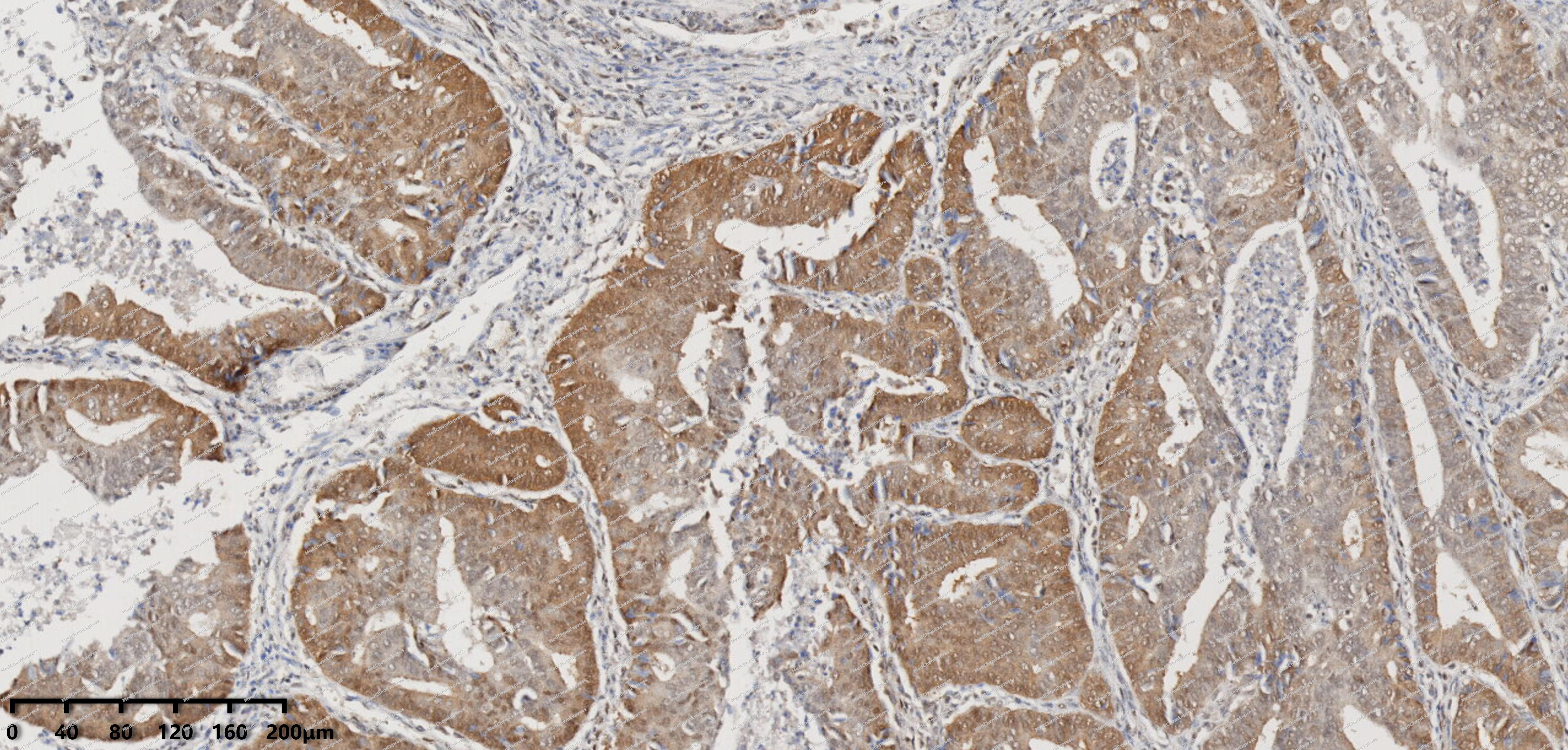 Immunohistochemical analysis of paraffin-embedded human Endometrial carcinoma tissue using PKC theta antibody was performed. The section was pretreated using high-temperature mediated EDTA antigen retrieval buffer (pH 9.0), for 20 minutes. The tissues were incubated with primary antibody (HY-P81361, 1:100 dilution) at room temperature for 20 minutes. Detection was performed using an HRP conjugated compact polymer system. DAB was used as the chromogen. The tissues were counterstained with hematoxylin and mounted with neutral balsam mounting medium.
 -
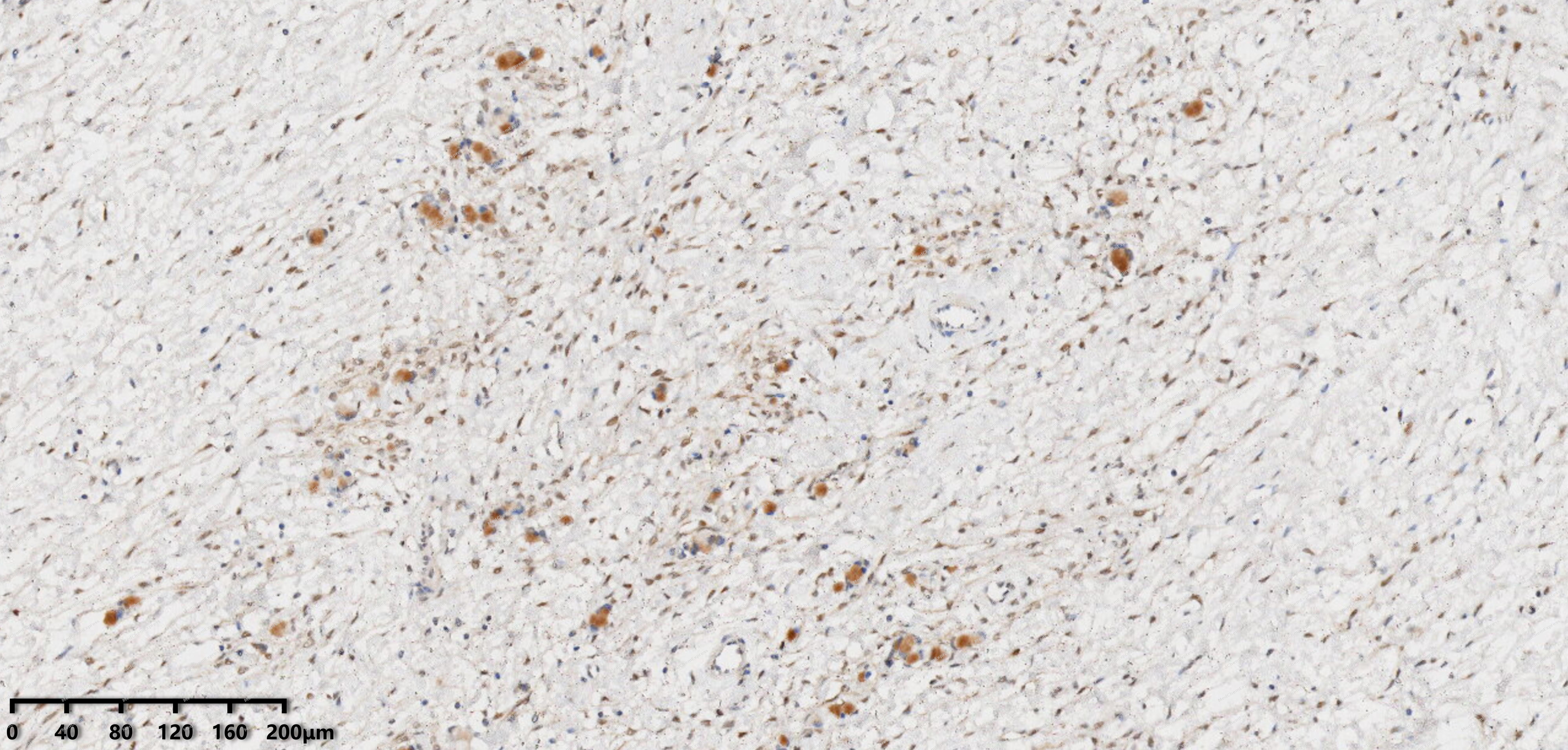 Immunohistochemical analysis of paraffin-embedded human uterine appendages tissue using PKC theta antibody was performed. The section was pretreated using high-temperature mediated EDTA antigen retrieval buffer (pH 9.0), for 20 minutes. The tissues were incubated with primary antibody (HY-P81361, 1:100 dilution) at room temperature for 20 minutes. Detection was performed using an HRP conjugated compact polymer system. DAB was used as the chromogen. The tissues were counterstained with hematoxylin and mounted with neutral balsam mounting medium.
 -
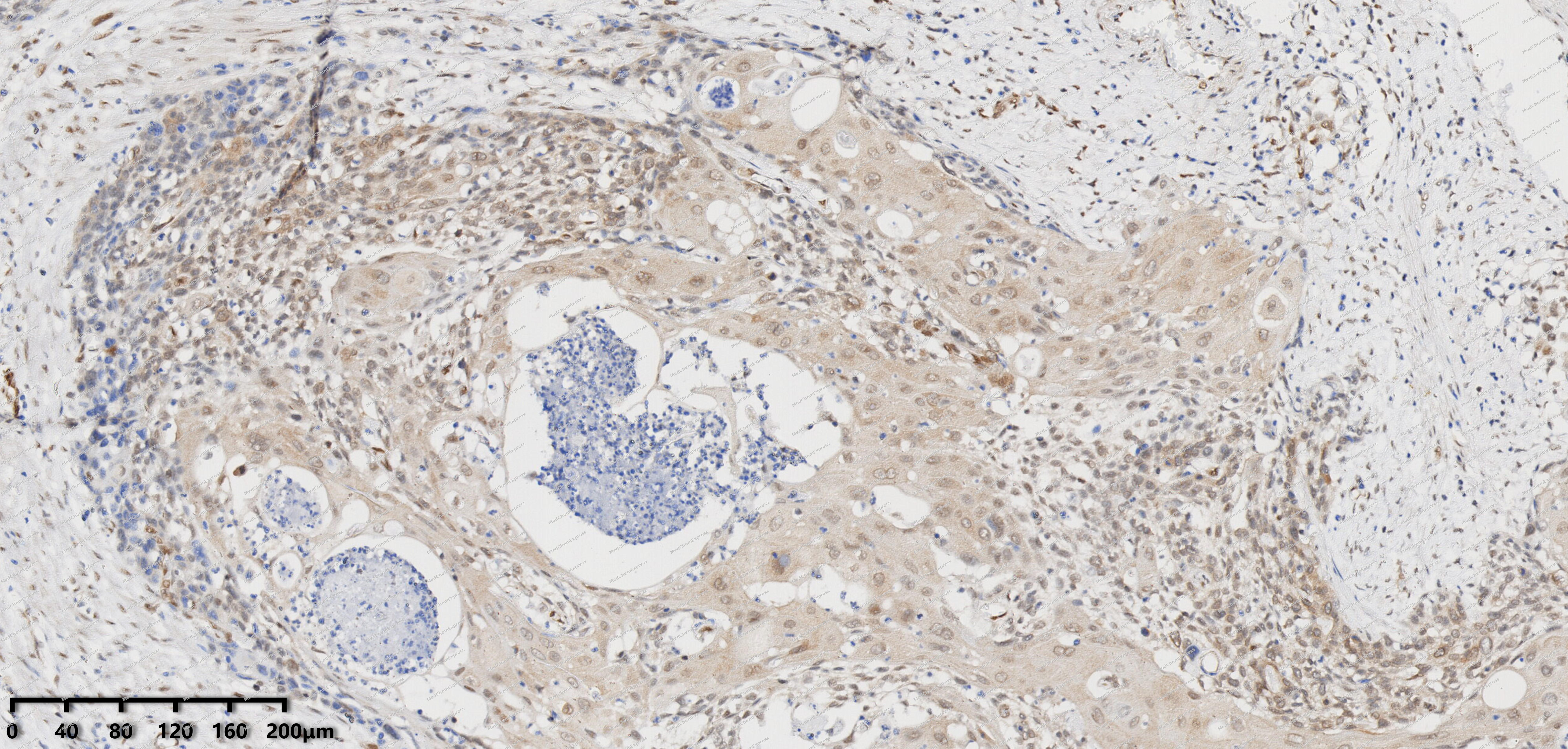 Immunohistochemical analysis of paraffin-embedded human esophagus tissue using PKC theta antibody was performed. The section was pretreated using high-temperature mediated EDTA antigen retrieval buffer (pH 9.0), for 20 minutes. The tissues were incubated with primary antibody (HY-P81361, 1:100 dilution) at room temperature for 20 minutes. Detection was performed using an HRP conjugated compact polymer system. DAB was used as the chromogen. The tissues were counterstained with hematoxylin and mounted with neutral balsam mounting medium.
 -
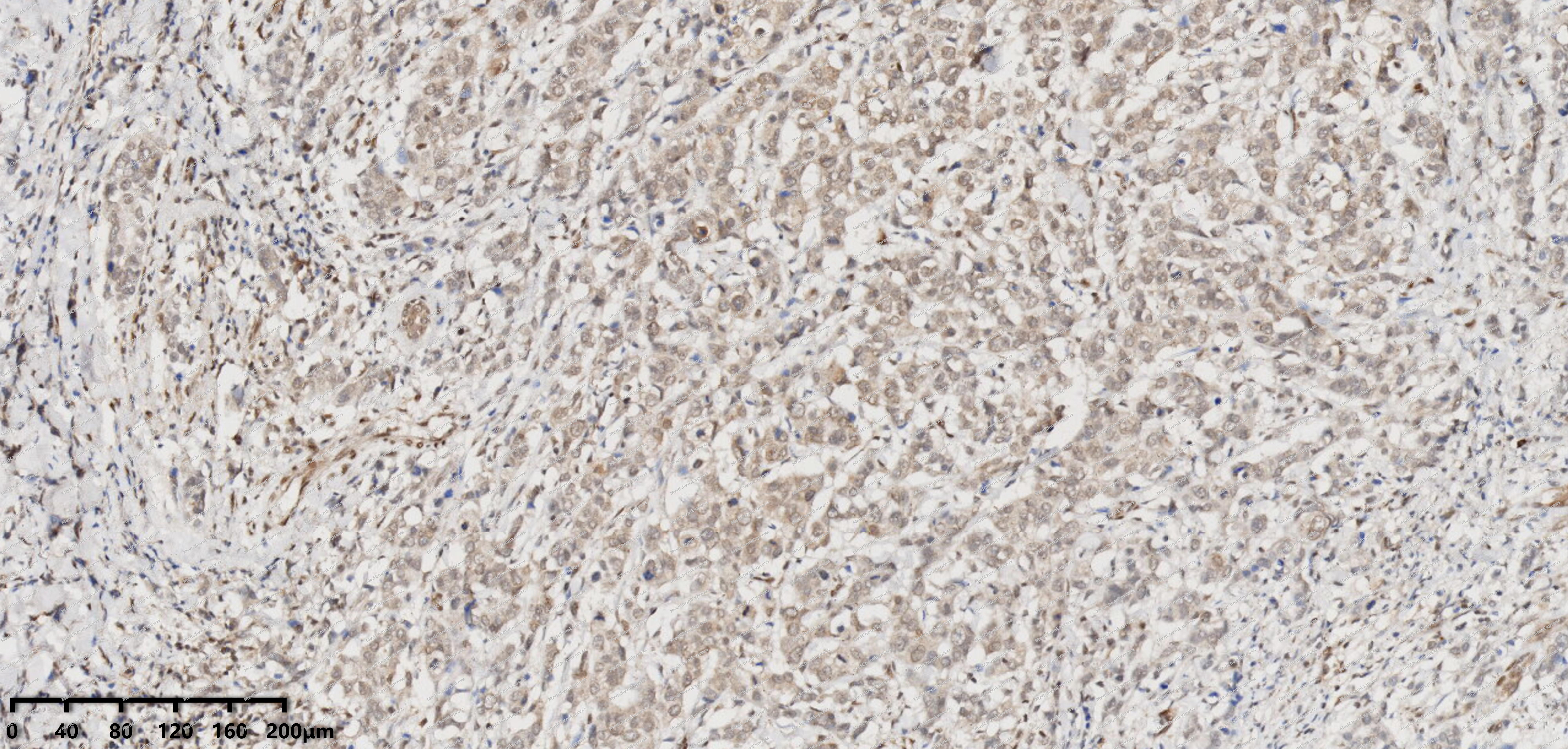 Immunohistochemical analysis of paraffin-embedded human breast cancer tissue using PKC theta antibody was performed. The section was pretreated using high-temperature mediated EDTA antigen retrieval buffer (pH 9.0), for 20 minutes. The tissues were incubated with primary antibody (HY-P81361, 1:100 dilution) at room temperature for 20 minutes. Detection was performed using an HRP conjugated compact polymer system. DAB was used as the chromogen. The tissues were counterstained with hematoxylin and mounted with neutral balsam mounting medium.
 -
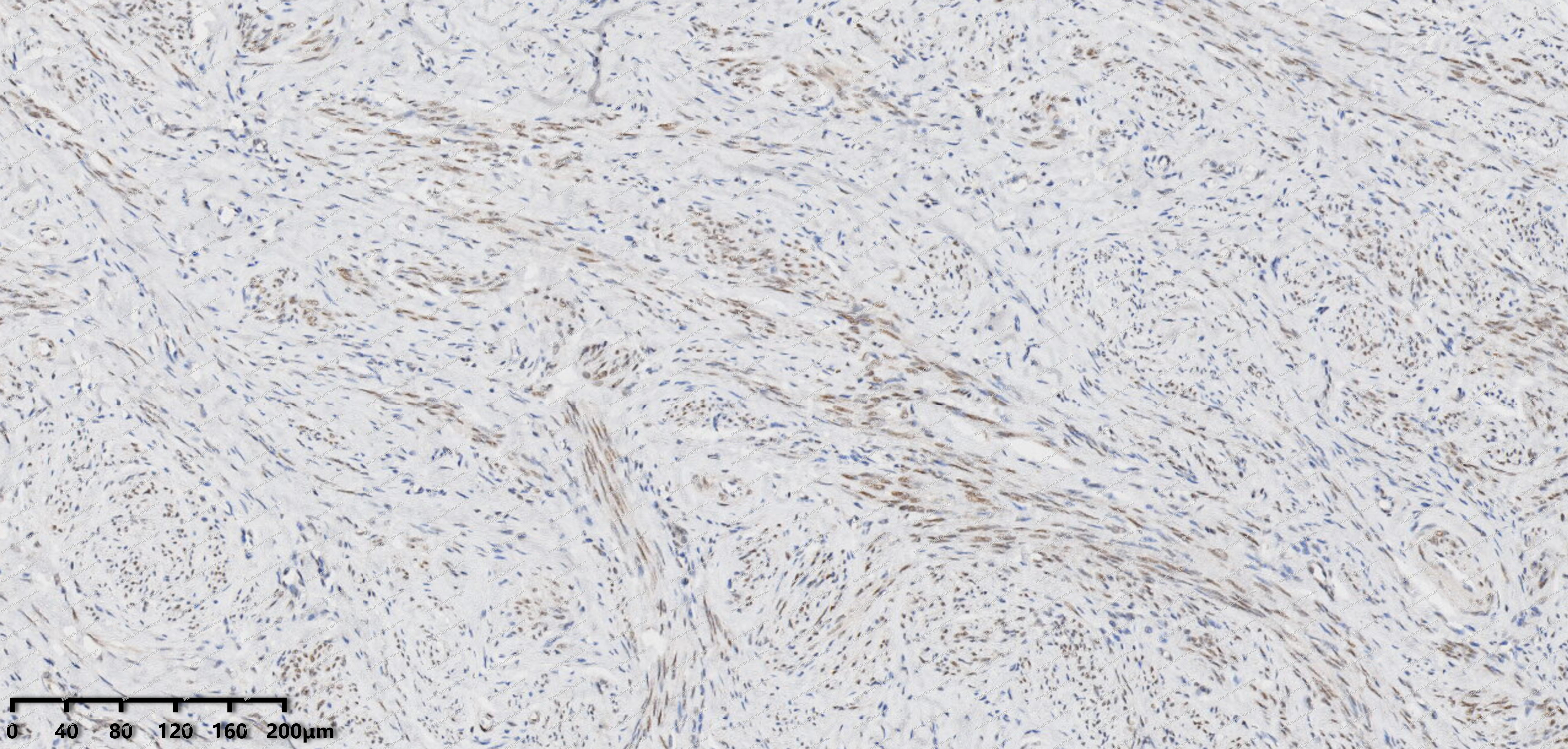 Immunohistochemical analysis of paraffin-embedded human cervix of uterus tissue using PKC theta antibody was performed. The section was pretreated using high-temperature mediated EDTA antigen retrieval buffer (pH 9.0), for 20 minutes. The tissues were incubated with primary antibody (HY-P81361, 1:100 dilution) at room temperature for 20 minutes. Detection was performed using an HRP conjugated compact polymer system. DAB was used as the chromogen. The tissues were counterstained with hematoxylin and mounted with neutral balsam mounting medium.
 -
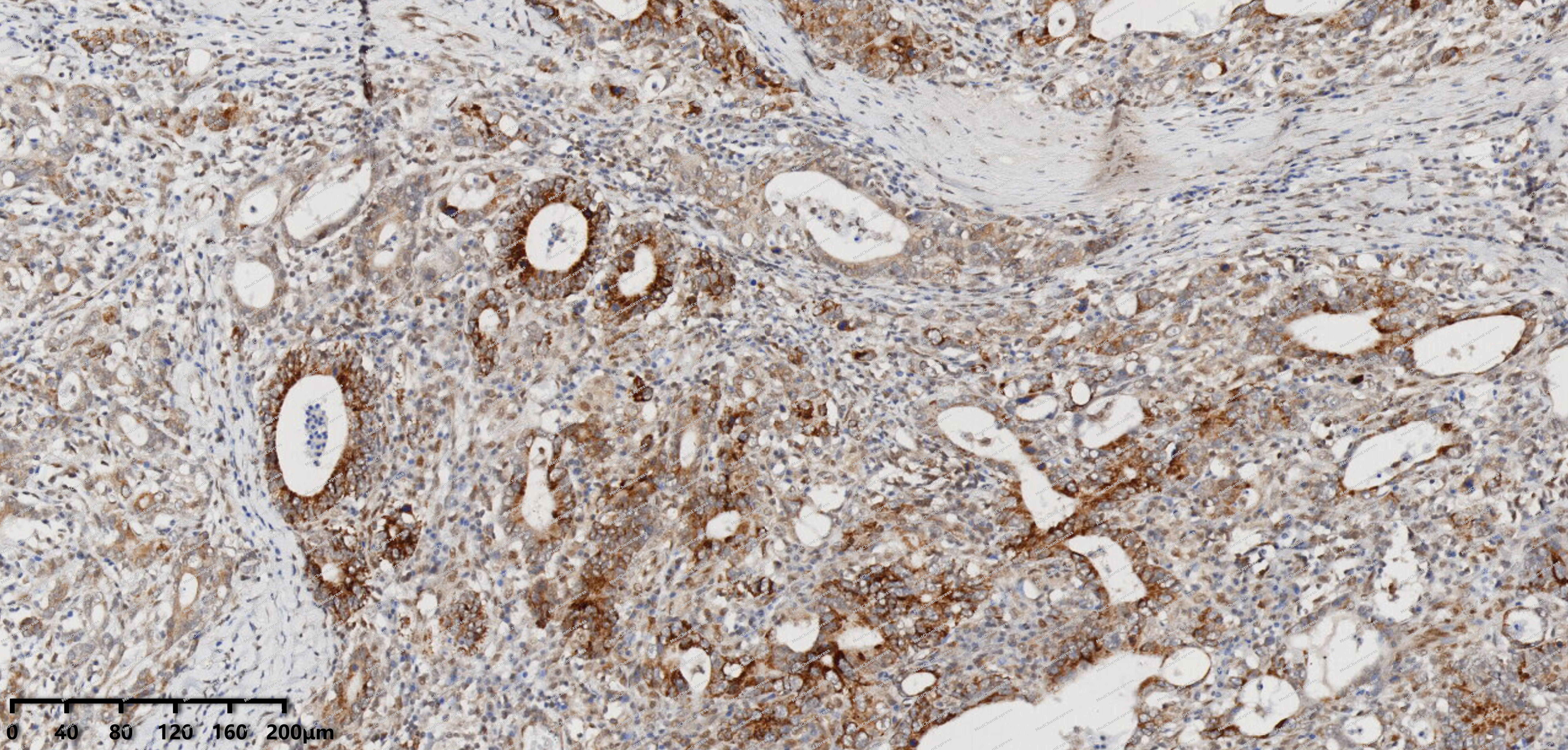 Immunohistochemical analysis of paraffin-embedded human gastric cancer tissue using PKC theta antibody was performed. The section was pretreated using high-temperature mediated EDTA antigen retrieval buffer (pH 9.0), for 20 minutes. The tissues were incubated with primary antibody (HY-P81361, 1:100 dilution) at room temperature for 20 minutes. Detection was performed using an HRP conjugated compact polymer system. DAB was used as the chromogen. The tissues were counterstained with hematoxylin and mounted with neutral balsam mounting medium.


Background
-
Function
PKC theta is a Calcium-independent, phospholipid- and diacylglycerol (DAG)-dependent serine/threonine-protein kinase that mediates non-redundant functions in T-cell receptor (TCR) signaling, including T-cells activation, proliferation, differentiation and survival, by mediating activation of multiple transcription factors such as NF-kappa-B, JUN, NFATC1 and NFATC2. In TCR-CD3/CD28-co-stimulated T-cells, is required for the activation of NF-kappa-B and JUN, which in turn are essential for IL2 production, and participates in the calcium-dependent NFATC1 and NFATC2 transactivation. Mediates the activation of the canonical NF-kappa-B pathway (NFKB1) by direct phosphorylation of CARD11 on several serine residues, inducing CARD11 association with lipid rafts and recruitment of the BCL10-MALT1 complex, which then activates IKK complex, resulting in nuclear translocation and activation of NFKB1. May also play an indirect role in activation of the non-canonical NF-kappa-B (NFKB2) pathway. In the signaling pathway leading to JUN activation, acts by phosphorylating the mediator STK39/SPAK and may not act through MAP kinases signaling. Plays a critical role in TCR/CD28-induced NFATC1 and NFATC2 transactivation by participating in the regulation of reduced inositol 1,4,5-trisphosphate generation and intracellular calcium mobilization. After costimulation of T-cells through CD28 can phosphorylate CBLB and is required for the ubiquitination and subsequent degradation of CBLB, which is a prerequisite for the activation of TCR. During T-cells differentiation, plays an important role in the development of T-helper 2 (Th2) cells following immune and inflammatory responses, and, in the development of inflammatory autoimmune diseases, is necessary for the activation of IL17-producing Th17 cells. May play a minor role in Th1 response. Upon TCR stimulation, mediates T-cell protective survival signal by phosphorylating BAD, thus protecting T-cells from BAD-induced apoptosis, and by up-regulating BCL-X(L)/BCL2L1 levels through NF-kappa-B and JUN pathways. In platelets, regulates signal transduction downstream of the ITGA2B, CD36/GP4, F2R/PAR1 and F2RL3/PAR4 receptors, playing a positive role in 'outside-in' signaling and granule secretion signal transduction. May relay signals from the activated ITGA2B receptor by regulating the uncoupling of WASP and WIPF1, thereby permitting the regulation of actin filament nucleation and branching activity of the Arp2/3 complex. May mediate inhibitory effects of free fatty acids on insulin signaling by phosphorylating IRS1, which in turn blocks IRS1 tyrosine phosphorylation and downstream activation of the PI3K/AKT pathway. Phosphorylates MSN (moesin) in the presence of phosphatidylglycerol or phosphatidylinositol. Phosphorylates PDPK1 at 'Ser-504' and 'Ser-532' and negatively regulates its ability to phosphorylate PKB/AKT1. Phosphorylates CCDC88A/GIV and inhibits its guanine nucleotide exchange factor activity. Phosphorylates and activates LRRK1, which phosphorylates RAB proteins involved in intracellular trafficking[1][2][3].
-
Subcellular Localization
Cytoplasm; Cell membrane; Peripheral membrane protein
-
Expression
Tissue_specificity:It is expressed in skeletal muscle, T cells, megakaryocytes, and platelets. -
Isoforms & Post-Translational Modification
Q04759 has 3 isomers: Q04759-1: 81865 Da (predicted); Q04759-2: 74287 Da (predicted); Q04759-3: 67560 Da (predicted).
Autophosphorylation at Thr-219 is required for targeting to the TCR and cellular function of PRKCQ upon antigen receptor ligation. Following TCR stimulation, phosphorylated at Tyr-90 and Ser-685 -
Subunit
Part of a lipid raft complex composed at least of BCL10, CARD11, MALT1 and IKBKB (PubMed:16356855). Interacts with GLRX3 (via N-terminus) (PubMed:10636891). Interacts with ECT2 (PubMed:15254234). Interacts with CCDC88A/GIV; the interaction leads to phosphorylation of CCDC88A and inhibition of its guanine nucleotide exchange factor activity (PubMed:23509302). Interacts with PRKCH upstream open reading frame 2; the interaction leads to inhibition of kinase activity (PubMed:34593629). Interacts with CD28 (PubMed:21964608)
-
SwissProt ID
-
Synonyms
PRKCT; PRKCQ; nPKC-theta; Protein kinase C theta type
-
Research Field
Signal Transduction


Documentation
-
Data Sheet (234 KB)
-
SDS (252 KB)
- English - EN (252 KB)
- Français - FR (252 KB)
- Deutsch - DE (252 KB)
- Norwegian - NO (252 KB)
- Español - ES (252 KB)
- Swedish - SV (252 KB)
- Italian - IT (252 KB)
- Korean - KR (252 KB)
- Portuguese - PT (252 KB)
-
User Guide for Antibodies (1077 KB)

 Powered by Bioz
