



REEP2 Antibody (YA1850)
(Synonyms: Receptor expression-enhancing protein 2)
Based on 1 Customer Validation
REEP2 Antibody (YA1850) is a Rabbit-derived and non-conjugated IgG monoclonal antibody, targeting to REEP2.
-
Host:
Rabbit
-
Isotype:
IgG
-
Application:
WB
-
Reactivity :
Human
-
Formulation:
Supplied in 50mM Tris-Glycine(pH 7.4), 0.15M NaCl, 40% Glycerol, 0.01% Sodium azide and 0.05% BSA
-
Conjugation:
Non-conjugated


To place orders, for customer services and technical support, please contact: MedChemExpress USA
Tel: 609-228-6898 E-mail: [email protected] [email protected]





Applications
| Application |
WB

WB: Western Blot
|
|---|---|
| Dilution Ratio | 1:1000-1:5000 |


Product Details
REEP2 Antibody (YA1850) is a Rabbit-derived and non-conjugated IgG monoclonal antibody, targeting to REEP2.
-
Host Rabbit

-
Clonality Recombinant,Monoclonal

-
Species ReactivityHuman
-
Observed Molecular WeightObserved band size: 28 kDa
Note: Due to possible protein modifications or aggregation, the molecular weight should be confirmed by actual measurement, and the predicted value is for reference only.

-
Calculated Molecular Weight Predicted band size: 28 kDa

A synthetic peptide of human REEP2 aa 161-174.
Endogenous
Affinity Purified
Non-conjugated
Unmodified
IgG


Product Properties
-
Appearance
Solution
-
Formulation
Supplied in 50mM Tris-Glycine(pH 7.4), 0.15M NaCl, 40% Glycerol, 0.01% Sodium azide and 0.05% BSA
-
Concentration
Batch-dependent, Please check the COA for the concentration of each lot. Check Lot Concentration
-
Storage & Stability
Stored at -20°C for 1 year. Avoid repeated freeze / thaw cycles.
-
Shipping
Shipping with blue ice.


Verification Images
-
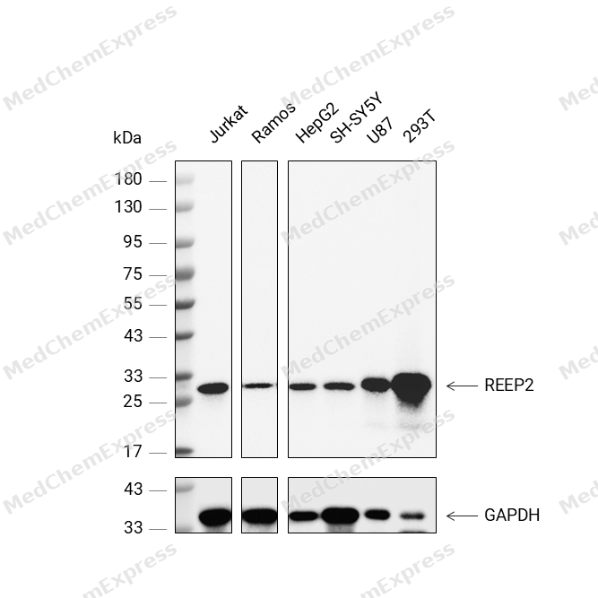 Western blot analysis was performed on protein extracts (25 μg) from Jurkat (lane 2), Ramos (lane 3), HepG2 (lane 4), SH-SY5Y (lane 5), U87 (lane 6), and 293T (lane 7) using REEP2 antibody. Proteins were transferred onto a 0.45 μm PVDF membrane using the Trans-Blot® Turbo™ system for 13 min. The membrane was then blocked with 5% nonfat milk in TBST (HY-K1025) for 1 h at room temperature. The primary antibody (1:1000) and loading control antibody GAPDH Antibody (HRP) (HY-P80954A) (1:5000) were diluted in 5% nonfat milk in TBST and incubated with the membrane overnight at 4°C. After washing, the membrane of primary antibody was incubated with HRP-conjugated goat anti-rabbit/mouse IgG secondary antibody (HY-P8001/HY-P8004) (1:5000) diluted in 5% nonfat milk in TBST for 1 h at room temperature. Protein bands were visualized using an Ultra High Sensitivity ECL detection kit (HY-K1005).


Background
-
Function
REEP2 is required for endoplasmic reticulum (ER) network formation, shaping and remodeling. May enhance the cell surface expression of odorant receptors
-
Subcellular Localization
Membrane; Multi-pass membrane protein
-
Expression
Tissue_specificity:Detected in brain, heart and skeletal muscle, and at low levels in placenta, kidney and pancreas (PubMed:11161817) . Expressed in circumvallate papillae (PubMed:16720576) -
Isoforms & Post-Translational Modification
Q9BRK0 has 2 isomers: Q9BRK0-1: 28261 Da (predicted); Q9BRK0-2: 28446 Da (predicted).
-
Subunit
Interacts with odorant receptor proteins
-
SwissProt ID
-
Synonyms
Receptor expression-enhancing protein 2
-
Research Field
Neuroscience


Documentation

 Powered by Bioz
