



RPS6 Antibody (YA677)
(Synonyms: RPS6; OK/SW-cl.2; 40S ribosomal protein S6; Phosphoprotein NP33)
Based on 1 Customer Validation
RPS6 Antibody (YA677) is a Mouse-derived and non-conjugated IgG1 monoclonal antibody, targeting to RPS6.
-
Host:
Mouse
-
Isotype:
IgG
-
Application:
WB, ICC/IF, IP
-
Reactivity :
Human, Mouse, Rat, Monkey
-
Formulation:
Supplied in 1*PBS (pH 7.3), 50% glycerol and 0.5% BSA. Preservative: 0.02% sodium azide.
-
Conjugation:
Non-conjugated


To place orders, for customer services and technical support, please contact: MedChemExpress USA
Tel: 609-228-6898 E-mail: [email protected] [email protected]





Applications
| Application |
WB

WB: Western Blot
|
ICC/IF
ICC/IF: Immunocytochemistry/
Immunofluorescence |
IP
IP: Immunoprecipitation
|
|---|---|---|---|
| Dilution Ratio | 1:500-1:1000 | 1:50-1:200 | 1:20 |


Product Details
RPS6 Antibody (YA677) is a Mouse-derived and non-conjugated IgG1 monoclonal antibody, targeting to RPS6.
-
Host Mouse

-
Clonality Monoclonal

-
Species ReactivityHuman, Mouse, Rat, Monkey
-
Observed Molecular WeightObserved band size: 32 kDa
Note: Due to possible protein modifications or aggregation, the molecular weight should be confirmed by actual measurement, and the predicted value is for reference only.

-
Calculated Molecular Weight Predicted band size: 29 kDa

Entrez Gene: 6194 Human ; 20104 Mouse ; 29304 Rat
SwissProt: P62753 Human ; P62754 Mouse ; P62755 Rat
OMIM: 180460 Human
Synthetic peptide corresponding to Human S6 Ribosomal.AA range:full length.
Endogenous
affinity purified
Non-conjugated
Unmodified
IgG


Product Properties
-
Appearance
Solution
-
Formulation
Supplied in 1*PBS (pH 7.3), 50% glycerol and 0.5% BSA. Preservative: 0.02% sodium azide.
-
Concentration
Batch-dependent, Please check the COA for the concentration of each lot. Check Lot Concentration
-
Storage & Stability
Stored at -20°C for 1 year. Avoid repeated freeze / thaw cycles.
-
Shipping
Shipping with blue ice.


Verification Images
-
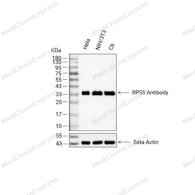 Western blot analysis of extracts from Hela(lane 2(20ug) ,NIH/3T3(lane 3(20ug) and C6(lane 4(20ug) using RPS6 Antibody (HY-P80885) Rabbit mAb. Proteins were transferred to a PVDF membrane and blocked with 5% non-fat milk in TBST for 2 hour at room temperature. The primary antibody (1/1000) and Loading control antibody (Beta Actin, HY-P83730, 1/10000) was used in 5% non-fat milk in TBST at 4°C overnight. Goat Anti-Mouse/Rabbit IgG-HRP Secondary Antibody (1/10000) was used for 1 hour at room temperature.
 -
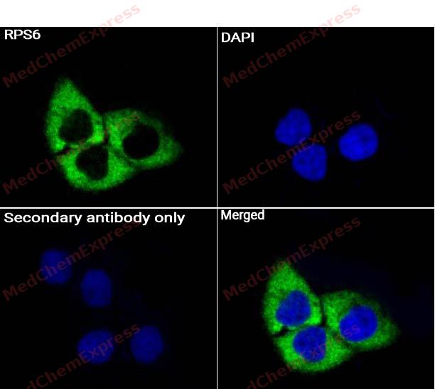 Immunocytochemistry analysis of Hela cells labeling RPS6 Antibody (HY-P80885) at 1/50 dilution. Cells were fixed in 4% paraformaldehyde for 15 minutes at room temperature, permeabilized with 0.1% Triton X-100 for 10 minutes at room temperature, then blocked with QuickBlock™ Blocking Buffer for Immunol Staining for 10 min at room temperature. Cells were then incubated with RPS6 Antibody (HY-P80885) at 1/50 dilution in QuickBlock™ Blocking Buffer for Immunol Staining at 4 ℃. Alexa Fluor® 488-conjugated AffiniPure Goat Anti-Rabbit IgG H&L(HY-P8002, Green) was used as the secondary antibody at 1/1,000 dilution. PBS instead of the primary antibody was used as the secondary antibody only control. The Nuclear counterstain was DAPI (Blue).
 -
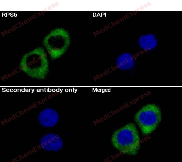 Immunocytochemistry analysis of Hela cells labeling RPS6 Antibody (HY-P80885) at 1/150 dilution. Cells were fixed in 4% paraformaldehyde for 15 minutes at room temperature, permeabilized with 0.1% Triton X-100 for 10 minutes at room temperature, then blocked with QuickBlock™ Blocking Buffer for Immunol Staining for 10 min at room temperature. Cells were then incubated with RPS6 Antibody (HY-P80885) at 1/150 dilution in QuickBlock™ Blocking Buffer for Immunol Staining at 4 ℃. Alexa Fluor® 488-conjugated AffiniPure Goat Anti-Rabbit IgG H&L(HY-P8002, Green) was used as the secondary antibody at 1/1,000 dilution. PBS instead of the primary antibody was used as the secondary antibody only control. The Nuclear counterstain was DAPI (Blue).


Background
-
Function
RPS6 component of the 40S small ribosomal subunit. Plays an important role in controlling cell growth and proliferation through the selective translation of particular classes of mRNA. Part of the small subunit (SSU) processome, first precursor of the small eukaryotic ribosomal subunit. During the assembly of the SSU processome in the nucleolus, many ribosome biogenesis factors, an RNA chaperone and ribosomal proteins associate with the nascent pre-rRNA and work in concert to generate RNA folding, modifications, rearrangements and cleavage as well as targeted degradation of pre-ribosomal RNA by the RNA exosome[1][2][3][4].
-
Subcellular Localization
Cytoplasm; Nucleus, nucleolus
-
Subunit
Component of the small ribosomal subunit. Part of the small subunit (SSU) processome, composed of more than 70 proteins and the RNA chaperone small nucleolar RNA (snoRNA) U3 (PubMed:34516797)
-
SwissProt ID
-
Synonyms
RPS6; OK/SW-cl.2; 40S ribosomal protein S6; Phosphoprotein NP33
-
Research Field
Epigenetics and Nuclear Signaling


Documentation
-
Data Sheet (262 KB)
-
SDS (251 KB)
- English - EN (251 KB)
- Français - FR (251 KB)
- Deutsch - DE (251 KB)
- Norwegian - NO (251 KB)
- Español - ES (251 KB)
- Swedish - SV (251 KB)
- Italian - IT (251 KB)
- Korean - KR (251 KB)
- Portuguese - PT (251 KB)
-
User Guide for Antibodies (1077 KB)
References
[1]. Anger AM, et al. Structures of the human and Drosophila 80S ribosome. Nature. 2013 May 2;497(7447):80-5. [Content Brief]
[2]. Vladimirov SN, et al. Characterization of the human small-ribosomal-subunit proteins by N-terminal and internal sequencing, and mass spectrometry. Eur J Biochem. 1996 Jul 1;239(1):144-9. [Content Brief]
[3]. Yamashita D, et al. hDREF regulates cell proliferation and expression of ribosomal protein genes. Mol Cell Biol. 2007 Mar;27(6):2003-13. [Content Brief]
[4]. Singh S, et al. Nucleolar maturation of the human small subunit processome. Science. 2021 Sep 10;373(6560):eabj5338. [Content Brief]

 Powered by Bioz
