



Tau Antibody (YA5461)
(Synonyms: MAPT)
Based on 1 Customer Validation
Tau Antibody (YA5461) is a Mouse-derived and non-conjugated monoclonal antibody, targeting to Tau.
-
Host:
Mouse
-
Application:
WB, IHC-P, ICC/IF
-
Reactivity :
Human, Rat, Mouse
-
Formulation:
Supplied in PBS containing 50% glycerol, 0.5% BSA and 0.02% sodium azide.
-
Conjugation:
Non-conjugated


To place orders, for customer services and technical support, please contact: MedChemExpress USA
Tel: 609-228-6898 E-mail: [email protected] [email protected]





Applications
| Application |
WB

WB: Western Blot
|
IHC-P
IHC-P: Immunohistochemistry-Paraffin
|
ICC/IF
ICC/IF: Immunocytochemistry/
Immunofluorescence |
|---|---|---|---|
| Dilution Ratio | 1:1000-2000 | 1:100-200 | 1:50-200 |


Product Details
Tau Antibody (YA5461) is a Mouse-derived and non-conjugated monoclonal antibody, targeting to Tau.
-
Host Mouse

-
Clonality Monoclonal

-
Species ReactivityHuman, Rat, Mouse
-
Observed Molecular WeightObserved band size: 50-85 kDa
Note: Due to possible protein modifications or aggregation, the molecular weight should be confirmed by actual measurement, and the predicted value is for reference only.

Synthetic Peptide of Tau
affinity chromatography.
Non-conjugated
Unmodified


Product Properties
-
Appearance
Solution
-
Formulation
Supplied in PBS containing 50% glycerol, 0.5% BSA and 0.02% sodium azide.
-
Concentration
Batch-dependent, Please check the COA for the concentration of each lot. Check Lot Concentration
-
Storage & Stability
Stored at -20°C for 1 year. Avoid repeated freeze / thaw cycles.
-
Shipping
Shipping with blue ice.


Verification Images
-
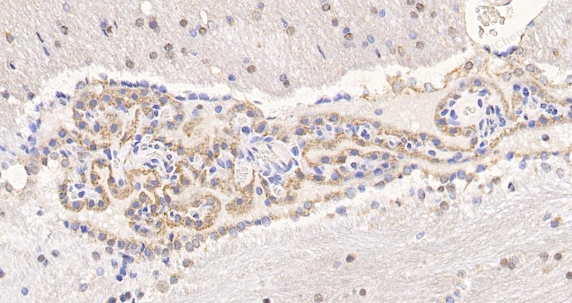 Immunohistochemical analysis of paraffin-embedded rat brain tissue using Tau Antibody. The section was pre-treated using heat mediated antigen retrieval with sodium citrate buffer (pH 6.0) for 8 minutes. The tissues were blocked in QuickBlock for 20 minutes at room temperature, washed with ddH2O and PBS, and then probed with the primary antibody at 1/100 dilution in 4℃ overnight. The detection was performed using an HRP conjugated compact polymer system. DAB was used as the chromogen. Tissues were counterstained with hematoxylin and mounted with DPX.
 -
Immunohistochemical analysis of paraffin-embedded rat brain tissue using Tau Antibody. The section was pre-treated using heat mediated antigen retrieval with sodium citrate buffer (pH 6.0) for 8 minutes. The tissues were blocked in QuickBlock for 20 minutes at room temperature, washed with ddH2O and PBS, and then probed with the primary antibody at 1/100 dilution in 4℃ overnight. The detection was performed using an HRP conjugated compact polymer system. DAB was used as the chromogen. Tissues were counterstained with hematoxylin and mounted with DPX.


Background
-
Function
Tau promotes microtubule assembly and stability, and might be involved in the establishment and maintenance of neuronal polarity. The C-terminus binds axonal microtubules while the N-terminus binds neural plasma membrane components, suggesting that tau functions as a linker protein between both. Axonal polarity is predetermined by TAU/MAPT localization (in the neuronal cell) in the domain of the cell body defined by the centrosome. The short isoforms allow plasticity of the cytoskeleton whereas the longer isoforms may preferentially play a role in its stabilization[1][2].
-
Subcellular Localization
Cytoplasm, cytosol; Cell membrane; Peripheral membrane protein; Cytoplasmic side; Cytoplasm, cytoskeleton; Cell projection, axon; Cell projection, dendrite; Secreted
-
Expression
Tissue_specificity:It is expressed in neurons. The PNS-tau subtype is expressed in the peripheral nervous system, while other subtypes are expressed in the central nervous system. -
Isoforms & Post-Translational Modification
P10636 has 9 isomers: P10636-1: 78928 Da (predicted); P10636-2: 36760 Da (predicted); P10636-3: 32944 Da (predicted); P10636-4: 39720 Da (predicted); P10636-5: 42603 Da (predicted); P10636-6: 40007 Da (predicted); P10636-7: 42967 Da (predicted); P10636-8: 45850 Da (predicted); P10636-9: 80941 Da (predicted).
Phosphorylation at serine and threonine residues in S-P or T-P motifs by proline-directed protein kinases (PDPK1, CDK1, CDK5, GSK3, MAPK) (only 2-3 sites per protein in interphase, seven-fold increase in mitosis, and in the form associated with paired helical filaments (PHF-tau)), and at serine residues in K-X-G-S motifs by MAP/microtubule affinity-regulating kinase (MARK1, MARK2, MARK3 or MARK4), causing detachment from microtubules, and their disassembly (PubMed:23666762, PubMed:7706316). Phosphorylation decreases with age. Phosphorylation within tau/MAP's repeat domain or in flanking regions seems to reduce tau/MAP's interaction with, respectively, microtubules or plasma membrane components (PubMed:7706316). Phosphorylation on Ser-610, Ser-622, Ser-641 and Ser-673 in several isoforms during mitosis. Phosphorylation at Ser-548 by GSK3B reduces ability to bind and stabilize microtubules. Phosphorylation at Ser-579 by BRSK1 and BRSK2 in neurons affects ability to bind microtubules and plays a role in neuron polarization. Phosphorylated at Ser-554, Ser-579, Ser-602, Ser-606 and Ser-669 by PHK. Phosphorylation at Ser-214 by SGK1 mediates microtubule depolymerization and neurite formation in hippocampal neurons. There is a reciprocal down-regulation of phosphorylation and O-GlcNAcylation. Phosphorylation on Ser-717 completely abolishes the O-GlcNAcylation on this site, while phosphorylation on Ser-713 and Ser-721 reduces glycosylation by a factor of 2 and 4 respectively. Phosphorylation on Ser-721 is reduced by about 41.5% by GlcNAcylation on Ser-717. Dephosphorylated at several serine and threonine residues by the serine/threonine phosphatase PPP5C;Polyubiquitinated. Requires functional TRAF6 and may provoke SQSTM1-dependent degradation by the proteasome (By similarity). PHF-tau can be modified by three different forms of polyubiquitination. 'Lys-48'-linked polyubiquitination is the major form, 'Lys-6'-linked and 'Lys-11'-linked polyubiquitination also occur;O-glycosylated. O-GlcNAcylation content is around 8.2%. There is reciprocal down-regulation of phosphorylation and O-GlcNAcylation. Phosphorylation on Ser-717 completely abolishes the O-GlcNAcylation on this site, while phosphorylation on Ser-713 and Ser-721 reduces O-GlcNAcylation by a factor of 2 and 4 respectively. O-GlcNAcylation on Ser-717 decreases the phosphorylation on Ser-721 by about 41.5%;Glycation of PHF-tau, but not normal brain TAU/MAPT. Glycation is a non-enzymatic post-translational modification that involves a covalent linkage between a sugar and an amino group of a protein molecule forming ketoamine. Subsequent oxidation, fragmentation and/or cross-linking of ketoamine leads to the production of advanced glycation endproducts (AGES). Glycation may play a role in stabilizing PHF aggregation leading to tangle formation in AD -
Subunit
Interacts with MARK1, MARK2, MARK3 and MARK4 (PubMed:23666762). Interacts with PSMC2 through SQSTM1 (By similarity). Interacts with SQSTM1 when polyubiquitinated (PubMed:15953362). Interacts with FKBP4 (By similarity). Binds to CSNK1D (PubMed:14761950). Interacts with SGK1 (PubMed:16982696). Interacts with EPM2A; the interaction dephosphorylates MAPT at Ser-396 (PubMed:19542233). Interacts with PIN1 (PubMed:11313338). Interacts with LRRK2 (PubMed:26014385). Interacts with LRP1, leading to endocytosis; this interaction is reduced in the presence of LRPAP1/RAP (PubMed:32296178)
-
SwissProt ID
-
Synonyms
MAPT


Documentation

 Powered by Bioz
