



ATTO 488 carboxylic acid

Based on 1 Customer Validation
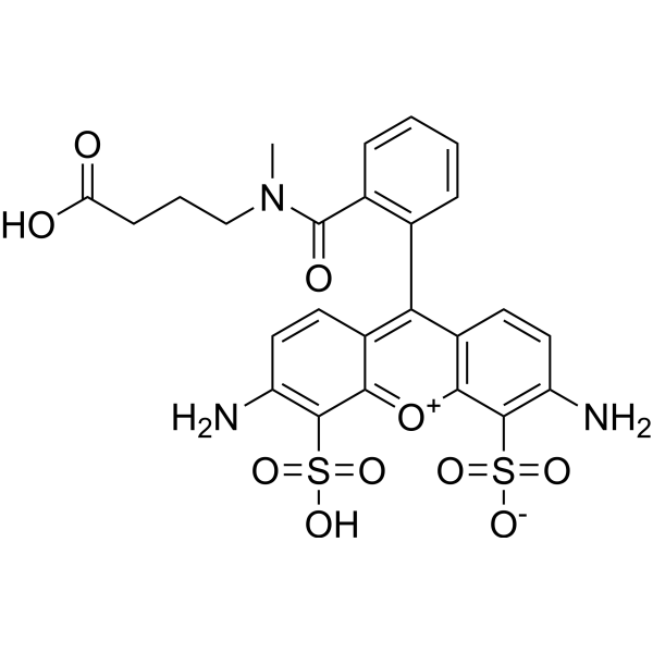
ATTO 488 carboxylic acid is a bright green fluorescent dye with high photostability, which is commonly used in protein fluorescent labeling, single-molecule fluorescence imaging and super-resolution microscopy studies. The carboxylic acid form of ATTO 488 can be covalently coupled to primary amines of proteins via activation, or prepared into a maleimide derivative for coupling with thiol groups; it emits green fluorescence after labeling. Ex/Em = 488/524 nm.
For research use only. We do not sell to patients.
- Purity : 97.24%
- CAS No.: 1443553-08-9
- Formula: C25H23N3O10S2
- Molecular Weight:589.59
-
Storage:
-20°C, protect from light
* In solvent : -80°C, 6 months; -20°C, 1 month (protect from light)

To place orders, for customer services and technical support, please contact: MedChemExpress USA
Tel: 609-228-6898 E-mail: [email protected] [email protected]


-
 Biological Activity
-
Chemical Information
-
Solvent & Solubility
- Purity & Documentation
- References
-
 Help & FAQs




Biological Activity
Description
In Vitro
Operating Instructions (The following is a recommended experimental protocol for reference only, and adjustments should be made according to actual requirements)
1. Stock Solution Preparation
1.1 Solvent: Anhydrous dimethyl sulfoxide (DMSO).
1.2 Concentration Recommendation: It is generally recommended to prepare a high-concentration stock solution of 1-10 mM.
2. Working Solution Preparation
2.1 Diluent: Phosphate-buffered saline (PBS) buffer (137 mM NaCl, 2.7 mM KCl, 10 mM Na2SO4, 1.8 mM KH2PO4, pH 7.4); alternatively, 20 mM HEPES buffer at pH 8.3 can be used.
2.2 Working Concentration: 1 nM for fluorescence correlation spectroscopy (FCS) experiments; 0.5-30 μM for single-molecule total internal reflection fluorescence (TIRF) imaging.
2.3 Notes: Adjust the working solution concentration as needed; prepare it immediately before use.
3. Staining Procedures
3.1 Sample Type Description
3.1.1 Protein Samples (BSA-labeled): Suitable for labeling proteins containing primary amines. BSA and ATTO 488 NHS ester are incubated at room temperature for 2 h in 20 mM HEPES buffer (pH 8.3) at a molar ratio of 1:1, with the N-terminal amino group being the main target (lysine residues may also be labeled). Unreacted dye is removed using a Centrispin-10 size exclusion column (5 kDa cutoff).
3.1.2 Cell Samples (Adherent Cells): U2OS cells are incubated with Lysosome-488 probe (a conjugate of Atto 488 NHS ester and cathepsin-targeting peptide) for 30 min, followed by a 4 h post-incubation in growth medium to reduce background fluorescence.
3.1.3 Liposome/Supported Lipid Bilayer Samples: 1 nM Atto 488 is incubated with liposomes of different concentrations in PBS before FCS measurement; 0.5-30 μM Atto 488 is incubated on DOPC-supported lipid bilayers (SLB) in the dark for approximately 10 min, followed by rinsing with PBS at least 3 times.
3.2 Incubation Conditions
3.2.1 Protein Labeling: Room temperature, 2 h, 20 mM HEPES pH 8.3.
3.2.2 Cell Staining: 37 °C, 5% CO2, 30 min probe incubation + 4 h post-incubation in medium.
3.2.3 SLB Imaging: Room temperature, dark condition, approximately 10 min.
3.3 Washing Procedures
3.3.1 Protein Labeling: Unreacted dye is removed by centrifugation using a Centrispin-10 size exclusion column (5 kDa cutoff).
3.3.2 Cell Samples: A 4 h post-incubation in growth medium is performed to reduce background.
3.3.3 SLB Samples: Rinse with PBS at least 3 times
4. Control Experiments
4.1 Unstained/Free Dye Control: FCS measurement of free Atto 488 (1 nM) in PBS is conducted to obtain the diffusion time and triplet state parameters of the free dye.
4.2 Covalent Binding Control: The same operation is carried out without the NHS linker. No single-molecule signal is detected after rinsing, which confirms that the protein binds via covalent bonds rather than non-specific adsorption.
4.3 pH Control: The pH of 100 nM ATTO 488 in MilliQ water is adjusted with HCl/NaOH, and the change in fluorescence intensity is measured to rule out the direct effect of pH on the fluorescence of the dye itself.
4.4 Colocalization Control: LysoTracker Green (50 nM) and GFP-LAMP1 are used as standard lysosome markers, and their colocalization with Lysosome-488 yields Pearson's coefficients of 0.776 and 0.811, respectively.
5. Detection and Analysis
5.1 Instrument Types: Fluorescence spectrophotometer, confocal microscope, TIRF microscope, SIM super-resolution microscope, fluorescence correlation spectroscopy (FCS) system.
5.2 Excitation/Emission Wavelengths: Excitation wavelength is 488 nm (alternatively 480 nm or 473 nm); emission wavelength is 524 nm, or detection is performed using a 505 nm long-pass filter / BLP01-488 long-pass + HQ 520/66 band-pass filter; excitation/emission slits are 2 nm.
5.3 Result Analysis
5.3.1 Fluorescence Intensity Changes: In the pH-responsive PAA brush system, fluorescence intensity is high at pH 3 (brush collapsed) and low at pH 10 (brush extended), with a switching amplitude of up to 80%, which is reversible and rapid. The decay of the FCS curve slows down after Atto 488 binds to liposomes; the Kp value is 253 in DOPC, 298 in DOTAP, and 73.7 in DOPG.
5.3.2 Fluorescence Localization: Lysosome-488 specifically labels lysosomes in U2OS cells, with Pearson's coefficients of 0.776 for colocalization with LysoTracker Green and 0.811 for colocalization with GFP-LAMP1; cell viability is >85%. The adsorption density of Atto 488 on DOPC-supported lipid bilayers is 30-40 molecules/104 μm, indicating weak interaction with the membrane.
5.3.3 Color Change: Green fluorescence. No significant changes in fluorescence lifetime and quantum yield are observed before and after binding to liposomes.
MedChemExpress (MCE) has not independently confirmed the accuracy of these methods. They are for reference only.


Chemical Information
-
CAS No. 1443553-08-9
-
Appearance Solid
-
Molecular Weight 589.59
-
Formula C25H23N3O10S2
-
Color Orange to red
-
SMILES
O=C(CCCN(C)C(C1=C(C2=C(C=CC(N)=C3S(=O)(O)=O)C3=[O+]C4=C2C=CC(N)=C4S(=O)([O-])=O)C=CC=C1)=O)O
-
Shipping
Room temperature in continental US; may vary elsewhere.
-
Storage
-20°C, protect from light
* In solvent : -80°C, 6 months; -20°C, 1 month (protect from light)


Solvent & Solubility
In Vitro:
H2O : ≥ 100 mg/mL (169.61 mM)
* "≥" means soluble, but saturation unknown.
Please refer to the solubility information to select the appropriate solvent. Once prepared, please aliquot and store the solution to prevent product inactivation from repeated freeze-thaw cycles.
Storage method and period of stock solution: -80°C, 6 months; -20°C, 1 month (protect from light). When stored at -80°C, please use it within 6 months. When stored at -20°C, please use it within 1 month.
* Note: If you choose water as the stock solution, please dilute it to the working solution, then filter and sterilize it with a 0.22 μm filter before use.
Please refer to the solubility information to select the appropriate solvent. Once prepared, please aliquot and store the solution to prevent product inactivation from repeated freeze-thaw cycles.
Storage method and period of stock solution: -80°C, 6 months; -20°C, 1 month (protect from light). When stored at -80°C, please use it within 6 months. When stored at -20°C, please use it within 1 month.
* Note: If you choose water as the stock solution, please dilute it to the working solution, then filter and sterilize it with a 0.22 μm filter before use.
Concentration (start) × Volume (start) = Concentration (final) × Volume (final)


Purity & Documentation
-
Data Sheet (295 KB)
-
SDS (251 KB)
- English - EN (251 KB)
- Français - FR (251 KB)
- Deutsch - DE (251 KB)
- Norwegian - NO (251 KB)
- Español - ES (251 KB)
- Swedish - SV (251 KB)
- Italian - IT (251 KB)
- Korean - KR (251 KB)
- Portuguese - PT (251 KB)
-
Handling Instructions (2659 KB)
References


Complete Stock Solution Preparation Table
Please refer to the solubility information to select the appropriate solvent. Once prepared, please aliquot and store the solution to prevent product inactivation from repeated freeze-thaw cycles.
Storage method and period of stock solution: -80°C, 6 months; -20°C, 1 month (protect from light). When stored at -80°C, please use it within 6 months. When stored at -20°C, please use it within 1 month.
| Optional Solvent | Concentration Solvent Mass | 1 mg | 5 mg | 10 mg | 25 mg |
|---|---|---|---|---|---|
| H2O | 1 mM | 1.6961 mL | 8.4805 mL | 16.9609 mL | 42.4023 mL |
| 5 mM | 0.3392 mL | 1.6961 mL | 3.3922 mL | 8.4805 mL | |
| 10 mM | 0.1696 mL | 0.8480 mL | 1.6961 mL | 4.2402 mL | |
| 15 mM | 0.1131 mL | 0.5654 mL | 1.1307 mL | 2.8268 mL | |
| 20 mM | 0.0848 mL | 0.4240 mL | 0.8480 mL | 2.1201 mL | |
| 25 mM | 0.0678 mL | 0.3392 mL | 0.6784 mL | 1.6961 mL | |
| 30 mM | 0.0565 mL | 0.2827 mL | 0.5654 mL | 1.4134 mL | |
| 40 mM | 0.0424 mL | 0.2120 mL | 0.4240 mL | 1.0601 mL | |
| 50 mM | 0.0339 mL | 0.1696 mL | 0.3392 mL | 0.8480 mL | |
| 60 mM | 0.0283 mL | 0.1413 mL | 0.2827 mL | 0.7067 mL | |
| 80 mM | 0.0212 mL | 0.1060 mL | 0.2120 mL | 0.5300 mL | |
| 100 mM | 0.0170 mL | 0.0848 mL | 0.1696 mL | 0.4240 mL |
* Note: If you choose water as the stock solution, please dilute it to the working solution, then filter and sterilize it with a 0.22 μm filter before use.


ATTO 488 carboxylic acid Related Classifications
HY-D1305 Related Classifications
 Powered by Bioz
Keywords
- ATTO 488 carboxylic acid
- 1443553-08-9
- Fluorescent Dye
- U2OS cells
- lysosomes
- cationic lipids
- supported lipid bilayers
- total internal reflection fluorescence microscopy
- single-molecule fluorescence imaging
- pH-responsive polymer brush systems
- fluorescence correlation spectroscopy
- structured illumination microscopy
- large unilamellar vesicles
- Inhibitor
- inhibitor
- inhibit