



Berzosertib hydrochloride
Based on 27 publication(s) in Google Scholar
Berzosertib (VE-822) hydrochloride is an orally active, CNS-penetrant, and selective ATR kinase inhibitor. Berzosertib hydrochloride blocks ATR kinase activity, abrogates G2/M cell cycle checkpoint, impairs DNA damage repair. Berzosertib hydrochloride induces apoptosis, inhibnits conlony migration, inhibits cell proliferation, and activates cGAS-STING axes in cancer cells. Berzosertib hydrochloride can be used for the research of cancers, such as head and neck squamous cell carcinoma, and colorectal cancer.
연구목적의 판매만을 진행합니다. 환자를 대상으로 한 판매는 하지 않습니다.
- Purity : 98.77%
- CAS No.: 1428935-04-9
- 화학식: C24H26ClN5O3S
- 분자량:500.01
-
보관:
4°C, sealed storage, away from moisture
* In solvent : -80°C, 6 months; -20°C, 1 month (sealed storage, away from moisture)


To place orders, for customer services and technical support, please contact: MedChemExpress USA
Tel: 609-228-6898 E-mail: [email protected] [email protected]


-
 Biological Activity
-
Chemical Information
-
용액&용해도
- Protocol
- 순도&문서
- References
-
 Help & FAQs


Publications Citing Use of MedChemExpress (MCE) Berzosertib hydrochloride
More- Cancer Commun (Lond). 2023 Apr;43(4):435-454. [Abstract]
- Nat Commun. 2019 Jul 2;10(1):2910. [Abstract]
- Sci Transl Med. 2020 Feb 19;12(531):eaax2625. [Abstract]
- Cell Death Dis. 2024 Dec 6;15(12):882. [Abstract]
- Cancer Lett. 2026 Feb 4;642:218300.
- Cancer Lett. 2026 Apr 1:642:218300. [Abstract]
- Clin Cancer Res. 2022 Jun 1;28(11):2397-2408. [Abstract]
- NPJ Breast Cancer. 2025 Dec 3;11(1):135. [Abstract]
- Cell Rep. 2021 Mar 2;34(9):108808. [Abstract]
- Cell Syst. 2018 Apr 25;6(4):424-443.e7. [Abstract]
- Mol Cancer Ther. 2026 Jul 15:10.1158/1535-7163.MCT-26-0182.
- Eur J Med Chem. 2017 Feb 15:127:691-702. [Abstract]
- Oncogenesis. 2025 Mar 1;14(1):4. [Abstract]
- Cancer Immunol Immunother. 2024 Nov 2;74(1):8. [Abstract]
- Mol Cancer Res. 2020 Jan;18(1):91-104. [Abstract]
- Cell Oncol (Dordr). 2022 Dec;45(6):1401-1419. [Abstract]
- J Mol Med (Berl). 2019 Aug;97(8):1183-1193. [Abstract]
- Mol Oncol. 2025 Jul 13. [Abstract]
- Bioengineering (Basel). 2025 Oct 19;12(10):1121. [Abstract]
- J Biol Chem. 2026 Jun;302(6):111461. [Abstract]
- Anticancer Res. 2019 Jul;39(7):3553-3563. [Abstract]
- Biomed Pharmacother. 2026 Feb:195:118974. [Abstract]
- Patent. US20240285616A1
- Research Square Preprint. 2024 Nov 06.
- Patent. US20240285616A1.
- bioRxiv. 2024 Mar 28.
- University of London. 2021 Sep.
 Customer Validation & Images

-
In Vivo Efficacy Study
-
Flow Cytometry
-
Cell Proliferation/Viability Assay
-
Flow Cytometry
-
IF
All Caspase Isoforms
More

Biological Activity
제품 설명
IC50 & Target
[1]|
Caspase-3 |
ATR 0.2 nM (Ki) |
ATM 34 nM (Ki) |
In Vitro
Berzosertib (0.031-1 µM; 72 h) hydrochloride reduces cell viability in Cal-27 and FaDu HNSCC cell lines with IC50 values of 0.285 µM and 0.252 µM, respectively[1].
Berzosertib (0.125-0.5 µM; 24-48 h) hydrochloride inhibits migration in Cal-27 and in FaDu HNSCC cells[1].
Berzosertib (0.25-0.5 µM; 48 h) hydrochloride induces apoptosis in Cal-27 and FaDu cells[1].
Berzosertib (48-72 h) hydrochloride inhibits proliferation of A549, NCI-H226, and NCI-H520 cells with IC50 values in the 1-4 μM range[2].
Berzosertib (40-80 nM; 1 h) hydrochloride enhances radiosensitivity of A549, NCI-H226, and NCI-H520 NSCLC cell lines[2].
Berzosertib (40 nM; 1 h) hydrochloride inhibits radiation-induced ATR (Thr1989) phosphorylation in A549, NCI-H226, and NCI-H520 NSCLC cell lines without affecting ATM (Ser1981) activation[2].
Berzosertib (40 nM; 1 h) hydrochloride abrogates the radiation-induced G2/M cell cycle checkpoint in A549 NSCLC cells, shifting cells into G1 phase[2].
Berzosertib (40-80 nM; 1 h) hydrochloride enhances radiation-induced apoptosis in A549 NSCLC cells when combined with 10 Gy, but not 2 Gy, irradiation[2].
Berzosertib (40 nM; 1 h) hydrochloride inhibits DNA double-strand break repair in A549 cells[2].
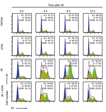
Berzosertib (1 µM; 2 h) hydrochloride impairs irradiation-induced G2/M checkpoint initiation and maintenance in HCT116 and CT26 cells, promoting mitotic entry after DNA damage[4].
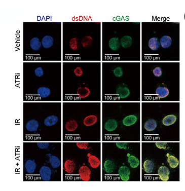
Berzosertib (1 µM; 2 h) hydrochloride combined with 5 Gy irradiation increases micronuclei formation and cytosolic dsDNA levels in HCT116 and CT26 colorectal cancer cell lines[4].
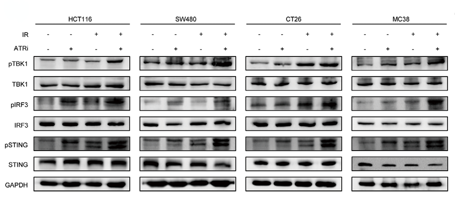
Berzosertib (1 µM; 2 h) hydrochloride combined with 5 Gy irradiation robustly activates the canonical cGAS-STING-pTBK1/pIRF3 pathway, and upregulates expression of interferon-stimulated genes CXCL10, CCL5, and IFNB in HCT116, SW480, CT26, and MC38 cells[4].
Berzosertib (1 µM; 2 h) hydrochloride combined with 5 Gy irradiation inhibits the recruitment of SHP1 to the TRAF6/STING complex in HCT116 cells, enhancing TRAF6-STING interaction[4].
MedChemExpress (MCE) has not independently confirmed the accuracy of these methods. They are for reference only.
-
Cell Line:Cal-27, FaDu cells
-
Concentration:0.031; 0.063; 0.125; 0.25; 0.5; 1 µM
-
Incubation Time:72 h
-
Result:Caused a dose-dependent decrease in cell viability in both cell lines.
Exhibited an IC50 value of 0.285 µM for Cal-27 cells.
Exhibited an IC50 value of 0.252 µM for FaDu cells.
-
Cell Line:Cal-27, FaDu cells
-
Concentration:0.125; 0.25; 0.5 µM
-
Incubation Time:24 h (Cal-27); 48 h (FaDu)
-
Result:Reduced Cal-27 cell gap closure to 82% at 0.25 µM and 50% at 0.5 µM at 24 h, compared to 98% in untreated cells.
Reduced FaDu cell gap closure to 24% at 0.25 µM and 0.5 µM at 24 h, compared to 41% in untreated cells.
Significantly inhibited FaDu cell gap closure at 0.5 µM at 48 h.
-
Cell Line:Cal-27, FaDu cells
-
Concentration:0.25 µM (Cal-27); 0.5 µM (FaDu)
-
Incubation Time:48 h
-
Result:Increased apoptosis levels to 279% of control in Cal-27 cells.
Increased apoptosis levels to 244% of control in FaDu cells.
-
Cell Line:A549, NCI-H226, and NCI-H520 cells
-
Concentration:40 nM
-
Incubation Time:1 h
-
Result:Did not affect radiation-induced ATM (Ser1981) phosphorylation.
Diminished radiation-induced activation of p-ATR (Thr1989) in all three cell lines even at 24 h post-radiation.
-
Cell Line:A549 cells
-
Concentration:40; 80 nM
-
Incubation Time:1 h
-
Result:Showed no increase in apoptosis when combined with 2 Gy radiation.
Caused a significant increase in apoptosis when combined with 10 Gy radiation, compared to radiation alone.
-
Cell Line:A549 cells
-
Concentration:40 nM
-
Incubation Time:1 h
-
Result:Combined with irradiation resulted in a statistically significant increase in the number of γH2AX foci at 8 h and 24 h compared with cells treated with irradiation alone.
-
Cell Line:HCT116, SW480, CT26, MC38
-
Concentration:1 µM
-
Incubation Time:2 h
-
Result:Caused a sharp, time-dependent increase in mRNA levels of CXCL10, CCL5, and IFNB in HCT116 and CT26 cells, with peak expression at 8-12 hours post-irradiation.
Induced significant increases in CXCL10, CCL5, and IFNB gene expressions in SW480 and MC38 cells compared to irradiation alone.
In Vivo
Berzosertib (60 mg/kg; p.o.; daily; 5 days; 1 h before 2.5 Gy whole brain irradiation) hydrochloride combined with daily 2.5 Gy whole brain irradiation significantly improves median overall survival and reduces intracranial tumor growth in mouse NSCLC brain metastasis xenograft model[2].
Berzosertib (60 mg/kg; i.g.; 2 h before Gy IR, then daily for 3 days) hydrochloride produces antitumor efficacy in mouse MC38 and CT26 models, when combined with 5 Gy irradiation and anti-PD-L1[4].
MedChemExpress (MCE) has not independently confirmed the accuracy of these methods. They are for reference only.
-
Animal Model:Female Hsd:athymic Nude-Foxn1 mice (6-8-week-old) subcutaneously implantated with UW-lung-16 and UW-lung-18[2]
-
Dosage:60 mg/kg
-
Administration:p.o.; daily; 10 days; 1 h before 2 Gy local tumor irradiation
-
Result:Delayed growth of UW-lung-16 and UW-lung-18 tumors.
Reduced estimated tumor growth curve slope for UW-lung-16 tumors significantly lower than alone, with a synergistic effect and dose enhancement factor (DEF) of 1.8.
Reduced estimated tumor growth curve slope for UW-lung-18 tumors significantly lower than alone, with a synergistic effect and DEF of 1.4.
Inhibited phospho-Chk1 (Ser345) alone and combined with irradiation.
Increased cleaved caspase-3 in the combination group.
Induced significantly more γH2AX foci in the combination group compared to other groups.
-
Animal Model:Athymic nude mice intracranial implantation of luciferase-transfected UW-Lung-16 cells[2]
-
Dosage:60 mg/kg
-
Administration:p.o.; daily; 5 days; 1 h before 2.5 Gy whole brain irradiation
-
Result:Reduced bioluminescence total flux significantly by days 32 and 40 post-implant compared to radiation alone.
Improved median overall survival to 95 days, compared to 67 days in the radiation alone group.
Showed no significant difference in body weight between groups during or 40 days post-treatment.
-
Animal Model:C57/B6J mice (female, 5-6 weeks old) subcutaneously injected with MC38 cells[4]
-
Dosage:60 mg/kg
-
Administration:i.g.; 2 h before Gy IR, then daily for 3 days
-
Result:Delayed tumor growth more effectively than dual or monotherapy regimens.
Led to complete tumor regression in some mice.
Extended mouse survival compared to other treatment groups.
Reduced tumor burden (as measured by bioluminescence total flux).
Decreased Ki67-positive proliferating cells.
Increased TUNEL-positive apoptotic cells.
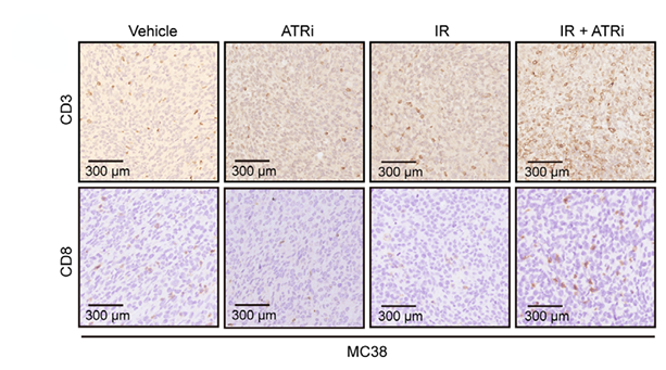
Increased CD3+ and CD8+ tumor-infiltrating lymphocyte counts.
Elevated levels of CD11c+ MHC-II+ dendritic cells and CD11c+ CD8+ tumor-infiltrating dendritic cells.
Increased CD86 maturation marker mean fluorescence intensity.
Activated the canonical cGAS-STING-pTBK1/pIRF3 axis.
Activated the non-canonical STING-p65 axis.
Upregulated mRNA expression of innate immune-related genes Cxcl10, Ccl5, and Ifnb.
-
Animal Model:Balb/C mice (female, 5-6 weeks old) subcutaneously injected with CT26 cells[4]
-
Dosage:60 mg/kg
-
Administration:i.g.; 2 h before Gy IR, then daily for 3 days
-
Result:Delayed tumor growth more effectively than dual or monotherapy regimens.
Extended mouse survival compared to other treatment groups.
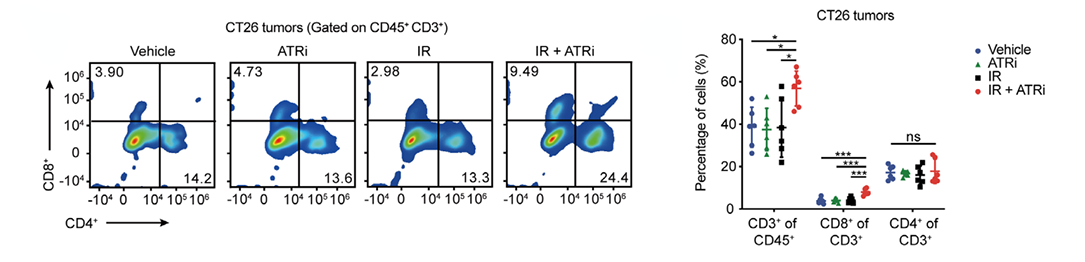
Increased CD3+ and CD8+ tumor-infiltrating lymphocyte counts.
Elevated levels of CD11c+ MHC-II+ dendritic cells and CD11c+ CD8+ tumor-infiltrating dendritic cells.
Increased CD86 maturation marker mean fluorescence intensity.
Activated the canonical cGAS-STING-pTBK1/pIRF3 axis.
Activated the non-canonical STING-p65 axis.
Upregulated mRNA expression of innate immune-related genes Cxcl10, Ccl5, and Ifnb.
Decreased SHP1 mRNA levels and SHP1 interaction with TRAF6/STING via promoting SHP1 SUMOylation at lysine 127.
- Annexin V plus membrane-impermeant dye apoptosis staining
- BrdU Incorporation Assay
- Cell migration
- Somatic Cell Culture
- CFSE Dye Dilution Proliferation Assay
- Dye-dilution cell tracking and proliferation staining
- Protocol for Cell Cycle
- CCK-8/WST-8 Cell Proliferation Assay
- Cell Counting-Based Growth Curve Assay
- Colony Formation (Clonogenic) Assay
- Cell Viability Determination by MTT Colorimetric Assay
- Kinase activity and phosphorylation assays
- EdU Incorporation Assay (Click Chemistry-Based DNA Synthesis Measurement)
- Detection of Apoptosis
- TUNEL staining for apoptotic DNA fragmentation
- Ki-67 Immunostaining Proliferation Assay
- Flow cytometric DNA-content cell-cycle staining
- PCNA Immunodetection Proliferation Assay
- Protocol for Cell Counting and Cell Density Analysis
- Apoptosis Solutions
- Protocol for Kinase activity and phosphorylation assays
- MTT Cell Proliferation Assay
- Genotoxicity/Mutagenicity Study


Chemical Information
-
CAS No. 1428935-04-9
-
Appearance Solid
-
분자량 500.01
-
화학식 C24H26ClN5O3S
-
Color Light yellow to yellow
-
SMILES
NC1=NC=C(C2=CC=C(S(=O)(C(C)C)=O)C=C2)N=C1C3=CC(C4=CC=C(CNC)C=C4)=NO3.Cl
-
Synonyms
VE-822 hydrochloride; VX-970 hydrochloride; M6620 hydrochloride
-
선적
Room temperature in continental US; may vary elsewhere.
-
보관
4°C, sealed storage, away from moisture
* In solvent : -80°C, 6 months; -20°C, 1 month (sealed storage, away from moisture)


Publications (27)
-
Journal Impact Factor
-
Most Recent
-
Cancer Commun (Lond)
Combining radiation and the ATR inhibitor berzosertib activates STING signaling and enhances immunotherapy via inhibiting SHP1 function in colorectal cancer. [Abstract]2023 Apr;43(4):435-454. PMID: 36855844
Berzosertib hydrochloride purchased from MedChemExpress. Usage Cited in: Cancer Commun (Lond). 2023 Apr;43(4):435-454. [Abstract]
Berzosertib (60 mg/kg) was administered by gavage 2 h before IR and consecutively for the next three days. Representative IHC images of CD3 staining of MC38 tumors were shown.
Berzosertib hydrochloride purchased from MedChemExpress. Usage Cited in: Cancer Commun (Lond). 2023 Apr;43(4):435-454. [Abstract]
Berzosertib (60 mg/kg) was administered by gavage 2 h before IR and consecutively for the next three days. Representative flow cytometry images and quantitative analysis of TILs in CT26 tumors were obtained.
Berzosertib hydrochloride purchased from MedChemExpress. Usage Cited in: Cancer Commun (Lond). 2023 Apr;43(4):435-454. [Abstract]
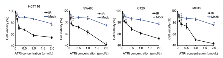
Berzosertib (0–2 μM; 24 h). Cell viability assay of multiple CRC cell lines treated with ATRi and IR + ATRi was performed.
Berzosertib hydrochloride purchased from MedChemExpress. Usage Cited in: Cancer Commun (Lond). 2023 Apr;43(4):435-454. [Abstract]
Berzosertib (1 μM). Cell cycle distribution analysis of HCT116 and CT26 cells treated with IR and IR + ATRi. Representative flow cytometry images of phospho-histone H3+ cells in HCT116 and CT26 cells were shown.
Berzosertib hydrochloride purchased from MedChemExpress. Usage Cited in: Cancer Commun (Lond). 2023 Apr;43(4):435-454. [Abstract]
Berzosertib (1 μM; 14 h). IF images of dsDNA and cGAS staining in HCT116 cells were shown.
Berzosertib hydrochloride purchased from MedChemExpress. Usage Cited in: Cancer Commun (Lond). 2023 Apr;43(4):435-454. [Abstract]
Berzosertib (1 μM; 14 h) was used. Immunoblotting of the key proteins from the canonical cGAS-STING axis in multiple CRC cell lines was performed.
-
Nat Commun
Myc targeted CDK18 promotes ATR and homologous recombination to mediate PARP inhibitor resistance in glioblastoma. [Abstract]2019 Jul 2;10(1):2910. PMID: 31266951 -
Sci Transl Med
BRCAness, SLFN11, and RB1 loss predict response to topoisomerase I inhibitors in triple-negative breast cancers. [Abstract]2020 Feb 19;12(531):eaax2625. PMID: 32075943 -
Cell Death Dis
KDM1A epigenetically enhances RAD51 expression to suppress the STING-associated anti-tumor immunity in esophageal squamous cell carcinoma. [Abstract]2024 Dec 6;15(12):882. PMID: 39638799 -
-
Cancer Lett
2026 Apr 1:642:218300. PMID: 41651400 -
Clin Cancer Res
Preclinical Modeling of Leiomyosarcoma Identifies Susceptibility to Transcriptional CDK Inhibitors through Antagonism of E2F-Driven Oncogenic Gene Expression. [Abstract]2022 Jun 1;28(11):2397-2408. PMID: 35325095 -
NPJ Breast Cancer
CDK2 inhibition enhances CDK4/6 inhibitor antitumor activity in comprehensive breast cancer PDX model screen. [Abstract]2025 Dec 3;11(1):135. PMID: 41339342 -
Cell Rep
2021 Mar 2;34(9):108808. PMID: 33657372 -
Cell Syst
A Library of Phosphoproteomic and Chromatin Signatures for Characterizing Cellular Responses to Drug Perturbations. [Abstract]2018 Apr 25;6(4):424-443.e7. PMID: 29655704 -
-
Eur J Med Chem
New approach of delivering cytotoxic drugs towards CAIX expressing cells: A concept of dual-target drugs. [Abstract]2017 Feb 15:127:691-702. PMID: 27823879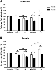
Berzosertib hydrochloride purchased from MedChemExpress. Usage Cited in: Eur J Med Chem. 2017 Feb 15:127:691-702. [Abstract]
Relative cell viability (%) in MDCK CAIX- and CAIX+ cells exposed to ATR inhibitors (VE-821 and VE-822) or the CAIXi conjugated derivatives in combination with radiation during normoxia (21% O2) and anoxia (≤0.02% O2). Normoxic cells are irradiated with 2 Gy and anoxic cells with 4 Gy to induce similar effects on cell viability.
-
Oncogenesis
STAG2 expression imparts distinct therapeutic vulnerabilities in muscle-invasive bladder cancer cells. [Abstract]2025 Mar 1;14(1):4. PMID: 40025053 -
Cancer Immunol Immunother
Combination of ataxia telangiectasia and Rad3-related inhibition with ablative radiotherapy remodels the tumor microenvironment and enhances immunotherapy response in lung cancer. [Abstract]2024 Nov 2;74(1):8. PMID: 39487895 -
Mol Cancer Res
Inhibition of the ATR-CHK1 Pathway in Ewing Sarcoma Cells Causes DNA Damage and Apoptosis via the CDK2-Mediated Degradation of RRM2. [Abstract]2020 Jan;18(1):91-104. PMID: 31649026 -
Cell Oncol (Dordr)
Novel preclinical gastroenteropancreatic neuroendocrine neoplasia models demonstrate the feasibility of mutation-based targeted therapy. [Abstract]2022 Dec;45(6):1401-1419. PMID: 36269546 -
J Mol Med (Berl)
2019 Aug;97(8):1183-1193. PMID: 31201471 -
Mol Oncol
Olaparib synergy screen reveals Exemestane induces replication stress in triple-negative breast cancer. [Abstract]2025 Jul 13. PMID: 40652528 -
Bioengineering (Basel)
Precision Oncology for High-Grade Gliomas: A Tumor Organoid Model for Adjuvant Treatment Selection. [Abstract]2025 Oct 19;12(10):1121. PMID: 41155119 -
J Biol Chem
2026 Jun;302(6):111461. PMID: 41999888 -
Anticancer Res
Schlafen11 Expression Is Associated With the Antitumor Activity of Trabectedin in Human Sarcoma Cell Lines. [Abstract]2019 Jul;39(7):3553-3563. PMID: 31262879 -
Biomed Pharmacother
Dihydroartemisinin enhances NKG2D CAR-T cell therapy against solid tumors by inducing NKG2D ligands and remodeling the tumor microenvironment. [Abstract]2026 Feb:195:118974. PMID: 41529511 -
-
-
-
-


용액&용해도
In Vitro:
DMSO : 40 mg/mL (80.00 mM; ultrasonic and warming and heat to 60°C; Hygroscopic DMSO has a significant impact on the solubility of product, please use newly opened DMSO)
Please refer to the solubility information to select the appropriate solvent. Once prepared, please aliquot and store the solution to prevent product inactivation from repeated freeze-thaw cycles.
Storage method and period of stock solution: -80°C, 6 months; -20°C, 1 month (sealed storage, away from moisture). When stored at -80°C, please use it within 6 months. When stored at -20°C, please use it within 1 month.
Please refer to the solubility information to select the appropriate solvent. Once prepared, please aliquot and store the solution to prevent product inactivation from repeated freeze-thaw cycles.
Storage method and period of stock solution: -80°C, 6 months; -20°C, 1 month (sealed storage, away from moisture). When stored at -80°C, please use it within 6 months. When stored at -20°C, please use it within 1 month.
Concentration (start) × Volume (start) = Concentration (final) × Volume (final)
In Vivo:
Select the appropriate dissolution method based on your experimental animal and administration route.
- For the following dissolution methods, please ensure to first prepare a clear stock solution using an In Vitro approach and then sequentially add co-solvents:
- To ensure reliable experimental results, the clarified stock solution can be appropriately stored based on storage conditions. As for the working solution for In Vivo experiments, it is recommended to prepare freshly and use it on the same day.
- The percentages shown for the solvents indicate their volumetric ratio in the final prepared solution. If precipitation or phase separation occurs during preparation, heat and/or sonication can be used to aid dissolution.
Add each solvent one by one: 10% DMSO 40% PEG300 5% Tween-80 45% Saline
Solubility: ≥ 4 mg/mL (8.00 mM); Clear solution
This protocol yields a clear solution of ≥ 4 mg/mL (saturation unknown).
Taking 1 mL working solution as an example, add 100 μL DMSO stock solution (40.0 mg/mL) to 400 μL PEG300, and mix evenly; then add 50 μL Tween-80 and mix evenly; then add 450 μL Saline to adjust the volume to 1 mL.
Preparation of Saline: Dissolve 0.9 g sodium chloride in ddH₂O and dilute to 100 mL to obtain a clear Saline solution.
Add each solvent one by one: 10% DMSO 90% (20% SBE-β-CD in Saline)
Solubility: ≥ 4 mg/mL (8.00 mM); Clear solution
This protocol yields a clear solution of ≥ 4 mg/mL (saturation unknown).
Taking 1 mL working solution as an example, add 100 μL DMSO stock solution (40.0 mg/mL) to 900 μL 20% SBE-β-CD in Saline, and mix evenly.
Preparation of 20% SBE-β-CD in Saline (4°C, storage for one week): 2 g SBE-β-CD powder is dissolved in 10 mL Saline, completely dissolve until clear.
In Vivo Dissolution Calculator
Please enter the basic information of animal experiments:
-
-
-
-
Recommended: Prepare an additional quantity of animals to account for potential losses during experiments.
Please enter your animal formula composition:
-
%DMSO +
Recommended: Keep the proportion of DMSO in working solution below 2% if your animal is weak.
-
%+
-
+%Tween-80 + +
-
%Saline +
The co-solvents required include: DMSO, . All of co-solvents are available by MedChemExpress (MCE). , Tween 80. All of co-solvents are available by MedChemExpress (MCE).
Working solution concentration: 0.22 mg/mL
Method for preparing stock solution: mg drug dissolved in μL DMSO. Stock solution concentration: mg/mL. * In solvent : -80°C, 6 months; -20°C, 1 month (sealed storage, away from moisture)
1. Take μL DMSO stock solution;
2. Add μL .
μL , mix evenly;
3. Then add μL Tween 80, mix evenly;
4. Then add μL
Please ensure that the stock solution in the first step is dissolved to a clear state, and add co-solvents in sequence. You can use ultrasonic heating (ultrasonic cleaner, recommended frequency 20-40 kHz), vortexing, etc. to assist dissolution.


순도&문서
-
Data Sheet (292 KB)
-
SDS (252 KB)
- English - EN (252 KB)
- Français - FR (252 KB)
- Deutsch - DE (252 KB)
- Norwegian - NO (252 KB)
- Español - ES (252 KB)
- Swedish - SV (252 KB)
- Italian - IT (252 KB)
- Korean - KR (252 KB)
- Portuguese - PT (252 KB)
-
Handling Instructions (2659 KB)
References
[1]. Schnoell J, et al. The ATR inhibitor berzosertib acts as a radio- and chemosensitizer in head and neck squamous cell carcinoma cell lines. Invest New Drugs. 2023;41(6):842-850. [Content Brief]
[2]. Baschnagel AM, et al. ATR Inhibitor M6620 (VX-970) Enhances the Effect of Radiation in Non-Small Cell Lung Cancer Brain Metastasis Patient-Derived Xenografts. Mol Cancer Ther. 2021;20(11):2129-2139. [Content Brief]
[3]. Fokas E, et al. Targeting ATR in vivo using the novel inhibitor VE-822 results in selective sensitization of pancreatic tumors to radiation. Cell Death Dis. 2012;3(12):e441. Published 2012 Dec 6. [Content Brief]
[4]. Liu C, et al. Combining radiation and the ATR inhibitor berzosertib activates STING signaling and enhances immunotherapy via inhibiting SHP1 function in colorectal cancer. Cancer Commun (Lond). 2023;43(4):435-454. [Content Brief]
[5]. Gorainow N, et al. Berzosertib enhances the sensitivity of pediatric diffuse midline glioma H3K27-altered cells to radiotherapy. Cell Death Dis. 2026;17(1):331. Published 2026 Mar 20. [Content Brief]
[6]. Kurmasheva RT, et al. Initial testing (stage 1) of M6620 (formerly VX-970), a novel ATR inhibitor, alone and combined with cisplatin and melphalan, by the Pediatric Preclinical Testing Program. Pediatr Blood Cancer. 2018;65(2):10.1002/pbc.26825. [Content Brief]


Complete Stock Solution Preparation Table
Please refer to the solubility information to select the appropriate solvent. Once prepared, please aliquot and store the solution to prevent product inactivation from repeated freeze-thaw cycles.
Storage method and period of stock solution: -80°C, 6 months; -20°C, 1 month (sealed storage, away from moisture). When stored at -80°C, please use it within 6 months. When stored at -20°C, please use it within 1 month.
| Optional Solvent | Concentration Solvent Mass | 1 mg | 5 mg | 10 mg | 25 mg |
|---|---|---|---|---|---|
| DMSO | 1 mM | 2.0000 mL | 9.9998 mL | 19.9996 mL | 49.9990 mL |
| 5 mM | 0.4000 mL | 2.0000 mL | 3.9999 mL | 9.9998 mL | |
| 10 mM | 0.2000 mL | 1.0000 mL | 2.0000 mL | 4.9999 mL | |
| 15 mM | 0.1333 mL | 0.6667 mL | 1.3333 mL | 3.3333 mL | |
| 20 mM | 0.1000 mL | 0.5000 mL | 1.0000 mL | 2.4999 mL | |
| 25 mM | 0.0800 mL | 0.4000 mL | 0.8000 mL | 2.0000 mL | |
| 30 mM | 0.0667 mL | 0.3333 mL | 0.6667 mL | 1.6666 mL | |
| 40 mM | 0.0500 mL | 0.2500 mL | 0.5000 mL | 1.2500 mL | |
| 50 mM | 0.0400 mL | 0.2000 mL | 0.4000 mL | 1.0000 mL | |
| 60 mM | 0.0333 mL | 0.1667 mL | 0.3333 mL | 0.8333 mL |


Berzosertib hydrochloride Related Classifications
HY-13902A Related Classifications
 Powered by Bioz
Keywords
- Berzosertib
- 1428935-04-9
- VE-822
- VX-970
- M6620
- VE822
- VE 822
- VX970
- VX 970
- M 6620
- M-6620
- ATM/ATR
- Apoptosis
- STING
- Caspase
- colorectal cancer
- non-small cell lung cancer brain metastases
- head and neck squamous cell carcinoma
- diffuse midline glioma H3K27-altered
- Cal-27
- A549
- FaDu
- ATR kinase
- pancreatic ductal adenocarcinoma
- SHP1
- Inhibitor
- inhibitor
- inhibit