



Chlorhexidine digluconate (20% in water)
Based on 13 publication(s) in Google Scholar
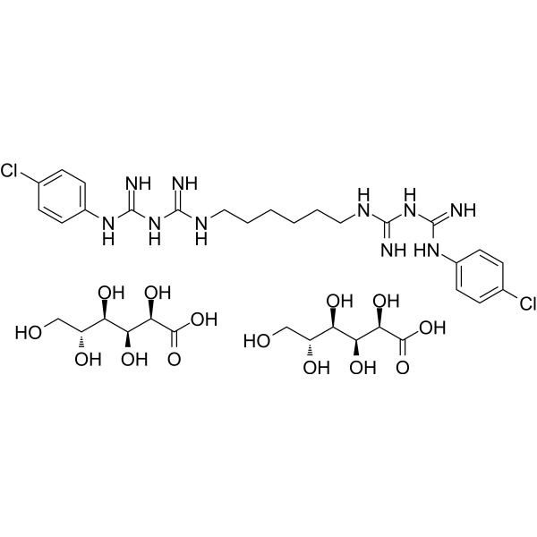
Chlorhexidine digluconate (20% in water) is a orally active cationic antimicrobial agent that targets microbial cell membranes. Chlorhexidine digluconate (20% in water) binds to cell membrane phospholipids non-specifically, destroys membrane structure and induces leakage of cell contents. Chlorhexidine digluconate (20% in water) has broad-spectrum bactericidal activity against both Gram-positive and Gram-negative bacteria. Chlorhexidine digluconate (20% in water) can interfere with membrane permeability, cause protein precipitation and energy metabolism disorders, such as rapid inhibition of microbial growth and induction of cell death (necrosis or apoptosis).
For research use only. We do not sell to patients.
- Purity : 98.88%
- CAS No.: 18472-51-0
- Formula: C34H54Cl2N10O14
- Molecular Weight:897.76
-
Storage:
Solution, -20°C, 2 years

To place orders, for customer services and technical support, please contact: MedChemExpress USA
Tel: 609-228-6898 E-mail: [email protected] [email protected]


-
 Biological Activity
-
Chemical Information
-
Solvent & Solubility
- Protocols
- Purity & Documentation
- References
-
 Help & FAQs





Publications Citing Use of MedChemExpress (MCE) Chlorhexidine digluconate (20% in water)
More- Nat Commun. 2021 Mar 29;12(1):1940. [Abstract]
- Cell Death Dis. 2022 Apr 22;13(4):396. [Abstract]
- Antioxidants (Basel). 2024 May 29;13(6):667. [Abstract]
- Dent Mater. 2025 Apr 17:S0109-5641(25)00607-4. [Abstract]
- Cell Oncol (Dordr). 2025 Jun;48(3):637-654. [Abstract]
- Front Cell Dev Biol. 2021 Mar 30:9:633259. [Abstract]
- Transl Oncol. 2024 Jul:45:101958. [Abstract]
- Cell Signal. 2021 Jul:83:110002. [Abstract]
- Mol Oncol. 2020 Feb;14(2):373-386. [Abstract]
- J Funct Foods. 2024 Jun.
- Genes (Basel). 2021 Apr 7;12(4):537. [Abstract]
- Biochem Biophys Res Commun. 2026 Jan 18:796:153158. [Abstract]
- bioRxiv. 2024 Nov 06.
 Customer Validation & Images

-
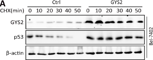
WB
All Antibiotic Isoforms
More

Biological Activity
Description
In Vitro
1. Cell death assay:
Chlorhexidine digluconate (0.000125%-0.016%; 24 h) induces cell death in L929 fibroblasts, inducing a pattern of cell necrosis and/or apoptosis, as well as cell stress. Furthermore, Chlorhexidine digluconate induces apoptosis at lower concentrations and necrosis at higher concentrations, and increases the expression of heat shock protein 70 (a marker of cell stress). Chlorhexidine digluconate may have an adverse effect on the resolution of apical periodontitis[1].
2. Cell viability experiment:
Chlorhexidine digluconate (0.0005%-0.5%; 30 min) causes the death of canine embryonic fibroblasts with concentration of ≥0.013%, while allows the cells to survive with concentration of ≤0.006%[2].
3. Bacterial survival experiment:
Chlorhexidine digluconate (0.0005%-0.5%; 30 min) has a concentration-dependent bactericidal effect on Staphylococcus aureus, and kills Staphylococcus aureus with concentration of ≥0.05%, while allows the bacteria to survive with concentration of ≤0.03%[2].
MedChemExpress (MCE) has not independently confirmed the accuracy of these methods. They are for reference only.
-
Cell Line:Chinese hamster ovary cells
-
Concentration:0.00001%, 0.00005%, 0.0001%, 0.0005%
-
Incubation Time:1, 2, 4 h
-
Result:Demonstrated a cytotoxic effect on CHO cells.
-
Cell Line:Chinese hamster ovary cells
-
Concentration:0.00005%, 0.0001%, 0.0005%
-
Incubation Time:1 h
-
Result:After a 1 hour treatment, the amount of cell apoptosis was about 1.62%, 5.51%, and 5.47%, respectively.
The amount of cell necrosis was 2.38%, 8.62%, and 22.50%, respectively.
In Vivo
Chlorhexidine (0.125%-1.0%; subcutaneous injection; single dose) digluconate causes toxic effects in the plantar space of the hind paw of Balb/c mice. Concentrations ≥0.5% induces coagulative necrosis of the epidermis, dermis and subcutaneous tissues and neutrophil-dominated inflammatory responses, while concentrations ≤0.25% induces moderate inflammation and interstitial edema[1].
2. Lung inhalation toxicity test:
Chlorhexidine (0.125%-1%; intratracheal instillation; single dose; 8-week observation) digluconate causes collagen fiber deposition and inflammatory cell infiltration in lung tissue in the C57BL/6J mouse model, leading to restrictive ventilation dysfunction. Transcriptome analysis shows that genes related to extracellular matrix deposition and mucus secretion (such as Muc5b and Muc5ac) are upregulated[3].
3. Genetic toxicity test:
0.12% Chlorhexidine dihydrochloride (0.5 mL; oral, twice a day for 8 days) causes DNA damage in peripheral blood and oral mucosal cells of rats, without causing chromosome breakage or loss in erythrocytes[4].
MedChemExpress (MCE) has not independently confirmed the accuracy of these methods. They are for reference only.
-
Animal Model:Male Imprinting Control Region (ICR) mice at 6 to 8-wk-old (body weight, 25g)[1]
-
Dosage:Chlorhexidine gluconate 0.05% and 0.025%
-
Administration:One-time intraperitoneal injection
-
Result:With 0.05% lavage had significantly lower incidence of postoperative intra-abdominal abscesses compared with the mice that had saline lavage only.
- Research Protocol for Infectious Diseases
- Detection of Apoptosis
- TUNEL staining for apoptotic DNA fragmentation
- Cell Cytotoxicity Assay
- Mammalian live/dead viability and cytotoxicity staining
- Annexin V plus membrane-impermeant dye apoptosis staining
- Transepithelial/transendothelial electrical resistance assay
- Apoptosis Solutions
- Gram Staining of Tissue Sections
- Bacterial live/dead nucleic-acid viability staining


Chemical Information
-
CAS No. 18472-51-0
-
Appearance Liquid
-
Molecular Weight 897.76
-
Formula C34H54Cl2N10O14
-
Color Colorless to light yellow
-
SMILES
N=C(NC(NCCCCCCNC(NC(NC1=CC=C(Cl)C=C1)=N)=N)=N)NC2=CC=C(Cl)C=C2.O[C@H]([C@@H](O)C(O)=O)[C@H](O)[C@H](O)CO.O[C@H]([C@@H](O)C(O)=O)[C@H](O)[C@H](O)CO
-
Shipping
Room temperature in continental US; may vary elsewhere.
-
Storage
Solution, -20°C, 2 years


Publications (13)
-
Journal Impact Factor
-
Most Recent
-
Nat Commun
The folate cycle enzyme MTHFD2 induces cancer immune evasion through PD-L1 up-regulation. [Abstract]2021 Mar 29;12(1):1940. PMID: 33782411 -
Cell Death Dis
HJURP regulates cell proliferation and chemo-resistance via YAP1/NDRG1 transcriptional axis in triple-negative breast cancer. [Abstract]2022 Apr 22;13(4):396. PMID: 35459269 -
Antioxidants (Basel)
Pitavastatin Calcium Confers Fungicidal Properties to Fluconazole by Inhibiting Ubiquinone Biosynthesis and Generating Reactive Oxygen Species. [Abstract]2024 May 29;13(6):667. PMID: 38929106 -
Dent Mater
Triple-modified PEEK surface via plasma treatment, polydopamine coating and chlorhexidine: Assessment of biocompatibility and antibacterial properties. [Abstract]2025 Apr 17:S0109-5641(25)00607-4. PMID: 40251086 -
Cell Oncol (Dordr)
UBE2Q2 promotes tumor progression and glycolysis of hepatocellular carcinoma through NF-κB/HIF1α signal pathway. [Abstract]2025 Jun;48(3):637-654. PMID: 39833608 -
Front Cell Dev Biol
2021 Mar 30:9:633259. PMID: 33859984 -
Transl Oncol
20-hydroxyecdysone suppresses bladder cancer progression via inhibiting USP21: A mechanism associated with deubiquitination and degradation of p65. [Abstract]2024 Jul:45:101958. PMID: 38663220 -
Cell Signal
2021 Jul:83:110002. PMID: 33823241 -
Mol Oncol
Loss of RDM1 enhances hepatocellular carcinoma progression via p53 and Ras/Raf/ERK pathways. [Abstract]2020 Feb;14(2):373-386. PMID: 31670863 -
-
Genes (Basel)
Autosomal Recessive Retinitis Pigmentosa Associated with Three Novel REEP6 Variants in Chinese Population. [Abstract]2021 Apr 7;12(4):537. PMID: 33917198 -
Biochem Biophys Res Commun
2026 Jan 18:796:153158. PMID: 41418346 -


Solvent & Solubility
In Vitro:
DMSO : 100 mg/mL (111.39 mM; Need ultrasonic; Hygroscopic DMSO has a significant impact on the solubility of product, please use newly opened DMSO)


Purity & Documentation
-
Data Sheet (280 KB)
-
SDS (393 KB)
- English - EN (393 KB)
- Français - FR (393 KB)
- Deutsch - DE (393 KB)
- Norwegian - NO (393 KB)
- Español - ES (393 KB)
- Swedish - SV (393 KB)
- Italian - IT (393 KB)
- Korean - KR (393 KB)
- Portuguese - PT (393 KB)
-
Handling Instructions (2659 KB)
References
[1]. Faria G, et al. Evaluation of chlorhexidine toxicity injected in the paw of mice and added to cultured l929 fibroblasts. J Endod. 2007 Jun;33(6):715-22. [Content Brief]
[2]. Sanchez IR, et al. Chlorhexidine diacetate and povidone-iodine cytotoxicity to canine embryonic fibroblasts and Staphylococcus aureus. Vet Surg. 1988;17(4):182-185. [Content Brief]
[3]. Zhang J, et al. Pulmonary Toxicity Assessment after a Single Intratracheal Inhalation of Chlorhexidine Aerosol in Mice. Toxics. 2023 Nov 7;11(11):910. [Content Brief]
[4]. Ribeiro DA, et al. Chlorhexidine induces DNA damage in rat peripheral leukocytes and oral mucosal cells. J Periodontal Res. 2004 Oct;39(5):358-61. [Content Brief]

Calculators
Concentration (start) × Volume (start) = Concentration (final) × Volume (final)


Complete Stock Solution Preparation Table
Please refer to the solubility information to select the appropriate solvent. Once prepared, please aliquot and store the solution to prevent product inactivation from repeated freeze-thaw cycles.
Storage method and period of stock solution: -80°C, 6 months; -20°C, 1 month. When stored at -80°C, please use it within 6 months. When stored at -20°C, please use it within 1 month.
| Optional Solvent | Concentration Solvent Mass | 1 mg | 5 mg | 10 mg | 25 mg |
|---|---|---|---|---|---|
| DMSO | 1 mM | 1.1139 mL | 5.5694 mL | 11.1388 mL | 27.8471 mL |
| 5 mM | 0.2228 mL | 1.1139 mL | 2.2278 mL | 5.5694 mL | |
| 10 mM | 0.1114 mL | 0.5569 mL | 1.1139 mL | 2.7847 mL | |
| 15 mM | 0.0743 mL | 0.3713 mL | 0.7426 mL | 1.8565 mL | |
| 20 mM | 0.0557 mL | 0.2785 mL | 0.5569 mL | 1.3924 mL | |
| 25 mM | 0.0446 mL | 0.2228 mL | 0.4456 mL | 1.1139 mL | |
| 30 mM | 0.0371 mL | 0.1856 mL | 0.3713 mL | 0.9282 mL | |
| 40 mM | 0.0278 mL | 0.1392 mL | 0.2785 mL | 0.6962 mL | |
| 50 mM | 0.0223 mL | 0.1114 mL | 0.2228 mL | 0.5569 mL | |
| 60 mM | 0.0186 mL | 0.0928 mL | 0.1856 mL | 0.4641 mL | |
| 80 mM | 0.0139 mL | 0.0696 mL | 0.1392 mL | 0.3481 mL | |
| 100 mM | 0.0111 mL | 0.0557 mL | 0.1114 mL | 0.2785 mL |


Chlorhexidine digluconate (20% in water) Related Classifications
HY-B0608 Related Classifications
 Powered by Bioz