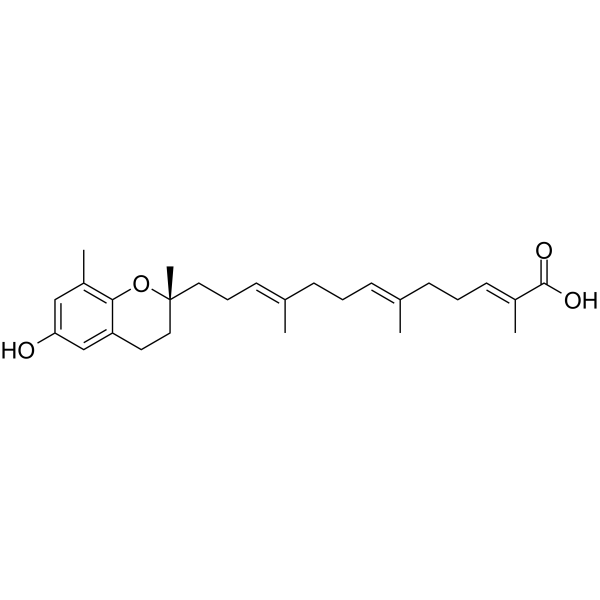
Garcinoic acid
Garcinoic acid is an orally active anti-inflammatory agent that crosses the blood-brain barrier. Garcinoic acid also enhances efferocytosis and enzyme/receptor regulation, and selectively inhibits human COX-2, porcine α-amylase, Saccharomyces cerevisiae α-glucosidase and human DNA polymerase β (IC50=11 μM), as well as activates human PXR. Garcinoic acid enhances macrophage efferocytosis via receptors such as MerTK and LRP-1, and promotes the production of pro-resolving lipid mediators. Garcinoic acid inhibits NF-κB activation and pro-inflammatory cytokine secretion, interferes with Aβ aggregation, downregulates NLRP3 inflammasome activity, and binds to targets including CD44 and EGFR to inhibit leukemia cell proliferation. The pharmacological activities of Garcinoic acid, such as antioxidant, anti-inflammatory and lipid metabolism-regulating effects, are widely used in studies related to various diseases including atherosclerosis, Alzheimer's disease, type 2 diabetes, inflammatory bowel disease and viral pneumonia.
For research use only. We do not sell to patients.
- CAS No.: 91893-83-3
- Formula: C27H38O4
- Molecular Weight:426.59
-
Storage:
Please store the product under the recommended conditions in the Certificate of Analysis.
All DNA/RNA Synthesis Isoforms
More
Biological Activity
Description
IC50 & Target
|
hCOX-2 |
In Vitro
Garcinoic acid (2.5 μM; 24 h, 14-24 h) inhibits LPS-induced Cox2 and iNos protein expression in murine RAW264.7 macrophages, reducing iNos levels to 17% of LPS control at 2.5 μM[1].
Garcinoic acid (0.02-100 μM; 72 h incubation with cells, following 24 h initial cell culture) inhibits proliferation of KASUMI-1, HL-60, THP-1, RS4;11, and MOLT-4 leukemia cell lines with IC50 values ranging from 18.04 μM to 49.01 μM, showing the strongest activity against HL-60 cells[2].
Garcinoic acid (0.25-5 μM; 24 h) does not reduce the viability of mouse bone marrow-derived macrophages[3].
Exogenous Lipoxin A4 (100 nM; 2 h) or Resolvin E1 (10 nM; 30 min) restores garcinoic acid (1 μM; 24 h)-enhanced efferocytosis in mouse bone marrow-derived macrophages with Nrf2 silenced or HO-1 inhibited[3].
Garcinoic acid (1-50 μM; 24 h) dose-dependently upregulates PXR, CYP3A4, and MDR1 expression in HepG2 cells, with the response dependent on functional PXR[5].
Garcinoic acid (25 μM; 24 h pre-incubation, 24 h incubation with Aβ(1-42)) restores and stimulates ApoE efflux in mouse cortical astrocytes via a PPARγ-independent mechanism, up-regulates PPARγ and PXR expression, and increases expression of the PXR-regulated genes CYP4F2 and MDR1 in these cells at 25 μM[6].
Garcinoic acid (5 μM; 6 h) suppresses cleavage of GSDMD into the pyroptosis-inducing NT-GSDMD fragment in LPS/ATP-stimulated murine J774A.1 macrophages after 6 hours of total incubation[8].
MedChemExpress (MCE) has not independently confirmed the accuracy of these methods. They are for reference only. Further protocols information, click here.
-
Cell Line:murine RAW264.7 macrophages
-
Concentration:2.5 μM
-
Incubation Time:24 h (pre-incubated); 14 h (co-incubated with LPS for Cox2); 24 h (co-incubated with LPS for iNos)
-
Result:Did not detectably modulate basal Cox2 or iNos protein levels.
Reduced Cox2 protein expression to 67% relative to LPS-only controls.
Reduced iNos protein expression to 17% relative to LPS-only controls.
-
Cell Line:KASUMI-1, HL-60, THP-1, RS4;11, MOLT-4
-
Concentration:0.02-100 μM
-
Incubation Time:72 h (incubation with cells, following 24 h initial cell culture)
-
Result:Inhibited proliferation of KASUMI-1 cells, with an IC50 value of 30.81 μM.
Inhibited proliferation of HL-60 cells, with an IC50 value of 18.04 μM.
Inhibited proliferation of THP-1 cells, with an IC50 value of 49.01 μM.
Inhibited proliferation of RS4;11 cells, with an IC50 value of 42.87 μM.
Inhibited proliferation of MOLT-4 cells, with an IC50 value of 26.73 μM.
-
Cell Line:mouse bone marrow-derived macrophages (BMDMs)
-
Concentration:0.25-5 μM
-
Incubation Time:24 h
-
Result:Showed no cytotoxic effect on BMDMs, with cell viability remaining near 100% across all groups.
In Vivo
Garcinoic acid (1 mg/kg; i.p.; daily; from 7 days pre-DSS exposure until study end) attenuates DSS (HY-116282C)-induced colitis in male C57BL/6 mice by restoring efferocytosis, increasing specialized pro-resolving lipid mediator levels, and activating Nrf2 signaling[3].
Garcinoic acid (5-100 mg; p.o.; single bolus) acts as a selective PXR agonist in mice, dose-dependently increasing PXR, CYP3A11, and MDR1 expression in liver and intestinal tissue at well-tolerated doses ≤25 mg, while higher doses cause mortality[5].
Garcinoic acid (5-25 mg; p.o.; single dose) dose-dependently up-regulates PPARγ, ApoE, and PXR expression in the brain of healthy C57Bl/6 mice, with significant effects observed at doses ≥5 mg[6].
Garcinoic acid (200 mg/kg; p.o.; daily; 10 days) significantly reduces amyloid β plaque number and deposition in the brain of TgCRND8 Alzheimer's disease model mice, while up-regulating ApoE and MDR1 expression[6].
MedChemExpress (MCE) has not independently confirmed the accuracy of these methods. They are for reference only.
-
Animal Model:C57Bl/6 background (8-week-old male, 25-28 g, apolipoprotein E knockout, high fat diet-fed for 8 weeks)[1]
-
Dosage:1 mg/kg
-
Administration:i.p.; weekly; 8 weeks
-
Result:Decreased intra-plaque nitrotyrosine levels to 50% of control levels.
Downregulated blood CD4 positive T cells by 14.8%.
Increased blood natural killer (NK) cells from 3.2% to 5.6%.
Increased blood natural killer T (NKT) cells from 0.2% to 0.6%.
Upregulated spleen NK cells from 2.0% to 2.8%.
Showed no significant changes in body/organ weight, plasma lipid profile, total lesion size, necrotic core area, lipid content, collagen content, VCAM-1 levels, CD68 levels, MCP-1 levels, IL1β levels, plasma NO levels, or plasma prostanoid levels.
-
Animal Model:C57BL/6 (5-week-old male; DSS-induced colitis)[3]
-
Dosage:1 mg/kg
-
Administration:i.p.; daily; from 7 days pre-DSS exposure until study end
-
Result:Prevented DSS-induced reduction in colon length.
Increased body weight compared to DSS-only mice.
Reduced Disease Activity Index (DAI) scores compared to DSS-only mice.
Restored colonic tissue architecture, reducing epithelial degeneration, inflammatory cell infiltration, and crypt loss compared to DSS-only mice.
Increased the ratio of macrophage-associated apoptotic cells to free apoptotic cells in colon tissue, restoring efferocytosis to control levels.
Increased plasma levels of Lipoxin A4, Resolvin D2, and Resolvin E1 compared to DSS-only mice.
Increased colon tissue levels of Lipoxin A4, Resolvin D2, and Resolvin E1 compared to DSS-only mice.
Increased colon tissue protein levels of Nrf2 and NAD(P)H:quinone oxidoreductase (NQO1) compared to control mice.
-
Animal Model:TgCRND8 transgenic (5-month-old, 14-15-month-old)[6]
-
Dosage:200 mg/kg
-
Administration:p.o.; daily; 10 days
-
Result:Reduced 10-50 μm2 amyloid β plaques from ~400 to ~300, 50-200 μm2 plaques from ~100 to ~50, and 200-400 μm2 plaques from ~50 to ~10 (all significant vs. untreated Tg mice, p < 0.05 or p < 0.005) in 14-15-month-old Tg mice.
Confirmed decreased amyloid β deposition in both 5-month-old and 14-15-month-old Tg mice via Congo Red staining.
Increased ApoE and MDR1 expression in the hippocampus of Tg mice significantly.
Reached concentrations between 0.8 and 10.3 ppm (ng/mg of proteins) in the parietal cortex and hippocampus of treated Tg mice.
Chemical Information
-
CAS No. 91893-83-3
-
Molecular Weight 426.59
-
Formula C27H38O4
-
SMILES
CC1=C2C(CC[C@](C)(O2)CC/C=C(C)/CC/C=C(C)/CC/C=C(C)/C(O)=O)=CC(O)=C1
-
Structure Classification
-
Initial Source
-
Shipping
Room temperature in continental US; may vary elsewhere.
-
Storage
Please store the product under the recommended conditions in the Certificate of Analysis.
Protocols
-
BrdU Incorporation Assay
Bromodeoxyuridine (BrdU) incorporation assay is based on the principle that BrdU, a thymidine analog, is incorporated into newly synthesized DNA during the S phase of the cell cycle, thereby serving as a marker of DNA replication and cellular proliferation. Incorporated BrdU can be detected using anti-BrdU antibodies following DNA denaturation, enabling visualization or quantification of proliferating cells through immunochemical detection methods such as immunofluorescence or immunohistochemistry.
-
Somatic Cell Culture
A method of simulating the in vivo environment in vitro to maintain the cell growth, differentation and main functions.
-
CFSE Dye Dilution Proliferation Assay
The CFSE (carboxyfluorescein diacetate succinimidyl ester) dye dilution proliferation assay is based on the covalent labeling of intracellular proteins by a cell-permeant fluorescent dye that becomes fluorescent upon intracellular ester cleavage and then is stably retained within cells. As labeled cells divide, the dye is partitioned equally between daughter cells, resulting in a stepwise halving of fluorescence intensity that can be quantified by flow cytometry to determine the number of cell divisions undergone by each cell population. This fluorescence dilution approach enables quantitative tracking of lymphocyte proliferation at the single-cell level over multiple rounds of division. CFSE-based proliferation analysis has been widely applied to measure antigen-driven lymphocyte expansion in vitro, where discrete fluorescence peaks correspond to successive cell divisions and allow reconstruction of proliferative history within heterogeneous populations.
-
Dye-dilution cell tracking and proliferation staining
Dye-dilution cell tracking assays quantify cell proliferation by covalently labeling intracellular proteins with a stable fluorescent dye that is equally partitioned between daughter cells during mitosis, resulting in stepwise halving of fluorescence intensity with each cell division as measured by flow cytometry histograms. Carboxyfluorescein diacetate succinimidyl ester (CFSE) is a prototypical dye that diffuses into cells, is enzymatically converted into a fluorescent compound, and then covalently binds intracellular amine groups, producing long-lived fluorescence suitable for tracking multiple rounds of division in vitro and in vivo. Successive generations of dividing cells form discrete peaks of decreasing fluorescence intensity, enabling estimation of proliferation history, precursor frequency, and division index within heterogeneous populations. Alternative dyes such as CellTrace Violet (CTV) and far-red membrane dyes (e. g. , PKH26) follow the same dilution principle but differ
-
CCK-8/WST-8 Cell Proliferation Assay
The CCK-8/WST-8 assay is based on the reduction of the water-soluble tetrazolium salt WST-8 to a water-soluble formazan product by cellular dehydrogenases in metabolically active cells, where the generated formazan amount is proportional to the number of living cells and is quantified by measuring absorbance in the visible range, providing a colorimetric readout for cell viability and proliferation assessment. This class of tetrazolium-based assays improves upon earlier MTT-based systems by producing a water-soluble formazan, eliminating the need for organic solubilization steps and enabling direct spectrophotometric measurement in culture medium.
-
Protocol for Cell Cycle
Cell-cycle analysis by flow cytometry measures DNA content in single cells to estimate the fraction of cells in G0/G1, S, and G2/M phases. Propidium iodide intercalates into DNA, and after RNA removal with RNase, fluorescence intensity reflects cellular DNA content: 2N cells are assigned to G0/G1, cells between 2N and 4N to S phase, and 4N cells to G2/M. DNA-content analysis alone cannot reliably separate G0 from G1 or G2 from M. Ki-67 can distinguish quiescent G0 cells from cycling cells, EdU or BrdU incorporation marks active DNA synthesis in S phase, and phospho-histone H3 staining identifies mitotic cells within the 4N population.
-
Research Protocol for Cardiovascular Diseases
Cardiovascular disease can be modeled as maladaptive cardiac remodeling, where ischemic injury or pressure overload activates inflammatory signaling, fibroblast activation, extracellular-matrix deposition, cardiomyocyte hypertrophy, vascular remodeling, and progressive ventricular dysfunction. The TGF-β/SMAD axis is a central profibrotic pathway after myocardial injury and pressure overload, while innate immune and cytokine pathways regulate leukocyte recruitment, scar formation, and adverse remodeling. Key unresolved questions include which inflammatory signals are reparative versus harmful, when fibrosis is protective versus maladaptive, and whether pathway inhibition improves function without weakening necessary infarct healing or compensatory remodeling.
-
Research Protocol for Inflammation-related Diseases
The NLRP3 inflammasome is a cytosolic innate immune signaling platform that integrates priming signals and danger-signal activation to promote caspase-1 activation, maturation of IL-1β and IL-18, and gasdermin D-mediated pyroptotic cell death. The core experimental logic is to determine whether inflammatory disease phenotypes are driven by increased NLRP3 expression, ASC-containing inflammasome assembly, caspase-1 cleavage, GSDMD cleavage, and extracellular release of IL-1β/IL-18 rather than by nonspecific cell injury alone. The pathway is strongly linked to inflammation-related disease phenotypes because monosodium urate crystals activate NALP3/NLRP3 inflammasome signaling in gout-like crystal inflammation, cholesterol crystals activate NLRP3 inflammasomes in atherogenesis models, and DSS-induced intestinal inflammation has been reported to involve NLRP3 inflammasome activity. However, experimental colitis studies also show context-dependent protective effects of NLRP3 inflammasome co
-
Cell Counting-Based Growth Curve Assay
Cell counting-based growth curve assays quantify cell proliferation by directly measuring changes in viable cell number over time using manual or automated counting methods such as hemocytometer-based counting or instrument-assisted cell enumeration, enabling construction of growth curves that reflect population expansion dynamics in response to culture conditions. A widely used approach is trypan blue exclusion with hemocytometer counting, where membrane-compromised (non-viable) cells take up the dye, allowing discrimination between viable and non-viable cells while simultaneously enabling total cell number quantification. Repeated sampling across time points allows estimation of proliferation rate, growth phases, and comparative growth kinetics between experimental conditions.
-
Colony Formation (Clonogenic) Assay
The clonogenic (colony formation) assay measures the ability of a single cell to retain reproductive viability and form a macroscopic colony, typically defined as a cluster derived from one progenitor cell after a defined growth period. This assay is widely used to evaluate cell survival after exposure to ionizing radiation or cytotoxic treatments and is considered a standard method in radiation biology for generating dose-response relationships of reproductive cell death. Colony formation reflects long-term proliferative capacity rather than short-term metabolic activity, and survival is quantified by comparing treated versus untreated conditions based on colony number and derived survival fractions.
-
Research Protocol for Metabolic Diseases
AMP-activated protein kinase, AMPK, is a conserved cellular energy sensor that responds to reduced cellular energy status and coordinates metabolism by increasing ATP-generating catabolic pathways while suppressing ATP-consuming anabolic processes. In metabolic disease research, the AMPK pathway is experimentally relevant because it regulates hepatic lipid synthesis, fatty acid oxidation, glucose production, skeletal-muscle glucose disposal, mTORC1-linked biosynthesis, autophagy, mitochondrial homeostasis, and whole-body energy balance. The central pathway logic is that energy stress, metformin, exercise-like stimulation, or direct AMPK activators increase AMPKα Thr172 phosphorylation and downstream substrate phosphorylation, including ACC and RAPTOR. Phosphorylation of ACC suppresses lipogenesis and supports fatty acid oxidation, whereas phosphorylation of RAPTOR suppresses mTORC1 signaling and links cellular energy status to growth and protein synthesis control. The pathway is linked
-
Alzheimer’s Disease Modeling
Alzheimer’s Disease (AD) is a neurodegenerative disorder characterized by a progressive decline in cognitive functions and loss of specific types of neurons and synapses. Alzheimer's symptoms can be simulated in mice by injecting drugs (such as Aβ) or genetically modified.
-
Cell Viability Determination by MTT Colorimetric Assay
The following protocol uses the MTT colorimetric assay as a classic literature-established method for assessing cell viability/metabolic activity in cultured mammalian cells. MTT[3-(4,5-dimethylthiazol-2-yl)-2,5-diphenyltetrazolium bromide] is reduced by metabolically active cells to a colored formazan product; the amount of formazan is quantified spectrophotometrically and provides an indirect measure of metabolically active viable cells. Importantly, MTT reduction reflects cellular oxidoreductase/metabolic activity rather than an absolute direct count of living cells, so changes in cellular metabolism can alter the signal independently of cell number.
-
EdU Incorporation Assay (Click Chemistry-Based DNA Synthesis Measurement)
The EdU incorporation assay measures DNA synthesis by adding the thymidine analog 5-ethynyl-2′-deoxyuridine to cells or tissues, where it is incorporated into newly synthesized DNA during S phase. Incorporated EdU is detected by copper-catalyzed azide-alkyne cycloaddition, in which a fluorescent azide covalently reacts with the ethynyl group on EdU, allowing S-phase cells to be detected by fluorescence microscopy, flow cytometry, or high-content imaging. EdU detection does not require DNA denaturation or anti-BrdU antibody access, which preserves sample structure and improves compatibility with immunostaining and multiparameter cytometry compared with BrdU-based detection. EdU can be cytotoxic in a cell-type- and exposure-dependent manner, so pulse duration, concentration, and continuous-labeling designs should be validated for each cell type.
-
Ki-67 Immunostaining Proliferation Assay
Ki-67 immunostaining measures the growth fraction of a cell population by detecting Ki-67, a nuclear antigen present in proliferating cells and absent in quiescent G0 cells. The readout is the percentage of Ki-67-positive nuclei among total counted cells, commonly called the Ki-67 labeling index or proliferation index.
-
Cell Cytotoxicity Assay
Cytotoxicity assays are usually based on the assessment of cell membrane damage, which can also be indirectly detected by measuring cell viability. Detection methods include MTT assay, CKK-8 assay, LDH assay and ATP assay, etc.
-
PCNA Immunodetection Proliferation Assay
PCNA immunodetection measures proliferative activity by detecting proliferating cell nuclear antigen, a nuclear protein associated with DNA polymerase δ function and DNA replication. The assay readout is the proportion of PCNA-positive nuclei among total counted cells, but PCNA labeling is not identical to BrdU labeling because PCNA can mark late G1/early S-associated replication competence and may persist beyond active DNA synthesis depending on fixation and extraction conditions.
-
Protocol for Cell Counting and Cell Density Analysis
Cell counting and cell-density analysis estimate the number of cells in a known volume or field area. Manual hemocytometer counting uses a chamber of defined geometry to convert counted cells into cells/mL, while automated counters and image-analysis workflows detect cell objects from optical, brightfield, fluorescence, impedance, or digital-image features. Trypan blue viability counting is based on dye exclusion: viable cells with intact membranes exclude dye, while non-viable cells with compromised membranes stain blue. The readout is total cell density, viable-cell density, dead-cell density, and percent viability. Cell density can also be estimated from microscopy images by counting objects per image area, from flow cytometry using calibrated volume or reference particles, or from in situ microscopy in bioreactors after calibration against reference methods such as hemocytometer or flow cytometry.
-
DSS-Induced Colitis
Dextran sulfate sodium (DSS)-induced colitis is generated by administering DSS in mouse drinking water, producing epithelial injury, barrier disruption, weight loss, diarrhea, fecal blood, colon shortening, histologic mucosal damage, and inflammatory mediator changes; the model is mainly used to study acute or chronic intestinal inflammation resembling selected features of ulcerative colitis. DSS injury is interpreted through clinical and tissue readouts rather than a single molecular endpoint: daily body weight, stool consistency, and bleeding are combined into a disease activity index, while colon length, histology, cytokines, myeloperoxidase activity, intestinal permeability, and tight-junction markers provide complementary measures of inflammation and barrier damage.
-
TNBS-Induced Colitis
TNBS-induced colitis is produced by intrarectal delivery of 2,4,6-trinitrobenzene sulfonic acid in ethanol, where ethanol disrupts the mucosal barrier and TNBS haptenates colonic proteins, generating immune-mediated colonic inflammation with weight loss, diarrhea, ulceration, transmural injury, inflammatory-cell infiltration, and cytokine responses. The model is used as an experimental intestinal inflammation model with Crohn’s disease–like features, especially when Th1-type responses, IL-12–dependent inflammation, chronic relapsing inflammation, or fibrosis-related endpoints are studied.
-
MTT Cell Proliferation Assay
The MTT assay is a colorimetric endpoint assay for estimating viable cell number, cell growth, cytotoxicity, or cell activation in cultured mammalian cells. Living cells reduce the yellow tetrazolium salt MTT into purple/blue formazan, while dead cells do not generate the same signal; the resulting color can be quantified with a multiwell spectrophotometer. MTT reduction is commonly interpreted as a readout of metabolic activity that often correlates with viable cell number, but it should not be treated as a direct cell-counting method unless the assay is optimized for the cell type and experimental condition. Studies show that MTT reduction can involve mitochondrial and non-mitochondrial reducing systems, and formazan may accumulate in intracellular lipid droplets rather than simply marking mitochondria.
-
Pyroptosis Solutions
Pyroptosis is a lytic inflammatory cell-death pathway executed by gasdermin pores, most classically through inflammasome-mediated activation of caspase-1, cleavage of gasdermin D, membrane pore formation, LDH release, and secretion of IL-1β and IL-18. The canonical pathway is commonly modeled by priming cells with an inflammatory signal such as LPS to induce pro-IL-1β and inflammasome components, followed by an activation signal such as ATP or nigericin to activate NLRP3, ASC speck formation, caspase-1 cleavage, GSDMD cleavage, cytokine release, and pyroptotic membrane rupture. The non-canonical pathway is triggered when cytosolic LPS activates mouse caspase-11 or human caspase-4/5, leading to GSDMD cleavage and pyroptosis, and this can secondarily activate NLRP3-dependent IL-1β release. Pyroptosis is linked to inflammatory injury, infection, cancer, liver disease, ocular disease, placental inflammation, and other disease phenotypes, but unresolved questions include which gasdermin fam
Purity & Documentation
References
[1]. Wallert M, et al. The vitamin E derivative garcinoic acid from Garcinia kola nut seeds attenuates the inflammatory response. Redox Biol. 2019;24:101166. [Content Brief]
[3]. Isot I, et al. Garcinoic acid enhances inflammation resolution against colitis by activating Nrf2 dependent efferocytosis. Free Radic Biol Med. 2025;237:37-45. [Content Brief]
[4]. Olajide OA, et al. Garcinia kola and garcinoic acid suppress SARS-CoV-2 spike glycoprotein S1-induced hyper-inflammation in human PBMCs through inhibition of NF-κB activation. Phytother Res. 2021;35(12):6963-6973. [Content Brief]
[5]. Bartolini D, et al. Garcinoic Acid Is a Natural and Selective Agonist of Pregnane X Receptor. J Med Chem. 2020;63(7):3701-3712. [Content Brief]
[6]. Marinelli R, et al. Garcinoic acid prevents β-amyloid (Aβ) deposition in the mouse brain. J Biol Chem. 2020;295(33):11866-11876. [Content Brief]
[7]. Sabbatini S, et al. Modulation of C. albicans-Induced Immune Response in Vaginal Epithelial Cells by Garcinoic Acid. Microorganisms. 2024 Nov 29;12(12):2455. [Content Brief]
[8]. Börmel L, et al. The Vitamin E Derivative Garcinoic Acid Suppresses NLRP3 Inflammasome Activation and Pyroptosis in Murine Macrophages. Inflammation. 2025;48(5):3340-3352. [Content Brief]
[9]. Gujarathi S, et al. A Facile Semisynthesis and Evaluation of Garcinoic Acid and Its Analogs for the Inhibition of Human DNA Polymerase β. Molecules. 2020;25(24):5847. Published 2020 Dec 11. [Content Brief]
[10]. Terashima K, et al. Powerful antioxidative agents based on garcinoic acid from Garcinia kola. Bioorg Med Chem. 2002;10(5):1619-1625. [Content Brief]
Calculators
Concentration (start) × Volume (start) = Concentration (final) × Volume (final)