



HDAC-IN-48
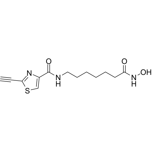
HDAC-IN-48 is a potent HDAC inhibitor. HDAC-IN-48 is a hybrid molecule with great cytotoxic profile (GI50~20 nM). HDAC-IN-48 consists of harmacophores of SAHA and CETZOLE molecules. HDAC-IN-48 induces ferroptosis and inhibits HDAC proteins. HDAC-IN-48 is a click chemistry reagent, it contains an Alkyne group and can undergo copper-catalyzed azide-alkyne cycloaddition (CuAAc) with molecules containing Azide groups.
Nur für Forschungszwecke. Wir verkaufen nicht an Patienten.
- CAS. Nr.: 3031411-05-6
- Formel: C13H17N3O3S
- Molecular Weight:295.36
-
Speicherung:
Please store the product under the recommended conditions in the Certificate of Analysis.

To place orders, for customer services and technical support, please contact: MedChemExpress USA
Tel: 609-228-6898 E-mail: [email protected] [email protected]


-
 Biologische Aktivität
-
Chemical Information
- Protokoll
- Reinheit & Dokumentation
- Verweise
-
 Help & FAQs




Biologische Aktivität
Beschreibung
Cellular Effect
|
Cell Line

|
Type | Value | Description | References |
|---|---|---|---|---|
| HCT-116 | IC50 |
0.61 μM
Compound: HY-1
|
Cytotoxicity against human HCT-116 cells assessed as reduction in cell viability incubated for 30 mins by methylene blue staining based analysis
Cytotoxicity against human HCT-116 cells assessed as reduction in cell viability incubated for 30 mins by methylene blue staining based analysis
|
[PMID: 36306372] |
| NCI-H23 | GI50 |
0.78 μM
Compound: HY-1
|
Cytotoxicity against human NCI-H23 cells assessed as cell growth inhibition at 10 uM incubated for 24 hrs by MTT assay
Cytotoxicity against human NCI-H23 cells assessed as cell growth inhibition at 10 uM incubated for 24 hrs by MTT assay
|
[PMID: 36306372] |
| NCI-H522 | IC50 |
0.5 μM
Compound: HY-1
|
Cytotoxicity against human NCI-H522 cells assessed as reduction in cell viability incubated for 30 mins by methylene blue staining based analysis
Cytotoxicity against human NCI-H522 cells assessed as reduction in cell viability incubated for 30 mins by methylene blue staining based analysis
|
[PMID: 36306372] |
| RPMI-8226 | GI50 |
0.35 μM
Compound: HY-1
|
Cytotoxicity against human RPMI-8226 cells assessed as cell growth inhibition incubated for 24 hrs by MTT assay
Cytotoxicity against human RPMI-8226 cells assessed as cell growth inhibition incubated for 24 hrs by MTT assay
|
[PMID: 36306372] |
| SF-539 | GI50 |
1 μM
Compound: HY-1
|
Cytotoxicity against human SF-539 cells assessed as cell growth inhibition incubated for 24 hrs by MTT assay
Cytotoxicity against human SF-539 cells assessed as cell growth inhibition incubated for 24 hrs by MTT assay
|
[PMID: 36306372] |
| WI-38 | IC50 |
8.37 μM
Compound: HY-1
|
Cytotoxicity against human WI-38 cells assessed as reduction in cell survival incubated for 30 mins by methylene blue staining based analysis
Cytotoxicity against human WI-38 cells assessed as reduction in cell survival incubated for 30 mins by methylene blue staining based analysis
|
[PMID: 36306372] |
In Vitro
HDAC-IN-48 (0-40 μM; 3 d) has superior antiproliferative activity on cancer cells (NCI-H522 and HCT-116) vs the normal cells (WI38 and RPE) with IC50s of 0.5 μM (NCI-H522), 0.61 μM (HCT-116), 8.37 μM (WI38, normal human lung fibroblasts), and 6.13 μM (RPE, retinal pigment epithelial cells), respectively[1].
HDAC-IN-48 (2.5 μM; 24 h) suppresses cell viability by inducing ferroptosis and HDAC inhibition[1].
HDAC-IN-48 (10 μM; 6 h) decreases the lipid peroxide level compared with SAHA[1].
HDAC-IN (0.58 μM, 1.16 μM, and 2.32 μM; 3 d) has no neurotoxic effects and (2.5, 5, and 10 μM; 3 d) leads to hyper acetylation of histones and tubulin[1].
Nondifferentiated PC-12 cells have stem-like properties, but when differentiated by a nerve growth factor, they demonstrate neuronal behavior. HDAC-IN-48 (0.58 μM, 1.16 μM, and 2.32 μM; 24 h) behaves as the HDAC control effect, shows few ferroptosis induction on both differentiated and undifferentiated PC-12 cells[1].
MedChemExpress (MCE) has not independently confirmed the accuracy of these methods. They are for reference only.
-
Cell Line:NCI-H522 cells
-
Concentration:2.5, 5, and 10 μM
-
Incubation Time:72 hours; at the corresponding concentrations in the presence of Liproxstatin-1 (0.25 μM)
-
Result:Led to hyperacetylation of histones and tubulin in a similar way to SAHA (pan-inhibitor).
-
Cell Line:NCI-H522, WI38, HCT-116, and RPE
-
Concentration:0-40 μM
-
Incubation Time:72 hours
-
Result:Showed selectivity among normal cells over cancer cells.
Inhibited cell survival with IC50s of 0.5 μM, 8.37 μM, 0.61 μM, and 6.13 μM.
-
Cell Line:NCI-H522 cells
-
Concentration:5 μM
-
Incubation Time:24 hours, 48 hours, and 72 hours
-
Result:Induced cell ferroptosis.


Chemical Information
-
CAS. Nr. 3031411-05-6
-
Molecular Weight 295.36
-
Formel C13H17N3O3S
-
SMILES
O=C(C1=CSC(C#C)=N1)NCCCCCCC(NO)=O
-
Versand
Room temperature in continental US; may vary elsewhere.
-
Speicherung
Please store the product under the recommended conditions in the Certificate of Analysis.


Reinheit & Dokumentation
Verweise

Calculators
Konzentration (Stammlösung) × Volumen (Stammlösung) = Konzentration (Ziellösung) × Volumen (Ziellösung)


HDAC-IN-48 Related Classifications
HY-151872 Related Classifications
 Powered by Bioz