



Hirsutenone

Based on 1 Customer Validation
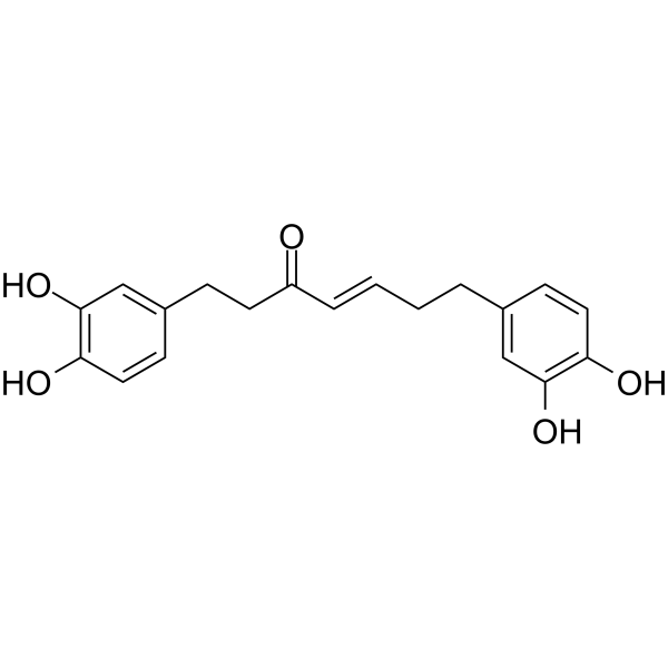
Hirsutenone is an active botanical diarylheptanoid present in Alnus species and exhibits many biological activities, including anti-inflammatory, anti-tumor promoting and anti-atopic dermatitis effects. Hirsutenone attenuates adipogenesis by binding directly to PI3K and ERK1 in a non-ATP competitive manner. Hirsutenone can be used for the study of obesity.
For research use only. We do not sell to patients.
- Purity : 99.74%
- CAS No.: 41137-87-5
- Formula: C19H20O5
- Molecular Weight:328.36
-
Storage:
4°C, protect from light
* In solvent : -80°C, 6 months; -20°C, 1 month (protect from light)

To place orders, for customer services and technical support, please contact: MedChemExpress USA
Tel: 609-228-6898 E-mail: [email protected] [email protected]


-
 Biological Activity
-
Chemical Information
- Protocols
- Purity & Documentation
- References
-
 Help & FAQs


-
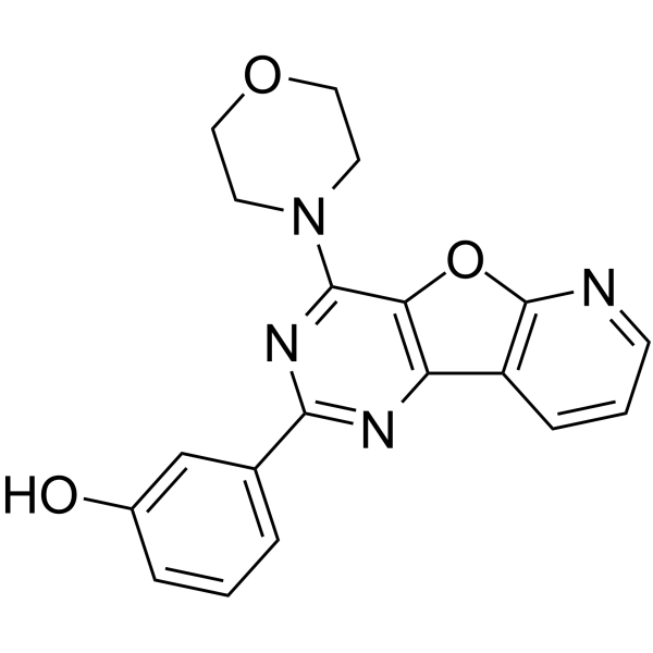
HY-10115
PI-103PI3K/mTOR Inhibitor -
HY-N0284
EsculetinPI3K Inhibitor -
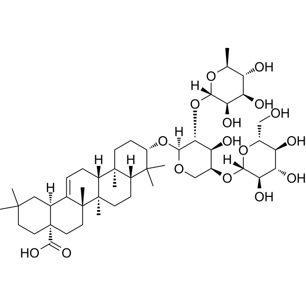
HY-N6950
Hederacolchiside A1Antischistosomal Agent -
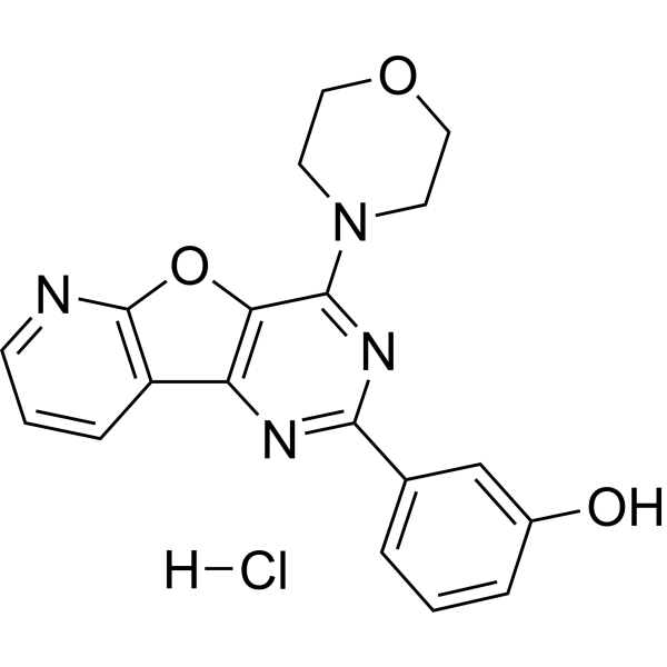
HY-10115A
PI-103 HydrochloridePI3K/mTOR Inhibitor -
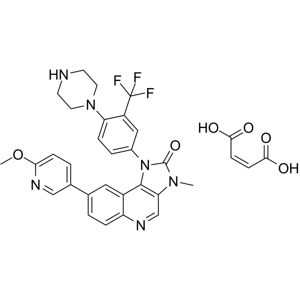
HY-13334
BGT226 maleate -
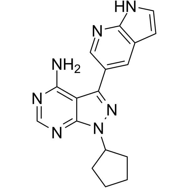
HY-10372
PP121Multi-targeted Kinase Inhibitor -
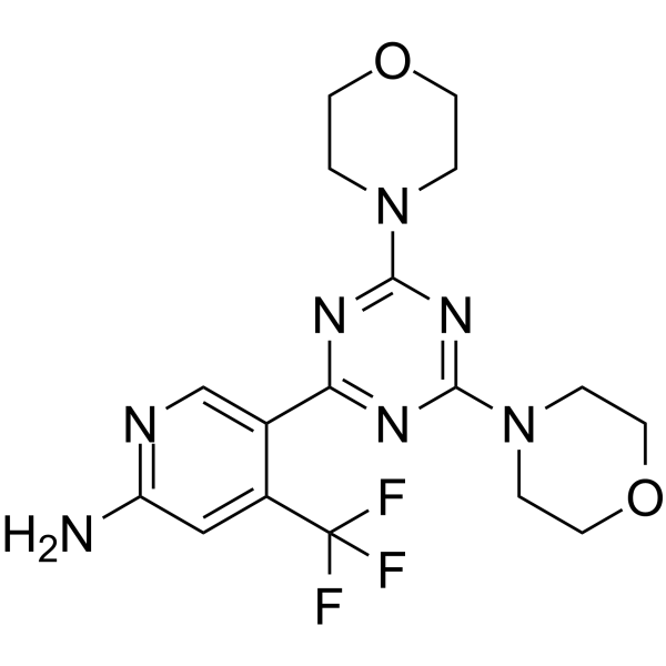
HY-12868
BimiralisibPI3K/mTOR Inhibitor -
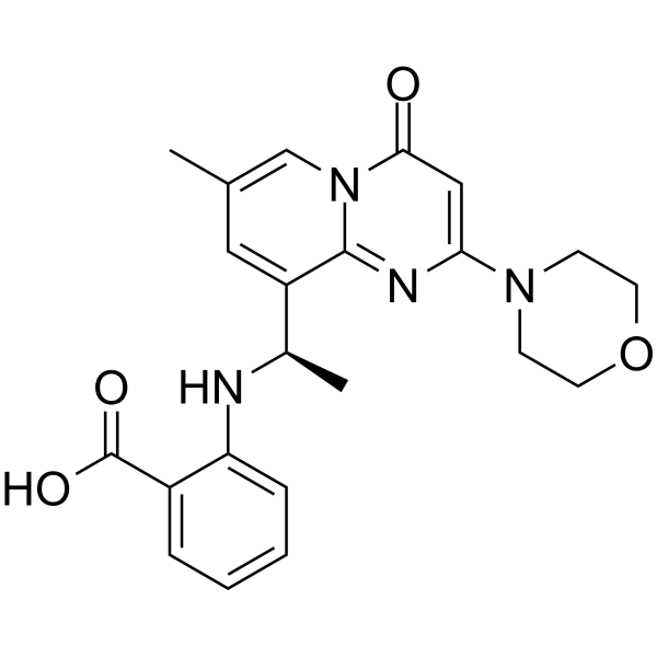
HY-10344
AZD 6482PI3Kβ Inhibitor -

HY-N0284R
Esculetin (Standard)PI3K Inhibitor -

HY-10115R
PI-103 (Standard)PI3K/mTOR Inhibitor -

HY-10372R
PP121 (Standard)Reference Standard -

HY-10115AR
PI-103 Hydrochloride (Standard)PI3K/mTOR Inhibitor


Biological Activity
Description
Cellular Effect
|
Cell Line

|
Type | Value | Description | References |
|---|---|---|---|---|
| HEK293 | EC50 |
220 μM
Compound: 42
|
Cytotoxicity against HEK293 cells after 16 hrs by alamar blue assay
Cytotoxicity against HEK293 cells after 16 hrs by alamar blue assay
|
[PMID: 20004045] |
| HSC-T6 | IC50 |
14 μM
Compound: 2
|
Antifibrotic activity against rat HSC-T6 cells assessed as inhibition of proliferation after 48 hrs by BrdU incorporation assay
Antifibrotic activity against rat HSC-T6 cells assessed as inhibition of proliferation after 48 hrs by BrdU incorporation assay
|
[PMID: 21504848] |
| RAW264.7 | IC50 |
>100 μM
Compound: 34
|
Antiinflammatory activity in mouse RAW264.7 cells assessed as inhibition of LPS-induced NF-kappaB activation incubated for 24 hrs by SEAP microplate reader analysis
Antiinflammatory activity in mouse RAW264.7 cells assessed as inhibition of LPS-induced NF-kappaB activation incubated for 24 hrs by SEAP microplate reader analysis
|
[PMID: 33422907] |
| RAW264.7 | IC50 |
>100 μM
Compound: 34
|
Antiinflammatory activity in mouse RAW264.7 cells assessed as inhibition of LPS-induced NO production incubated for 24 hrs by Griess reagent based analysis
Antiinflammatory activity in mouse RAW264.7 cells assessed as inhibition of LPS-induced NO production incubated for 24 hrs by Griess reagent based analysis
|
[PMID: 33422907] |
| RAW264.7 | IC50 |
>100 μM
Compound: 34
|
Antiinflammatory activity in mouse RAW264.7 cells assessed as inhibition of LPS-induced TNFalpha production preincubated for 1 hr followed by LPS stimulation and measured after 18 hrs by immunoassay
Antiinflammatory activity in mouse RAW264.7 cells assessed as inhibition of LPS-induced TNFalpha production preincubated for 1 hr followed by LPS stimulation and measured after 18 hrs by immunoassay
|
[PMID: 33422907] |
| RAW264.7 | IC50 |
0.8 μM
Compound: 34
|
Antiinflammatory activity in mouse RAW264.7 cells assessed as inhibition of LPS-induced NO production incubated for 20 hrs by Griess method
Antiinflammatory activity in mouse RAW264.7 cells assessed as inhibition of LPS-induced NO production incubated for 20 hrs by Griess method
|
[PMID: 33422907] |
| RAW264.7 | IC50 |
14.1 μM
Compound: 34
|
Antiinflammatory activity in mouse RAW264.7 cells assessed as inhibition of LPS-induced NO production incubated for 24 hrs by Griess method
Antiinflammatory activity in mouse RAW264.7 cells assessed as inhibition of LPS-induced NO production incubated for 24 hrs by Griess method
|
[PMID: 33422907] |
| RAW264.7 | IC50 |
25 μg/mL
Compound: 34
|
Antiinflammatory activity against mouse RAW264.7 cells assessed as inhibition of IFN-gamma and LPS-induced COX2 production preincubated for 2 hrs followed by IFN-gamma and LPS stimulation and measured after 16 hrs by spectrophotometric analysis
Antiinflammatory activity against mouse RAW264.7 cells assessed as inhibition of IFN-gamma and LPS-induced COX2 production preincubated for 2 hrs followed by IFN-gamma and LPS stimulation and measured after 16 hrs by spectrophotometric analysis
|
[PMID: 33422907] |
| RAW264.7 | IC50 |
27.2 μg/mL
Compound: 34
|
Antiinflammatory activity in mouse RAW264.7 cells assessed as inhibition of IFN-gamma and LPS-induced NO production incubated for 24 hrs by Griess assay
Antiinflammatory activity in mouse RAW264.7 cells assessed as inhibition of IFN-gamma and LPS-induced NO production incubated for 24 hrs by Griess assay
|
[PMID: 33422907] |
| RAW264.7 | IC50 |
39 μM
Compound: 34
|
Cytotoxicity against mouse RAW264.7 cells assessed as reduction in cell viability by MTT assay
Cytotoxicity against mouse RAW264.7 cells assessed as reduction in cell viability by MTT assay
|
[PMID: 33422907] |
| RAW264.7 | IC50 |
5 μM
Compound: 34
|
Antiinflammatory activity in mouse RAW264.7 cells assessed as inhibition of LPS-induced NO production preincubated for 0.5 hrs followed by LPS stimulation and measured after 24 hrs by Griess reagent based analysis
Antiinflammatory activity in mouse RAW264.7 cells assessed as inhibition of LPS-induced NO production preincubated for 0.5 hrs followed by LPS stimulation and measured after 24 hrs by Griess reagent based analysis
|
[PMID: 33422907] |
| RAW264.7 | IC50 |
5.8 μM
Compound: 34
|
Antiinflammatory activity in mouse RAW264.7 cells assessed as inhibition of LPS-induced NO production preincubated for 1 hrs followed by LPS stimulation and measured after 24 hrs by Griess reagent based analysis
Antiinflammatory activity in mouse RAW264.7 cells assessed as inhibition of LPS-induced NO production preincubated for 1 hrs followed by LPS stimulation and measured after 24 hrs by Griess reagent based analysis
|
[PMID: 33422907] |
In Vitro
Hirsutenone (0-100 μM; 48 hours) inhibits adipogenesis in 3T3-L1 preadipocytes and exhibits non-toxicity at 20-100 μM[1]. Hirsutenone (0-100 μM; 48 hours) attenuates MDI-induced lipid accumulation in 3T3-L1 preadipocytes in a dose-dependent manner. In particular, HST at 40 and 80 μM significantly reduces MDI-induced adipogenesis in 3T3-L1 preadipocytes[1].Hirsutenone (0-100 μM; 48 hours) reduces the protein expression levels of PPARg, C/EBPa, and FAS in a dose-dependent manner in 3T3-L1 preadipocytes[1].Hirsutenone (80 μM; 20-24 hours) suppresses the cell cycle entry to S and G2/M phases occurrs at 20 hours when compares with at 20 hours. At 24 h, more cells are arrested in G1 phase (53% of total cells) when compares with the MDI-induced group which contains the majority of cells (56% of total cells) in G2/M phase[1].
MedChemExpress (MCE) has not independently confirmed the accuracy of these methods. They are for reference only. Further protocols information, click here.


Chemical Information
-
CAS No. 41137-87-5
-
Appearance Solid
-
Molecular Weight 328.36
-
Formula C19H20O5
-
Color White to off-white
-
SMILES
OC1=C(O)C=CC(CC/C=C/C(CCC2=CC(O)=C(O)C=C2)=O)=C1
-
Structure Classification
-
Initial Source
-
Shipping
Room temperature in continental US; may vary elsewhere.
-
Storage
4°C, protect from light
* In solvent : -80°C, 6 months; -20°C, 1 month (protect from light)

Protocols
-
3T3-L1 preadipocyte-to-adipocyte differentiation
3T3-L1 preadipocytes are induced to differentiate after growth arrest using adipogenic media containing insulin, dexamethasone, and IBMX; differentiation is assessed by lipid-droplet accumulation, triglyceride increase, Oil Red O staining, and adipocyte-marker induction such as PPARγ and C/EBPα.
-
TPA/Croton Oil Ear Edema and Dermatitis
The TPA (12-O-tetradecanoylphorbol-13-acetate) and croton oil-induced mouse ear edema model is a well-established acute cutaneous inflammation system used to evaluate topical anti-inflammatory activity by measuring edema formation, neutrophil infiltration, vascular permeability, and cytokine-mediated skin responses in vivo. The inflammatory response is triggered by topical application of phorbol esters (TPA) or croton oil constituents, leading to rapid activation of protein kinase C signaling, leukocyte recruitment, and increased vascular permeability, which can be quantified by ear thickness, weight, dye extravasation, and biochemical markers such as myeloperoxidase (MPO) activity and pro-inflammatory mediators in ear tissue homogenates. This model is widely used for screening anti-inflammatory agents, where reductions in edema and inflammatory biomarkers reflect suppression of acute dermal inflammation and immune cell infiltration. Histological evaluation typically confirms epidermal
-
Research Protocol for Inflammation-related Diseases
The NLRP3 inflammasome is a cytosolic innate immune signaling platform that integrates priming signals and danger-signal activation to promote caspase-1 activation, maturation of IL-1β and IL-18, and gasdermin D-mediated pyroptotic cell death. The core experimental logic is to determine whether inflammatory disease phenotypes are driven by increased NLRP3 expression, ASC-containing inflammasome assembly, caspase-1 cleavage, GSDMD cleavage, and extracellular release of IL-1β/IL-18 rather than by nonspecific cell injury alone. The pathway is strongly linked to inflammation-related disease phenotypes because monosodium urate crystals activate NALP3/NLRP3 inflammasome signaling in gout-like crystal inflammation, cholesterol crystals activate NLRP3 inflammasomes in atherogenesis models, and DSS-induced intestinal inflammation has been reported to involve NLRP3 inflammasome activity. However, experimental colitis studies also show context-dependent protective effects of NLRP3 inflammasome co
-
Research Protocol for Metabolic Diseases
AMP-activated protein kinase, AMPK, is a conserved cellular energy sensor that responds to reduced cellular energy status and coordinates metabolism by increasing ATP-generating catabolic pathways while suppressing ATP-consuming anabolic processes. In metabolic disease research, the AMPK pathway is experimentally relevant because it regulates hepatic lipid synthesis, fatty acid oxidation, glucose production, skeletal-muscle glucose disposal, mTORC1-linked biosynthesis, autophagy, mitochondrial homeostasis, and whole-body energy balance. The central pathway logic is that energy stress, metformin, exercise-like stimulation, or direct AMPK activators increase AMPKα Thr172 phosphorylation and downstream substrate phosphorylation, including ACC and RAPTOR. Phosphorylation of ACC suppresses lipogenesis and supports fatty acid oxidation, whereas phosphorylation of RAPTOR suppresses mTORC1 signaling and links cellular energy status to growth and protein synthesis control. The pathway is linked
-
Lipid Droplets: Oil Red O/Sudan Dye Lipid Staining
Lipid droplets are intracellular organelles with a neutral-lipid core that stores triacylglycerols and sterol esters, and Oil Red O or Sudan dyes detect these hydrophobic lipid deposits by partitioning into retained lipids in fresh or frozen specimens. Oil Red O stains neutral triglycerides and lipids in frozen tissue sections or air-dried cytologic preparations, while Sudan Black B has also been used as a histochemical fat stain for lipid-rich tissue structures.


Purity & Documentation
-
Data Sheet (264 KB)
-
SDS (398 KB)
- English - EN (398 KB)
- Français - FR (398 KB)
- Deutsch - DE (398 KB)
- Norwegian - NO (398 KB)
- Español - ES (398 KB)
- Swedish - SV (398 KB)
- Italian - IT (398 KB)
- Korean - KR (398 KB)
- Portuguese - PT (398 KB)
-
Handling Instructions (2659 KB)
References

Calculators
Concentration (start) × Volume (start) = Concentration (final) × Volume (final)


Hirsutenone Related Classifications
HY-N4042 Related Classifications
 Powered by Bioz