



Hydroxycitric acid
Based on 1 publication(s) in Google Scholar
Hydroxycitric acid is an orally active, multi-target, multi-bioactive organic acid. activates Nrf2 and its downstream molecule GPX4, increases glutathione levels, and thereby inhibits ferroptosis. Hydroxycitric acid activates the Nrf2/Keap1 and ACLY/NF-κB signaling pathways, upregulates the activities of antioxidant enzymes such as superoxide dismutase, reduces MDA content, thereby alleviating oxidative stress and renal tubular epithelial cell apoptosis, and improves pulmonary vascular and right ventricular remodeling. Hydroxycitric acid activates both the AMPK and mTORC1/S6K pathways, triggers the unfolded protein response, arrests the cancer cell cycle, and induces DNA fragmentation.
For research use only. We do not sell to patients.
- Purity : 98.0%
- CAS No.: 6205-14-7
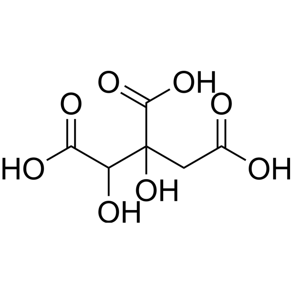
- Formula: C6H8O8
- Molecular Weight:208.12
-
Storage:Powder -20°C, 3 years ; In solvent -80°C, 6 months , -20°C, 1 month

To place orders, for customer services and technical support, please contact: MedChemExpress USA
Tel: 609-228-6898 E-mail: [email protected] [email protected]


-
 Biological Activity
-
Chemical Information
-
Solvent & Solubility
- Protocols
- Purity & Documentation
- References
-
 Help & FAQs


-
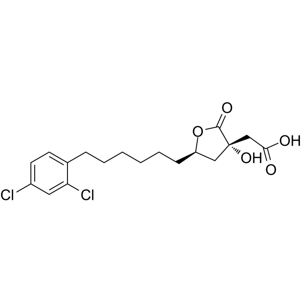
HY-16450
SB 204990ATP Citrate Lyase Inhibitor -
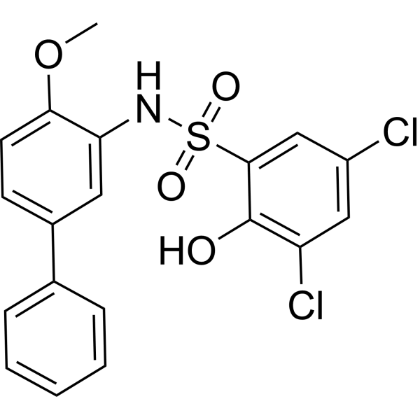
HY-16107
BMS-303141ATP Citrate Lyase Inhibitor -
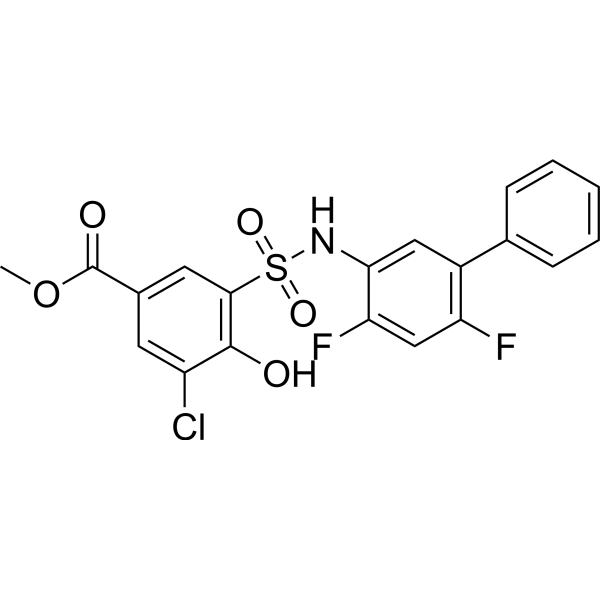
HY-127111
NDI-091143ATP Citrate Lyase Inhibitor -
HY-W009156
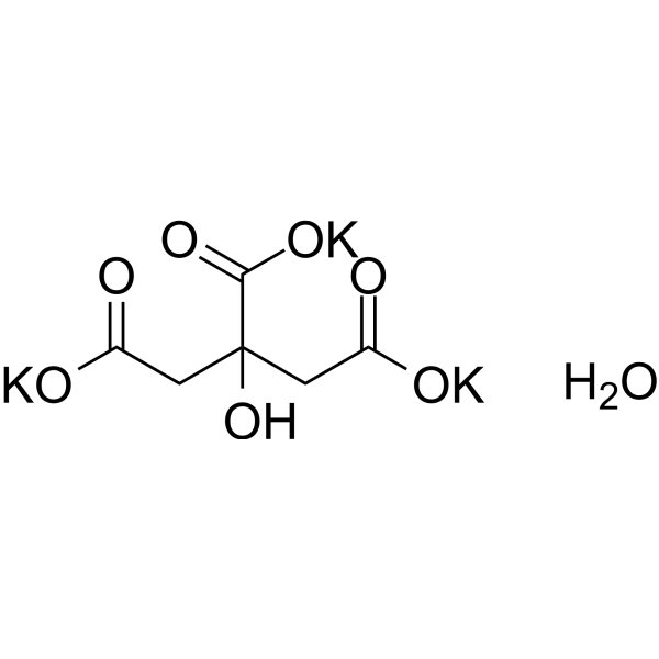
Citric acid tripotassium hydrateOrganic Acid -
HY-N7347
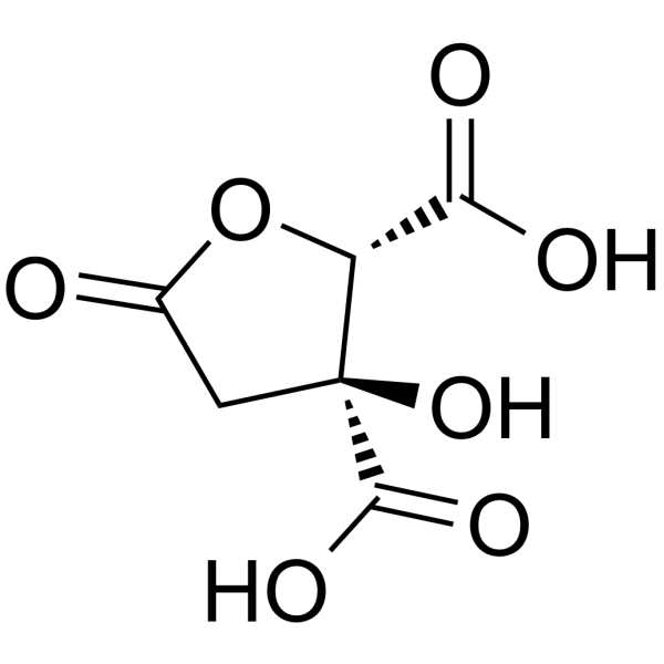
(-)-Hydroxycitric acid lactoneATP-citrate Lyase Inhibitor -
HY-W012946
2-Furoic acid -

HY-W009156R
Citric acid tripotassium hydrate (Standard)Standard of Citric acid tripotassium hydrate -

HY-N7347R
(-)-Hydroxycitric acid lactone (Standard)ATP-citrate Lyase Inhibitor -

HY-W012946R
2-Furoic acid (Standard)
Publications Citing Use of MedChemExpress (MCE) Hydroxycitric acid
MoreAll DNA/RNA Synthesis Isoforms
MoreAll AMPK Isoforms
More

Biological Activity
Description
In Vitro
Hydroxycitric acid (1-100 mM; 72 h) potently inhibits the proliferation of K562, MEG-01, CML-T1, KYO-1 and SKH-1 chronic myeloid leukemia (CML) cell lines, with IC50 values ranging from 3.73 to 11.34 mM; in contrast, it exerts no effect on normal mouse embryonic fibroblasts even at concentrations as high as 100 mM[1].
Hydroxycitric acid (0.5-5 mM) induces DNA fragmentation in K562 chronic myeloid leukemia (CML) cells[1].
Hydroxycitric acid (0.5-10 mM; 48-72 h) induces G2/M cell cycle arrest in K562 chronic myeloid leukemia (CML) cells[1].
Hydroxycitric acid (0.5-5 mM, 24-48 h) activates AMPK by increasing the phosphorylation level of T172 in K562, MEG-01, KYO-1 and SKH-1 chronic myeloid leukemia (CML) cells, without altering the total protein level of AMPK[1].
Hydroxycitric acid (0.5-1 mM; 24-48 h) activates both the AMPK and mTORC1 pathways, and triggers the unfolded protein response in K562 chronic myeloid leukemia (CML) cells by upregulating the phosphorylation level of eIF2α and the expression level of ATF4[1].
Hydroxycitric acid (5-10 mM) significantly inhibits hypoxia-induced proliferation of human pulmonary artery smooth muscle cells at concentrations of 5 mM and 10 mM[3].
Hydroxycitric acid (matched to hypoxic/normoxic exposure conditions) inhibits the proliferation of human pulmonary artery smooth muscle cells under both normoxic and hypoxic conditions, with a stronger inhibitory effect on cells in a hypoxic environment[3].
Hydroxycitric acid (24 h) significantly inhibits hypoxia-induced migration of human pulmonary artery smooth muscle cells[3].
Hydroxycitric acid (overnight) significantly reduces hypoxia-induced reactive oxygen species production in human pulmonary artery smooth muscle cells[3].
Hydroxycitric acid (combined with hypoxic exposure) significantly reduces the expression of hypoxia-inducible factor-1 (HIF-1) mRNA in hypoxia-induced human pulmonary artery smooth muscle cells[3].
MedChemExpress (MCE) has not independently confirmed the accuracy of these methods. They are for reference only.
-
Cell Line:K562 CML cells
-
Concentration:0.5, 5 and 10 mM
-
Incubation Time:48 h; 72 h
-
Result:Caused a significant accumulation of K562 cells in the G2/M phase of the cell cycle after 48 h treatment with 10 mM.
Caused significant accumulation of cells in the G2/M phase after 72 h treatment with 5 mM and 10 mM.
-
Cell Line:CML cell lines K562, MEG-01, KYO-1, SKH-1
-
Concentration:0.5 and 1 mM (K562); 0.5-5 mM (MEG-01, KYO-1, SKH-1)
-
Incubation Time:24 and 48 h (K562); 24 h (MEG-01, KYO-1, SKH-1)
-
Result:Significantly increased phosphorylation of AMPKα at T172 in all tested CML cell lines without altering total AMPKα protein levels.
Increased p-AMPKα levels by ~2-, ~6-, and ~4-fold in MEG-01 cells at 0.5 mM, 1 mM, and 5 mM doses, respectively.
Increased p-AMPKα levels by ~1.5-, ~1.2-, and ~1-fold in KYO-1 cells at 0.5 mM, 1 mM, and 5 mM doses, respectively.
Increased p-AMPKα levels by ~30-fold in SKH-1 cells at 5 mM dose.
-
Cell Line:K562 CML cells
-
Concentration:0.5 and 1 mM
-
Incubation Time:24 and 48 h
-
Result:Increased phosphorylation of AMPKα, ACC, p70 S6 kinase, S6 ribosomal protein, and eIF2α, and increased ATF4 protein levels.
Indicated concurrent activation of the AMPK and mTORC1 pathways, plus activation of the unfolded protein response.\nShowed AMPK and ACLY interacted in K562 cells via co-immunoprecipitation.
Reduced the total level of immunoprecipitated AMPK-ACLY complex, but the ratio of immunoprecipitated ACLY to AMPK remained unchanged compared to untreated controls, indicating no effect on the interaction between the two proteins.
-
Cell Line:K562 CML cells
-
Concentration:1 mM
-
Incubation Time:24 and 48 h
-
Result:Confirmed a fraction of AMPK co-eluted with ACLY, showing they are part of a common protein complex.
Had no major effect on the migration of this protein complex.
In Vivo
Hydroxycitric acid (250 mg/kg; i.p.; administered daily for 4 consecutive weeks) significantly reduces monocrotaline-induced pulmonary arterial hypertension in rats, decreases RVSP by 28.97% via anti-inflammatory and anti-fibrotic effects, and ameliorates right ventricular and pulmonary vascular remodeling[3].
hydroxycitrate (250 mg/kg; i.p.; administered daily for 4 consecutive weeks) significantly alleviates hypoxia-induced pulmonary arterial hypertension in rats, reduces RVSP by 17.5% via anti-inflammatory and antioxidant effects, and improves right ventricular and pulmonary vascular remodeling[3].
MedChemExpress (MCE) has not independently confirmed the accuracy of these methods. They are for reference only.
-
Animal Model:NOD.Cg-PrkdcscidIl2rgtm1WjI/SzJ (NSG) (male, 8-10 weeks old)[1]
-
Dosage:3 mg/kg
-
Administration:p.o.; daily; 25 days
-
Result:Reduced final average tumor volume.
-
Animal Model:Sprague-Dawley (male, 8-10 weeks old, hypobaric hypoxia-induced model)[3]
-
Dosage:250 mg/kg/day
-
Administration:i.p.; daily; 4 weeks
-
Result:Reduced RVSP from 42.30 mmHg to 34.9 mmHg.
Decreased right ventricular index from 32.62% to 26.11%.
Reduced right ventricular fibrotic area from 8.94% to 5.80%.
Attenuated upregulated collagen-1 mRNA levels.
Reduced pulmonary arteriole medium film layer thickness from 33.57 μm to 22.29 μm.
Decreased vascular wall area ratio from 0.71% to 0.45%.
Reduced serum IL-1β levels from 66.50 pg/mL to 55.80 pg/mL.
Increased cardiac SOD concentration from 7.31 U/mg to levels significantly higher than hypoxia-only controls.
Reduced cardiac H2O2 concentration from 8.76 mM to levels significantly lower than hypoxia-only controls.
-
Animal Model:Sprague-Dawley (male, 8-10 weeks old, monocrotaline-induced model)[3]
-
Dosage:250 mg/kg/day
-
Administration:i.p.; daily; 4 weeks
-
Result:Reduced right ventricular systolic pressure (RVSP) from 47.60 mmHg to 33.81 mmHg .
Decreased right ventricular index from 39.7% to 26.48%.
Decreased right ventricular weight/body weight ratio from 0.87 mg/g to 0.63 mg/g.
Reduced right ventricular fibrotic area from 15.40% to 8.82%.
Attenuated upregulated mRNA levels of α-SMA, collagen-1, and collagen-3.
Reduced pulmonary arteriole medium film layer thickness from 30.14 μm to 21.86 μm.
Decreased vascular wall area ratio from 0.65% to 0.51%.
Reduced serum levels of IL-6 from 121.20 pg/mL to 59.73 pg/mL, and TNF-α from 19.13 pg/mL to 11.82 pg/mL.
Suppressed elevated p-IκBα and p65 protein expressions in lung tissue.
- Detection of Apoptosis
- TUNEL staining for apoptotic DNA fragmentation
- Flow cytometric DNA-content cell-cycle staining
- Annexin V plus membrane-impermeant dye apoptosis staining
- ROS/oxidative-stress fluorescent staining
- BrdU Incorporation Assay
- Apoptosis Solutions
- Protocol for Cell Cycle
- Ferroptosis Solutions
- Research Protocol for Inflammation-related Diseases
Clinical Trial
| NCT Number | Sponsor | Condition | Start Date |
Phase

|
|---|---|---|---|---|
| NCT01329991 | Plexxikon| | 2011-05 | PHASE1 |


Chemical Information
-
CAS No. 6205-14-7
-
Appearance Solid
-
Molecular Weight 208.12
-
Formula C6H8O8
-
Color White to off-white
-
SMILES
OC(C(C(O)=O)O)(C(O)=O)CC(O)=O
-
Structure Classification
-
Initial Source
-
Shipping
Room temperature in continental US; may vary elsewhere.
-
Storage
Powder -20°C 3 years In solvent -80°C 6 months -20°C 1 month


Publications (1)
-
Journal Impact Factor
-
Most Recent
-
Polymers
High-Expansion Natural Composite Films for Controlled Delivery of Hydroxycitric Acid in Obesity Therapy. [Abstract]2025 Jun 18;17(12):1697. PMID: 40574227


Solvent & Solubility
In Vitro:
DMSO : 1.92 mg/mL (9.23 mM; ultrasonic and warming and adjust pH to 7 with 1 M HCL and heat to 60°C; Hygroscopic DMSO has a significant impact on the solubility of product, please use newly opened DMSO)
Please refer to the solubility information to select the appropriate solvent. Once prepared, please aliquot and store the solution to prevent product inactivation from repeated freeze-thaw cycles.
Storage method and period of stock solution: -80°C, 6 months; -20°C, 1 month. When stored at -80°C, please use it within 6 months. When stored at -20°C, please use it within 1 month.
Please refer to the solubility information to select the appropriate solvent. Once prepared, please aliquot and store the solution to prevent product inactivation from repeated freeze-thaw cycles.
Storage method and period of stock solution: -80°C, 6 months; -20°C, 1 month. When stored at -80°C, please use it within 6 months. When stored at -20°C, please use it within 1 month.
Concentration (start) × Volume (start) = Concentration (final) × Volume (final)


Purity & Documentation
-
Data Sheet (287 KB)
-
SDS (394 KB)
- English - EN (394 KB)
- Français - FR (394 KB)
- Deutsch - DE (394 KB)
- Norwegian - NO (394 KB)
- Español - ES (394 KB)
- Swedish - SV (394 KB)
- Italian - IT (394 KB)
- Korean - KR (394 KB)
- Portuguese - PT (394 KB)
-
Handling Instructions (2659 KB)
References
[1]. Verrelli D, et al. Hydroxycitric Acid Inhibits Chronic Myelogenous Leukemia Growth through Activation of AMPK and mTOR Pathway. Nutrients. 2022;14(13):2669. Published 2022 Jun 27. [Content Brief]
[2]. Yang D, et al. Hydroxycitric acid inhibits ferroptosis and ameliorates benign prostatic hyperplasia by upregulating the Nrf2/GPX4 pathway. World J Urol. 2025;43(1):318. Published 2025 May 20. [Content Brief]
[3]. Wang S, et al. Hydroxycitric Acid Tripotassium Hydrate Attenuates Monocrotaline and Hypoxia-Induced Pulmonary Hypertension in Rats. Int Heart J. 2024;65(2):318-328. [Content Brief]
[4]. Yang B, et al. Hydroxycitric acid prevents hyperoxaluric-induced nephrolithiasis and oxidative stress via activation of the Nrf2/Keap1 signaling pathway. Cell Cycle. 2023;22(17):1884-1899. [Content Brief]


Complete Stock Solution Preparation Table
Please refer to the solubility information to select the appropriate solvent. Once prepared, please aliquot and store the solution to prevent product inactivation from repeated freeze-thaw cycles.
Storage method and period of stock solution: -80°C, 6 months; -20°C, 1 month. When stored at -80°C, please use it within 6 months. When stored at -20°C, please use it within 1 month.
| Optional Solvent | Concentration Solvent Mass | 1 mg | 5 mg | 10 mg | 25 mg |
|---|---|---|---|---|---|
| DMSO | 1 mM | 4.8049 mL | 24.0246 mL | 48.0492 mL | 120.1230 mL |
| 5 mM | 0.9610 mL | 4.8049 mL | 9.6098 mL | 24.0246 mL |


Hydroxycitric acid Related Classifications
HY-N1437 Related Classifications
 Powered by Bioz